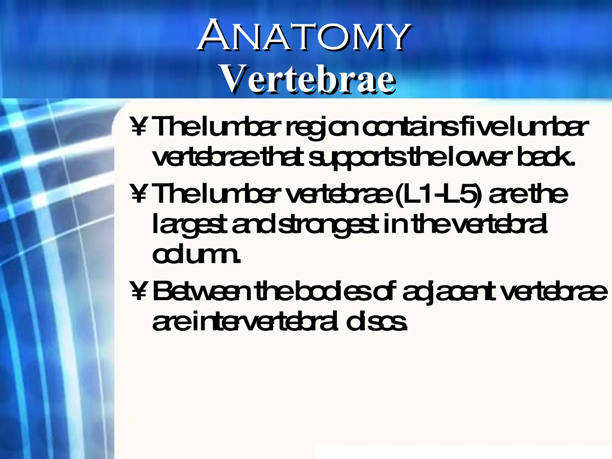
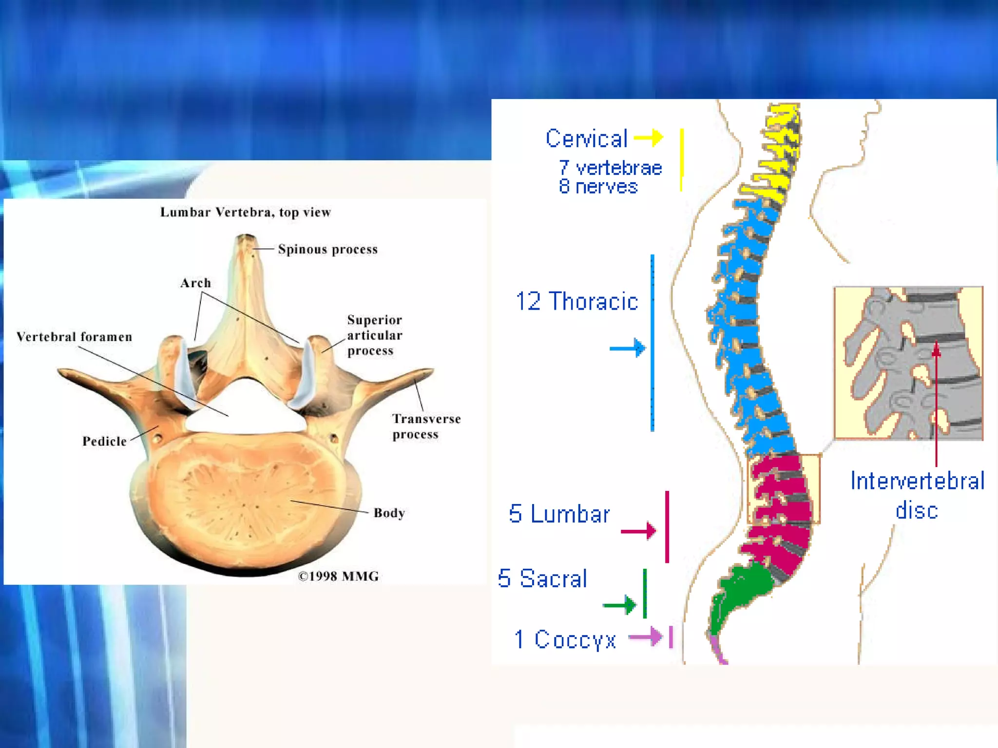
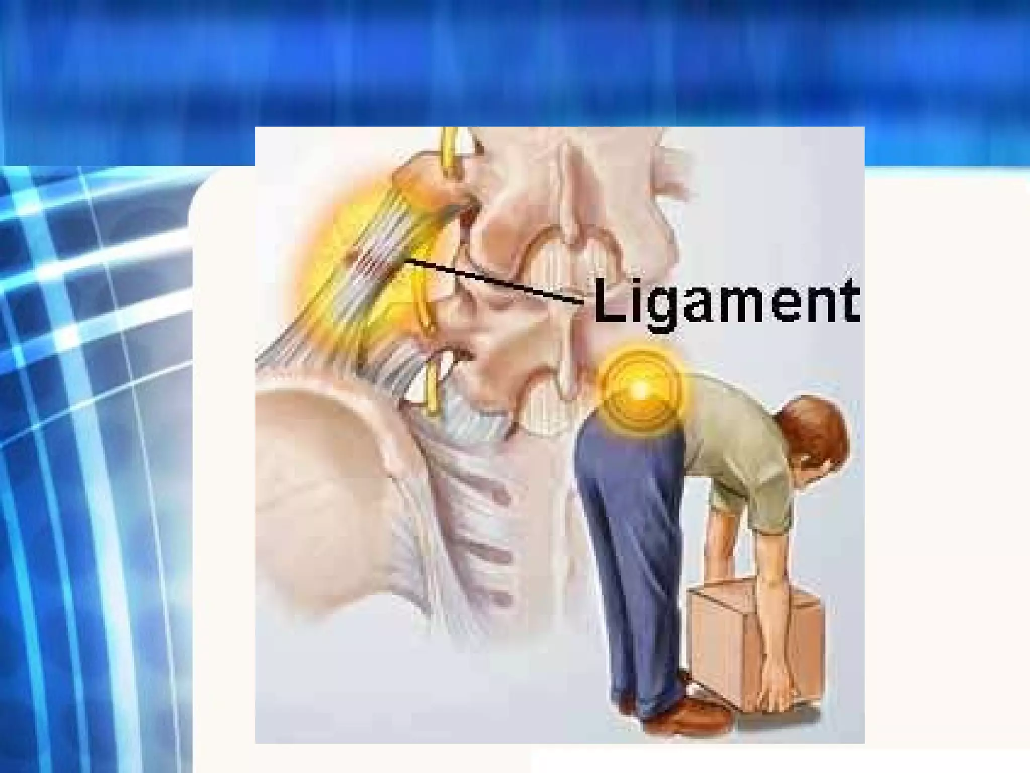
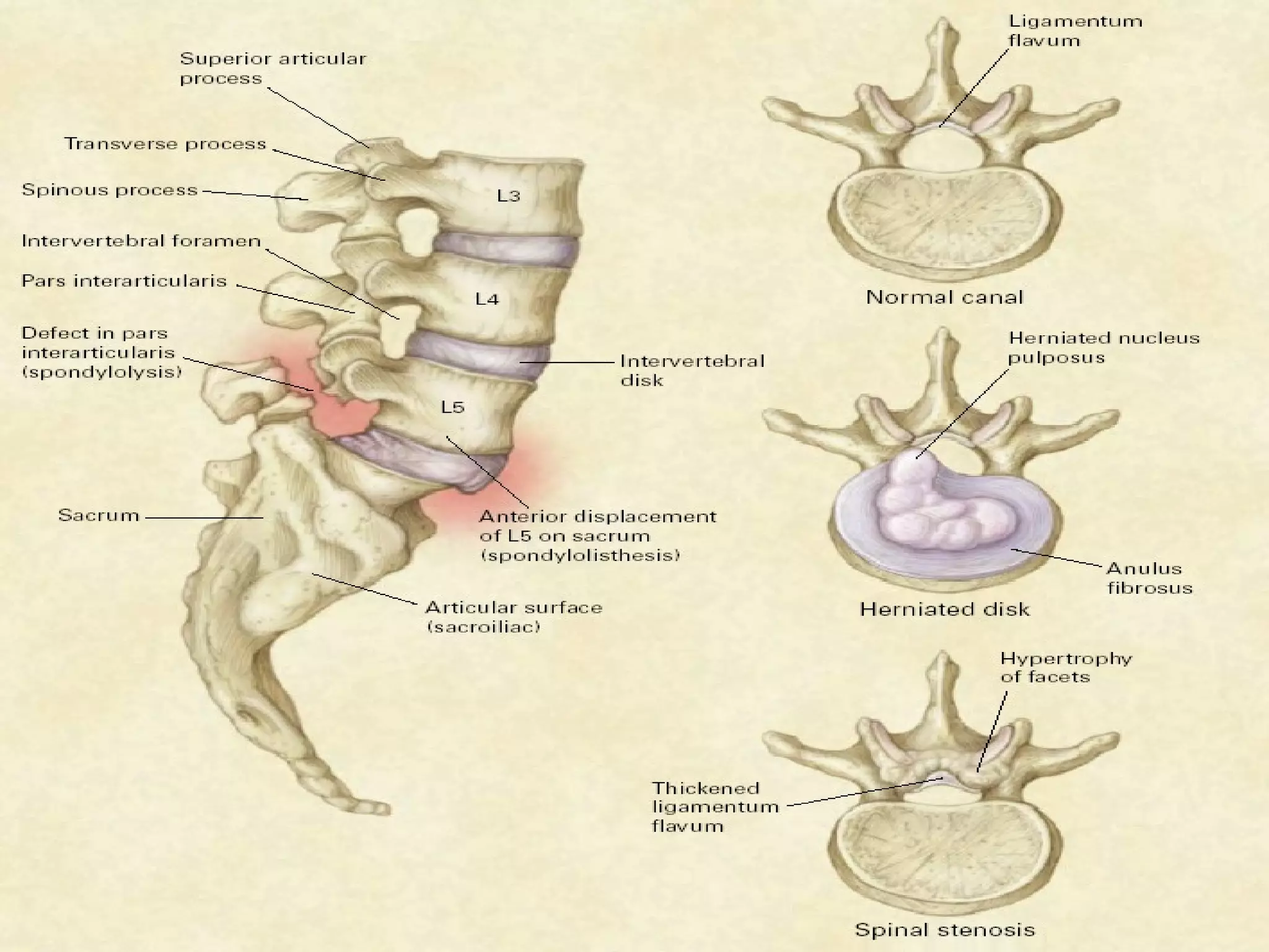
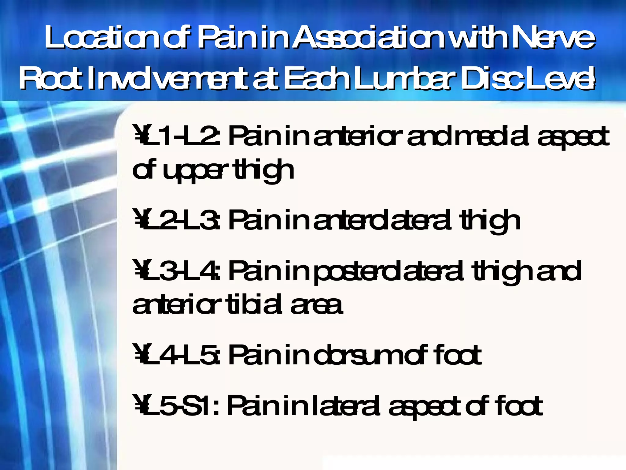
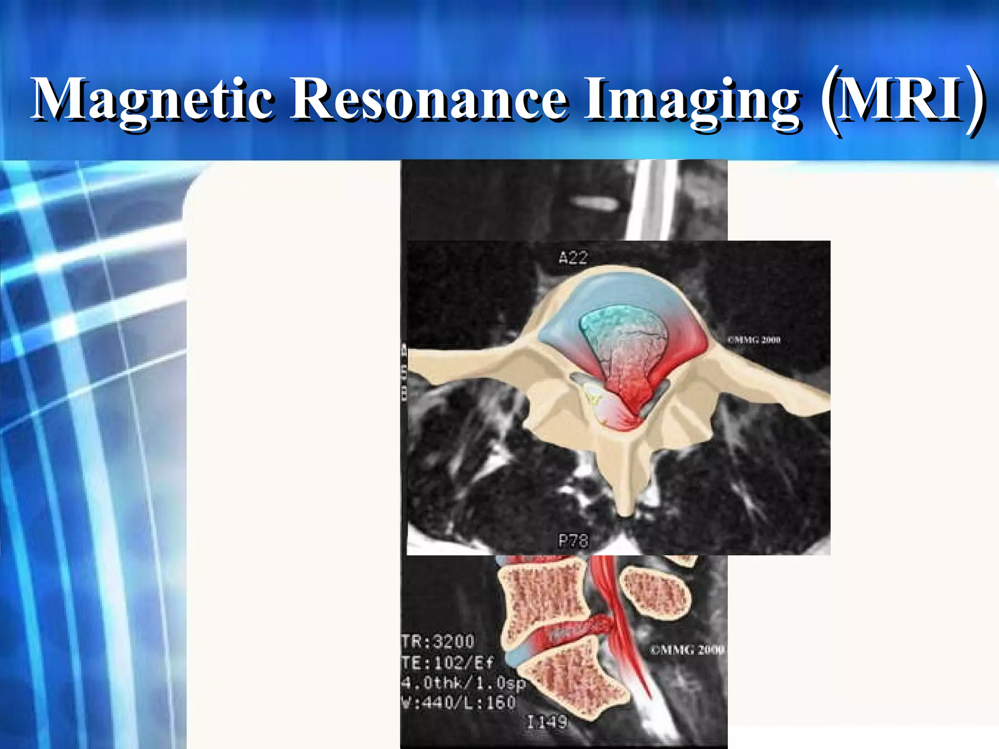
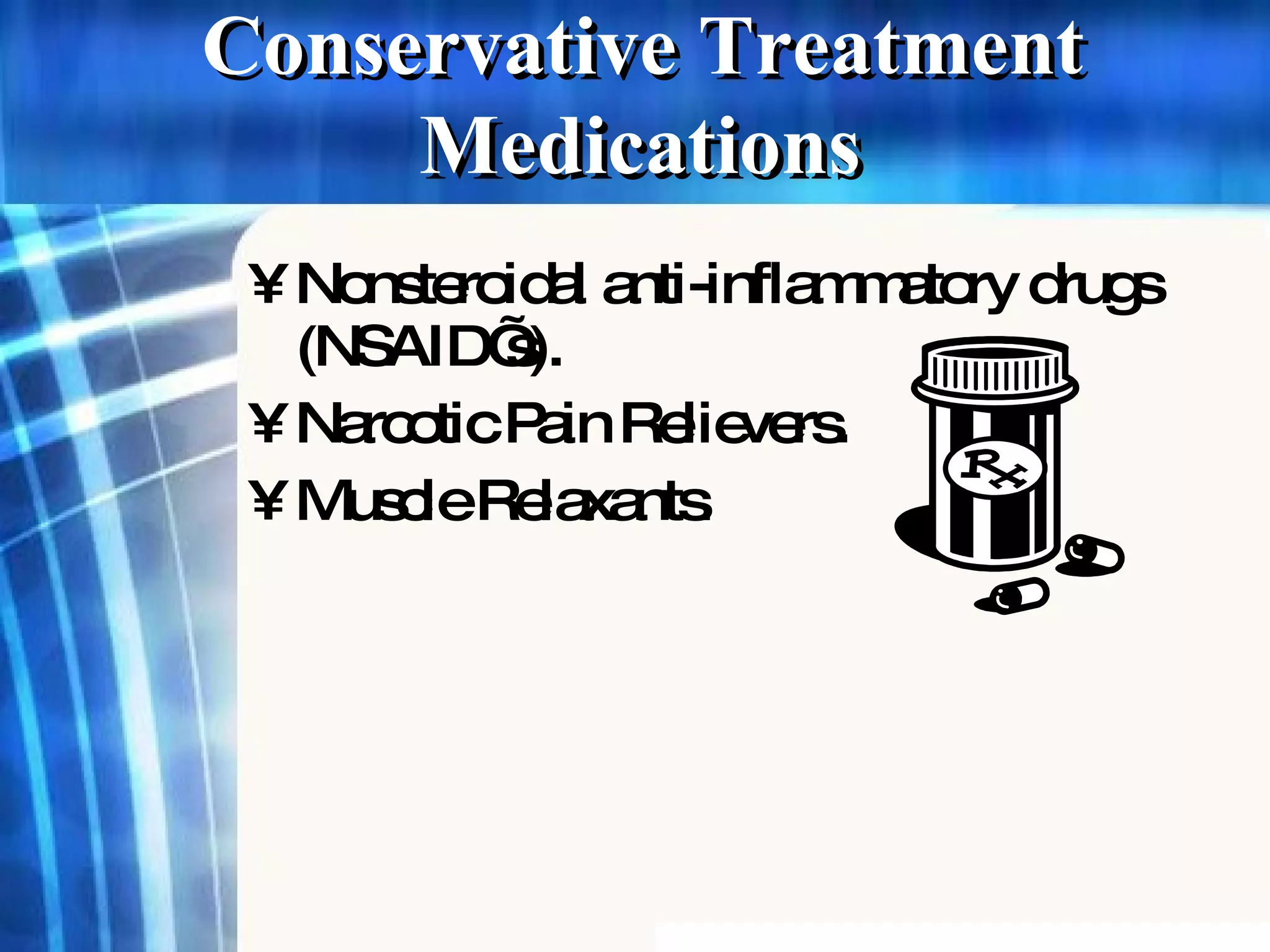
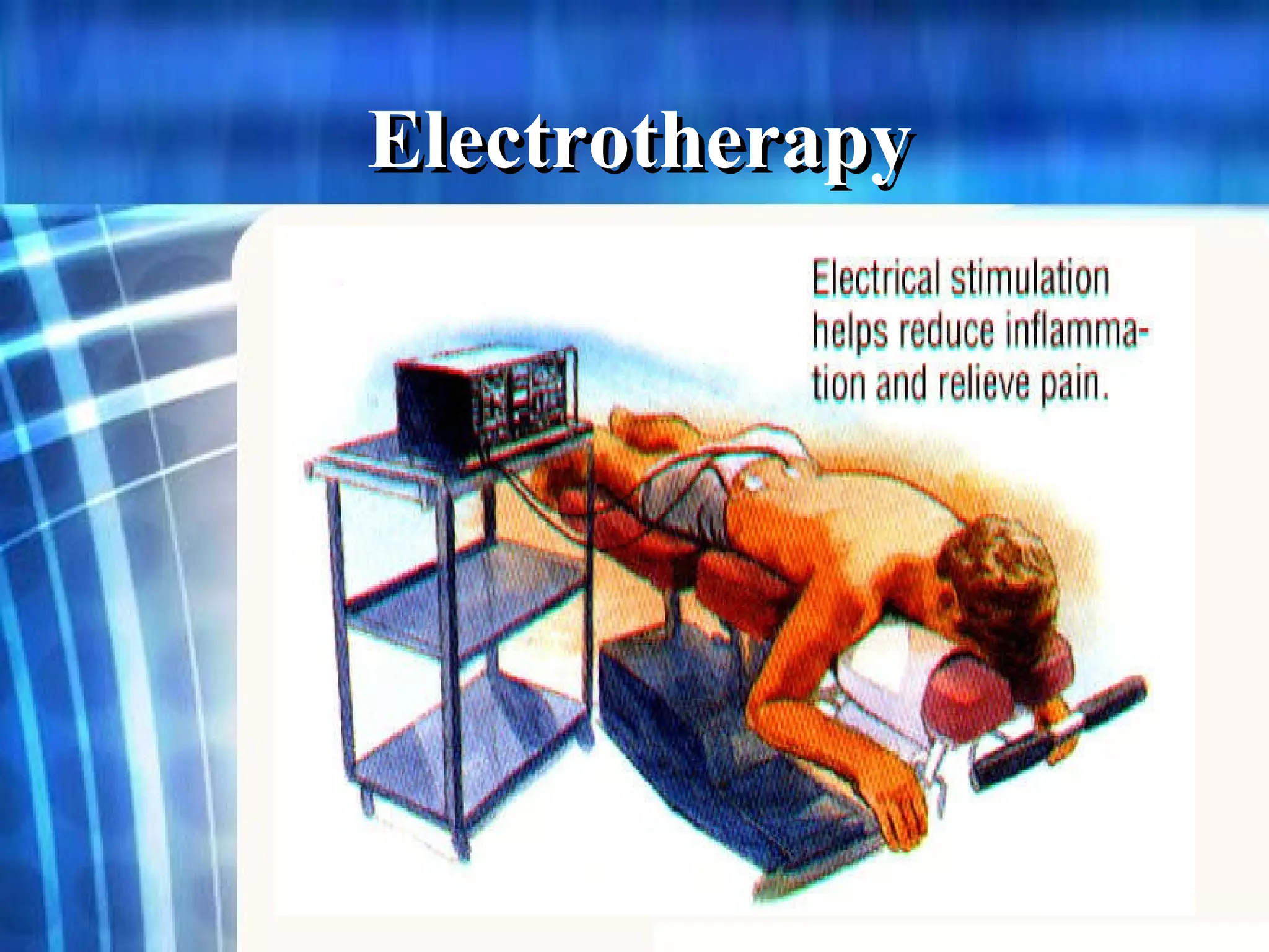
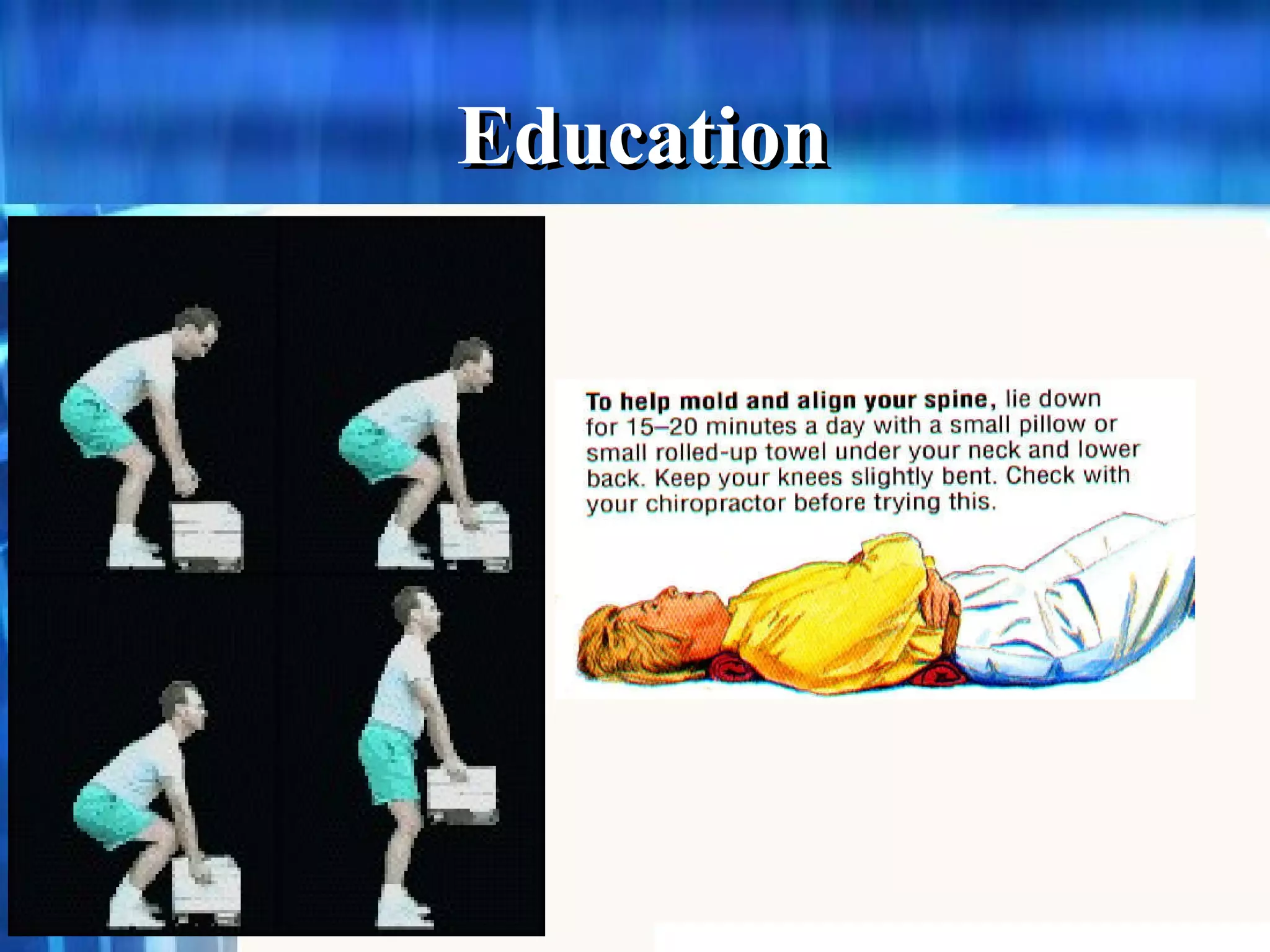
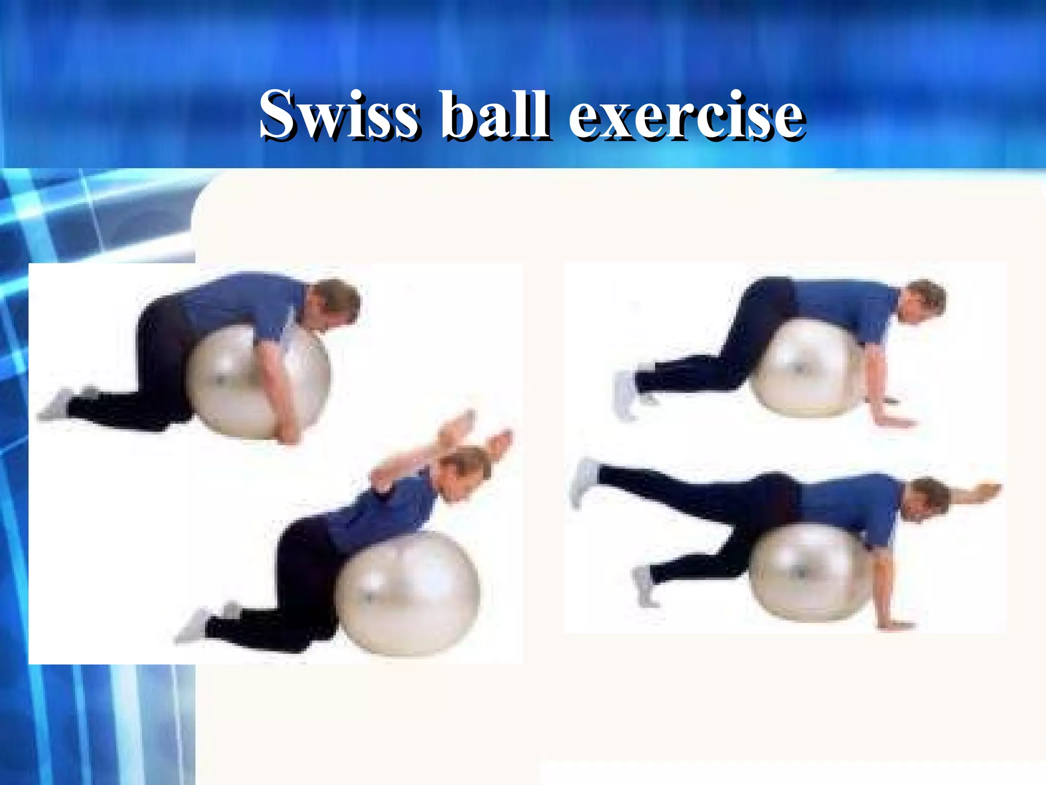
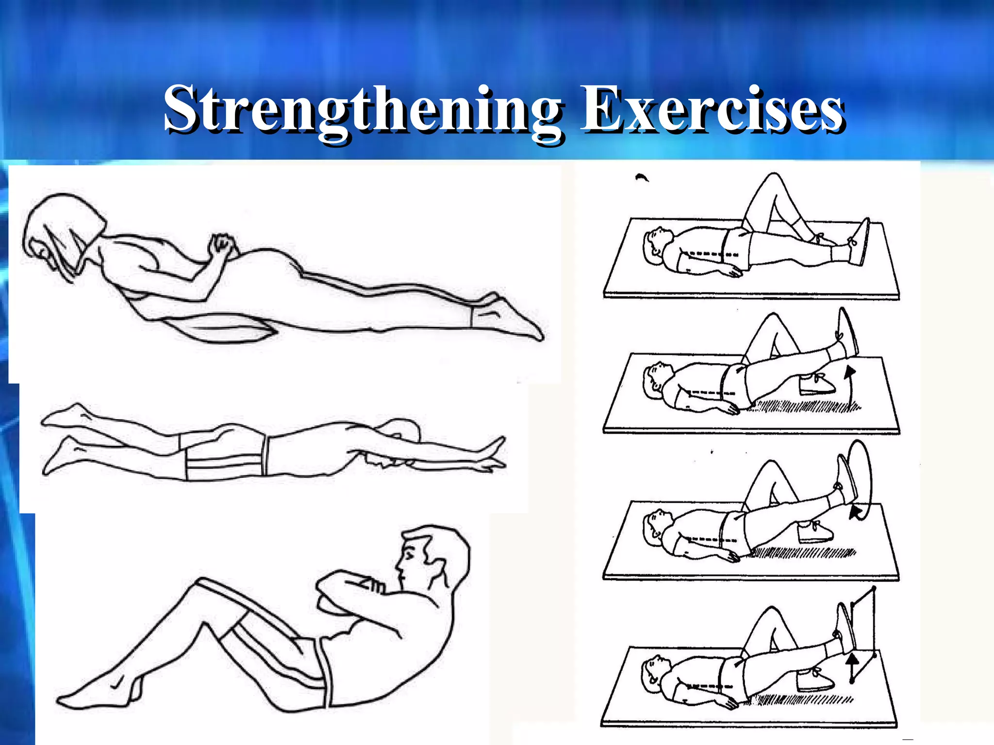
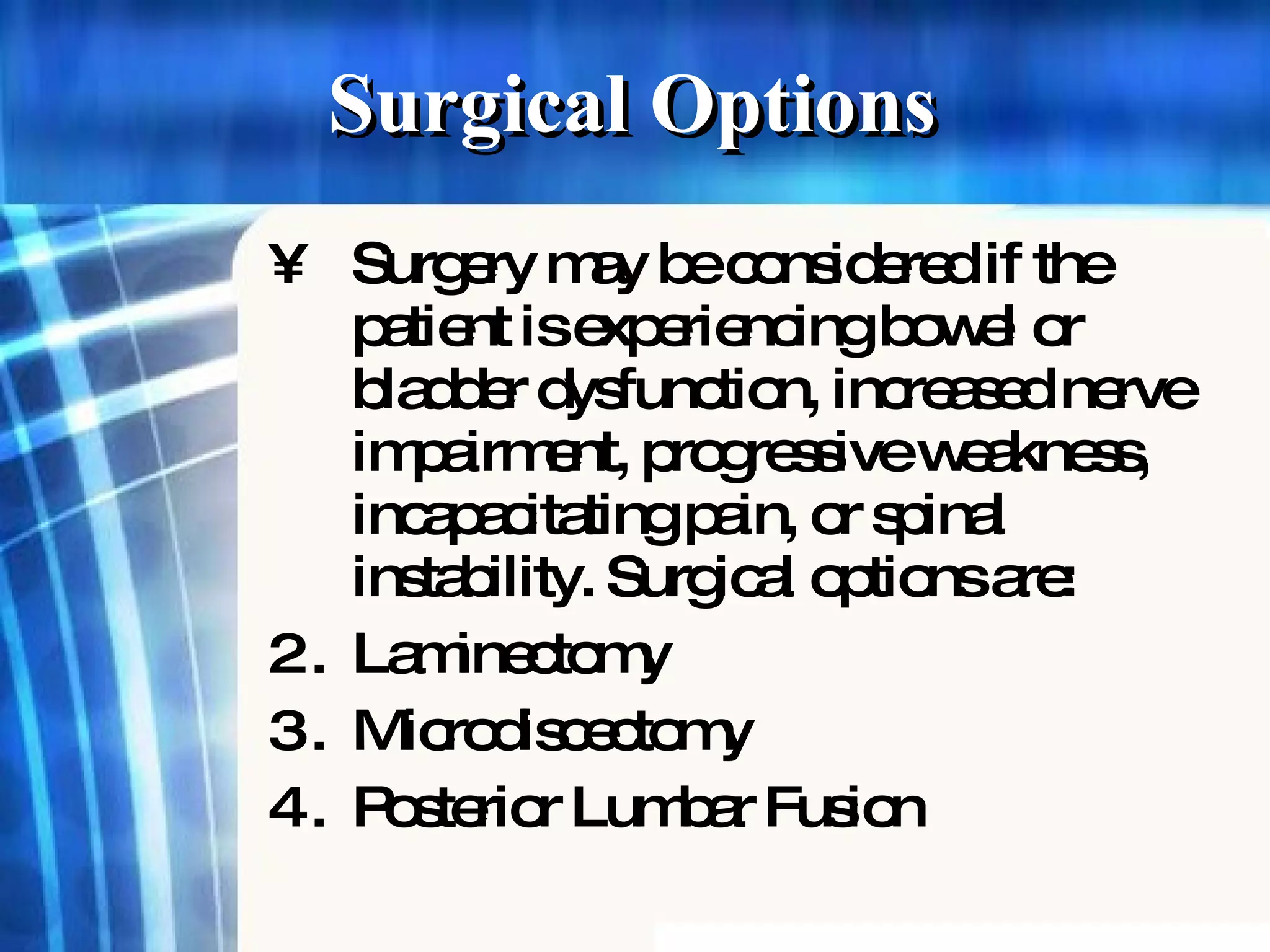
Pathomechanics of thoracic spine diseaseranjan mishra
The thoracic spine is the longest region of the spine, and by some measures it is also the most complex. Connecting with the cervical spine above and the lumbar spine below, the thoracic spine runs from the base of the neck down to the abdomen. It is the only spinal region attached to the rib cage.
Musculoskeletal system:Anatomy of the Lumbar region MuhamadAfif7
This is a brief presentation of the anatomy of the lumbar region.It covers the gross anatomy of the lumbar region including the lumbar vertebrae,the blood supply,muscles and nerve supply to this region.Some clinical aspects are also covered in this presentation
MUSCLES OF THE VERTEBRAL COLUMN- The system of ligaments in the vertebral column, combined with the tendons and muscles, provides a natural brace to help protect the spine from injury. For More Online Medical Resource, Visit at http://gisurgery.info
anatomy of lumbar spine, biomechanics of lumbar spine, movements at lumbar region, muscles of lumbar region, lumbar vertebra, kinetics and kinematics of lumbar spine
What is structure of lumber disc? What is disc bulge/prolapse/herniation? What is difference between disc bulge, disc prolapse, disc herniation or disc extrusion? What is criteria to diagnose lumber disc prolapse? How lumber disc herniation is treated medically or surgically? How lumber disc herniation is treated by conservative method? How lumber disc herniation is treated through physical therapy? What is physiotherapy after various disc surgeries? What is radiological method to diagnose disc prolapse?
Sacroiliac(SI) Joint Dysfunction,Evaluation and Treatment Dr.Md.Monsur Rahman
Dr.MD.Monsur Rahman,PT
MPT-Musculoskeletal Disorders
Maharishi Markandeshwar Institute Of Physiotherapy And Rehabilitation, Maharishi Markandeshwar (Deemed to be University), Mullana - Ambala,133-207 (Haryana)
USMLE MSK L002 Back Ligamnets and muscles of back.pdfAHMED ASHOUR
The anatomy of the back is complex and involves a combination of bones, muscles, nerves, and other structures that provide support, protection, and mobility.
The back is generally divided into several regions, including the cervical, thoracic, lumbar, sacral, and coccygeal regions.
Understanding the anatomy of the back is essential for healthcare professionals, including orthopedic specialists, physical therapists, and chiropractors, as well as for individuals interested in maintaining back health and preventing injuries.
21. Relative increases and decreases in intradiscal pressure in relation to different body positions
22.
23.
24.
25.
26.
27.
28. Anger/ anxiety/ stress Depression Back Pain Low mood Psychological Cycle Muscles further weakened Fear of pain/ damage (activities avoided) Muscles weakened by lack of activities Pain progresses Activities further avoided Physical cycle The Cycles of Back Pain
At night while we're sleeping the discs "reconstitute" and the nucleus fills with fluid which increases the pressure on the outer annulus. This is possibly why we feel stiff and are prone to injuries on sudden movements in the morning.
The discs form strong joints
In the normal, healthy disc, the nucleus distributes the load equally throughout the anulus. As the disc undergoes degeneration, the nucleus loses some of its cushioning ability and transmits the load unequally to the anulus. In the severely degenerated disc, the nucleus has lost all of its ability to cushion the load, which can lead to disc herniation.
Anterior longitudinal ligament limits extension Posterior longitudinal ligament limits flexion, supports to the discs except lumbar spine Interspinous limits flexion Supraspinous ligament limits flexion and rotation Ligamentum flavum exerts pull on joint capsule
Protrusion the disc bulges posteriorly without rupture of the annulus fibrosus proplase only the outer most fibers of the annulus fibrosus contain the nucleus Extrusion: rupture of the annulus fibrosus and part of the nucleus palposus moves into the epidural space Sequestration formation of discal fragments from the nucleus palposus outside the disc proper As a disc degenerates, it can herniate (the inner core extrudes) back into the spinal canal, which is known as a disc herniation (or a herniated disc) A herniation may develop suddenly or gradually over weeks or months. The four stages to a herniated disc include: Approximately 90% of disc herniations will occur at L4- L5 (lumbar segments 4 and 5) ,which causes pain in the L5 nerve or L5- S1 (lumbar segment 5 and sacral segment1), which causes pain in the S1 nerve.
spondylosis degeneration of the IVD spondylolysis defect in the pars interarticularis or the arch Sponylolisthesis forward displacement of one vertebra over another
The examiner should looks for limitation of movement and its possible causes, such as pain, spasm, stiffness, or blocking.
Prone knee bending test. examiner is pointing to where pain may be expected in the lumbar spine with positive test Sequence of subject postures in the slump test
radiographs have limited diagnostic value because degenerative changes are age-related and are equally present in asymptomatic and symptomatic persons.
The McKenzie exercise program is believed to be one of the most beneficial. Strengthening of the abdominal and back muscles Extension and isometric exercises are performed first and, after sufficient strength and pain relief are achieved, flexion exercises are allowed. Flexion exercises are delayed because flexion motions apply the greatest load to the intervertebral disc.