This document describes the radiological features of follicular nodular hyperplasia (FNH) seen on ultrasound, CT, MRI, and other imaging modalities. Key points include:
- On ultrasound, FNH lesions appear similar in reflectivity to the liver but can cause mass effect. A central scar is rarely seen. Doppler signals may be seen within and at the edge of lesions.
- On CT, lesions appear well-defined with mass effect. Attenuation matches the liver. A central low attenuation scar is seen. Arterial phase shows uniform enhancement except for the scar. Portal and delayed phases show isoattenuation with the liver and slow scar enhancement.
- MRI shows similar enhancement
Side of simple renal cysts usually solitary , but also showed a small but multiple or atrial rarely occurs in bilateral , compared with polycystic kidney disease, the clinical manifestations and pathological manifestations are different.
i made this ppt for presentation in class............i have added some already prepared ppts...
i think it wil be useful to some residents out there who dont find time in busy work schedules....all the best
Report Back from SGO 2024: What’s the Latest in Cervical Cancer?bkling
Are you curious about what’s new in cervical cancer research or unsure what the findings mean? Join Dr. Emily Ko, a gynecologic oncologist at Penn Medicine, to learn about the latest updates from the Society of Gynecologic Oncology (SGO) 2024 Annual Meeting on Women’s Cancer. Dr. Ko will discuss what the research presented at the conference means for you and answer your questions about the new developments.
Title: Sense of Smell
Presenter: Dr. Faiza, Assistant Professor of Physiology
Qualifications:
MBBS (Best Graduate, AIMC Lahore)
FCPS Physiology
ICMT, CHPE, DHPE (STMU)
MPH (GC University, Faisalabad)
MBA (Virtual University of Pakistan)
Learning Objectives:
Describe the primary categories of smells and the concept of odor blindness.
Explain the structure and location of the olfactory membrane and mucosa, including the types and roles of cells involved in olfaction.
Describe the pathway and mechanisms of olfactory signal transmission from the olfactory receptors to the brain.
Illustrate the biochemical cascade triggered by odorant binding to olfactory receptors, including the role of G-proteins and second messengers in generating an action potential.
Identify different types of olfactory disorders such as anosmia, hyposmia, hyperosmia, and dysosmia, including their potential causes.
Key Topics:
Olfactory Genes:
3% of the human genome accounts for olfactory genes.
400 genes for odorant receptors.
Olfactory Membrane:
Located in the superior part of the nasal cavity.
Medially: Folds downward along the superior septum.
Laterally: Folds over the superior turbinate and upper surface of the middle turbinate.
Total surface area: 5-10 square centimeters.
Olfactory Mucosa:
Olfactory Cells: Bipolar nerve cells derived from the CNS (100 million), with 4-25 olfactory cilia per cell.
Sustentacular Cells: Produce mucus and maintain ionic and molecular environment.
Basal Cells: Replace worn-out olfactory cells with an average lifespan of 1-2 months.
Bowman’s Gland: Secretes mucus.
Stimulation of Olfactory Cells:
Odorant dissolves in mucus and attaches to receptors on olfactory cilia.
Involves a cascade effect through G-proteins and second messengers, leading to depolarization and action potential generation in the olfactory nerve.
Quality of a Good Odorant:
Small (3-20 Carbon atoms), volatile, water-soluble, and lipid-soluble.
Facilitated by odorant-binding proteins in mucus.
Membrane Potential and Action Potential:
Resting membrane potential: -55mV.
Action potential frequency in the olfactory nerve increases with odorant strength.
Adaptation Towards the Sense of Smell:
Rapid adaptation within the first second, with further slow adaptation.
Psychological adaptation greater than receptor adaptation, involving feedback inhibition from the central nervous system.
Primary Sensations of Smell:
Camphoraceous, Musky, Floral, Pepperminty, Ethereal, Pungent, Putrid.
Odor Detection Threshold:
Examples: Hydrogen sulfide (0.0005 ppm), Methyl-mercaptan (0.002 ppm).
Some toxic substances are odorless at lethal concentrations.
Characteristics of Smell:
Odor blindness for single substances due to lack of appropriate receptor protein.
Behavioral and emotional influences of smell.
Transmission of Olfactory Signals:
From olfactory cells to glomeruli in the olfactory bulb, involving lateral inhibition.
Primitive, less old, and new olfactory systems with different path
Pulmonary Thromboembolism - etilogy, types, medical- Surgical and nursing man...VarunMahajani
Disruption of blood supply to lung alveoli due to blockage of one or more pulmonary blood vessels is called as Pulmonary thromboembolism. In this presentation we will discuss its causes, types and its management in depth.
These simplified slides by Dr. Sidra Arshad present an overview of the non-respiratory functions of the respiratory tract.
Learning objectives:
1. Enlist the non-respiratory functions of the respiratory tract
2. Briefly explain how these functions are carried out
3. Discuss the significance of dead space
4. Differentiate between minute ventilation and alveolar ventilation
5. Describe the cough and sneeze reflexes
Study Resources:
1. Chapter 39, Guyton and Hall Textbook of Medical Physiology, 14th edition
2. Chapter 34, Ganong’s Review of Medical Physiology, 26th edition
3. Chapter 17, Human Physiology by Lauralee Sherwood, 9th edition
4. Non-respiratory functions of the lungs https://academic.oup.com/bjaed/article/13/3/98/278874
HOT NEW PRODUCT! BIG SALES FAST SHIPPING NOW FROM CHINA!! EU KU DB BK substit...GL Anaacs
Contact us if you are interested:
Email / Skype : kefaya1771@gmail.com
Threema: PXHY5PDH
New BATCH Ku !!! MUCH IN DEMAND FAST SALE EVERY BATCH HAPPY GOOD EFFECT BIG BATCH !
Contact me on Threema or skype to start big business!!
Hot-sale products:
NEW HOT EUTYLONE WHITE CRYSTAL!!
5cl-adba precursor (semi finished )
5cl-adba raw materials
ADBB precursor (semi finished )
ADBB raw materials
APVP powder
5fadb/4f-adb
Jwh018 / Jwh210
Eutylone crystal
Protonitazene (hydrochloride) CAS: 119276-01-6
Flubrotizolam CAS: 57801-95-3
Metonitazene CAS: 14680-51-4
Payment terms: Western Union,MoneyGram,Bitcoin or USDT.
Deliver Time: Usually 7-15days
Shipping method: FedEx, TNT, DHL,UPS etc.Our deliveries are 100% safe, fast, reliable and discreet.
Samples will be sent for your evaluation!If you are interested in, please contact me, let's talk details.
We specializes in exporting high quality Research chemical, medical intermediate, Pharmaceutical chemicals and so on. Products are exported to USA, Canada, France, Korea, Japan,Russia, Southeast Asia and other countries.
ARTIFICIAL INTELLIGENCE IN HEALTHCARE.pdfAnujkumaranit
Artificial intelligence (AI) refers to the simulation of human intelligence processes by machines, especially computer systems. It encompasses tasks such as learning, reasoning, problem-solving, perception, and language understanding. AI technologies are revolutionizing various fields, from healthcare to finance, by enabling machines to perform tasks that typically require human intelligence.
TEST BANK for Operations Management, 14th Edition by William J. Stevenson, Ve...kevinkariuki227
TEST BANK for Operations Management, 14th Edition by William J. Stevenson, Verified Chapters 1 - 19, Complete Newest Version.pdf
TEST BANK for Operations Management, 14th Edition by William J. Stevenson, Verified Chapters 1 - 19, Complete Newest Version.pdf
micro teaching on communication m.sc nursing.pdfAnurag Sharma
Microteaching is a unique model of practice teaching. It is a viable instrument for the. desired change in the teaching behavior or the behavior potential which, in specified types of real. classroom situations, tends to facilitate the achievement of specified types of objectives.
Tom Selleck Health: A Comprehensive Look at the Iconic Actor’s Wellness Journeygreendigital
Tom Selleck, an enduring figure in Hollywood. has captivated audiences for decades with his rugged charm, iconic moustache. and memorable roles in television and film. From his breakout role as Thomas Magnum in Magnum P.I. to his current portrayal of Frank Reagan in Blue Bloods. Selleck's career has spanned over 50 years. But beyond his professional achievements. fans have often been curious about Tom Selleck Health. especially as he has aged in the public eye.
Follow us on: Pinterest
Introduction
Many have been interested in Tom Selleck health. not only because of his enduring presence on screen but also because of the challenges. and lifestyle choices he has faced and made over the years. This article delves into the various aspects of Tom Selleck health. exploring his fitness regimen, diet, mental health. and the challenges he has encountered as he ages. We'll look at how he maintains his well-being. the health issues he has faced, and his approach to ageing .
Early Life and Career
Childhood and Athletic Beginnings
Tom Selleck was born on January 29, 1945, in Detroit, Michigan, and grew up in Sherman Oaks, California. From an early age, he was involved in sports, particularly basketball. which played a significant role in his physical development. His athletic pursuits continued into college. where he attended the University of Southern California (USC) on a basketball scholarship. This early involvement in sports laid a strong foundation for his physical health and disciplined lifestyle.
Transition to Acting
Selleck's transition from an athlete to an actor came with its physical demands. His first significant role in "Magnum P.I." required him to perform various stunts and maintain a fit appearance. This role, which he played from 1980 to 1988. necessitated a rigorous fitness routine to meet the show's demands. setting the stage for his long-term commitment to health and wellness.
Fitness Regimen
Workout Routine
Tom Selleck health and fitness regimen has evolved. adapting to his changing roles and age. During his "Magnum, P.I." days. Selleck's workouts were intense and focused on building and maintaining muscle mass. His routine included weightlifting, cardiovascular exercises. and specific training for the stunts he performed on the show.
Selleck adjusted his fitness routine as he aged to suit his body's needs. Today, his workouts focus on maintaining flexibility, strength, and cardiovascular health. He incorporates low-impact exercises such as swimming, walking, and light weightlifting. This balanced approach helps him stay fit without putting undue strain on his joints and muscles.
Importance of Flexibility and Mobility
In recent years, Selleck has emphasized the importance of flexibility and mobility in his fitness regimen. Understanding the natural decline in muscle mass and joint flexibility with age. he includes stretching and yoga in his routine. These practices help prevent injuries, improve posture, and maintain mobilit
Title: Sense of Taste
Presenter: Dr. Faiza, Assistant Professor of Physiology
Qualifications:
MBBS (Best Graduate, AIMC Lahore)
FCPS Physiology
ICMT, CHPE, DHPE (STMU)
MPH (GC University, Faisalabad)
MBA (Virtual University of Pakistan)
Learning Objectives:
Describe the structure and function of taste buds.
Describe the relationship between the taste threshold and taste index of common substances.
Explain the chemical basis and signal transduction of taste perception for each type of primary taste sensation.
Recognize different abnormalities of taste perception and their causes.
Key Topics:
Significance of Taste Sensation:
Differentiation between pleasant and harmful food
Influence on behavior
Selection of food based on metabolic needs
Receptors of Taste:
Taste buds on the tongue
Influence of sense of smell, texture of food, and pain stimulation (e.g., by pepper)
Primary and Secondary Taste Sensations:
Primary taste sensations: Sweet, Sour, Salty, Bitter, Umami
Chemical basis and signal transduction mechanisms for each taste
Taste Threshold and Index:
Taste threshold values for Sweet (sucrose), Salty (NaCl), Sour (HCl), and Bitter (Quinine)
Taste index relationship: Inversely proportional to taste threshold
Taste Blindness:
Inability to taste certain substances, particularly thiourea compounds
Example: Phenylthiocarbamide
Structure and Function of Taste Buds:
Composition: Epithelial cells, Sustentacular/Supporting cells, Taste cells, Basal cells
Features: Taste pores, Taste hairs/microvilli, and Taste nerve fibers
Location of Taste Buds:
Found in papillae of the tongue (Fungiform, Circumvallate, Foliate)
Also present on the palate, tonsillar pillars, epiglottis, and proximal esophagus
Mechanism of Taste Stimulation:
Interaction of taste substances with receptors on microvilli
Signal transduction pathways for Umami, Sweet, Bitter, Sour, and Salty tastes
Taste Sensitivity and Adaptation:
Decrease in sensitivity with age
Rapid adaptation of taste sensation
Role of Saliva in Taste:
Dissolution of tastants to reach receptors
Washing away the stimulus
Taste Preferences and Aversions:
Mechanisms behind taste preference and aversion
Influence of receptors and neural pathways
Impact of Sensory Nerve Damage:
Degeneration of taste buds if the sensory nerve fiber is cut
Abnormalities of Taste Detection:
Conditions: Ageusia, Hypogeusia, Dysgeusia (parageusia)
Causes: Nerve damage, neurological disorders, infections, poor oral hygiene, adverse drug effects, deficiencies, aging, tobacco use, altered neurotransmitter levels
Neurotransmitters and Taste Threshold:
Effects of serotonin (5-HT) and norepinephrine (NE) on taste sensitivity
Supertasters:
25% of the population with heightened sensitivity to taste, especially bitterness
Increased number of fungiform papillae
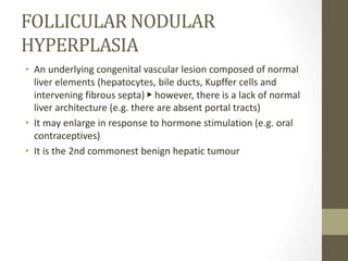
1. FOLLICULAR NODULAR
HYPERPLASIA
• An underlying congenital vascular lesion composed of normal
liver elements (hepatocytes, bile ducts, Kupffer cells and
intervening fibrous septa) ▶ however, there is a lack of normal
liver architecture (e.g. there are absent portal tracts)
• It may enlarge in response to hormone stimulation (e.g. oral
contraceptives)
• It is the 2nd commonest benign hepatic tumour
2. CLINICAL PRESENTATION
•It is usually asymptomatic (it may present with pain or
hepatomegaly)
•It occurs most commonly in women aged 20–50 years (and is
multiple in 20% of cases)
FOLLICULAR NODULAR
HYPERPLASIA
3. RADIOLOGICALFEATURES
• A central stellate fibrovascular scar is seen in 50% of cases
• there is no true capsule
• calcification, necrosis and haemorrhage are extremely rare (even large
lesions do not usually outgrow their blood supply)
ULTRASOUND
• There are non-specific features with lesions demonstrating a similar
reflectivity to the adjacent liver (but demonstrating mass effect)
• the central scar is rarely seen
• Doppler signals can be seen within and at the edge of the lesion
4. US appearance of FNH. (a) Sagittal US scan shows FNH that is slightly
hypoechoic relative to the surrounding liver tissue (arrows) and causes
slight distortion of the outer liver contour.
5. Figure 5b. Sagittal oblique US scan of another patient
shows FNH that is well differentiated from the
surrounding liver tissue (arrow). There is a suggestion
of radiating septa within the lesion.
6. Figure 5c. Axial US scan of another patient shows
FNH that is profoundly hypoechoic (arrow) due to
diffuse fatty infiltration of the surrounding liver tissue.
7. RADIOLOGICALFEATURES
NECT
• A well-defined mass which often exhibits a mass effect (with vessel
displacement)
• the lesion demonstrates the same attenuation as the surrounding liver
• there is a central low attenuation scar
CECT
• Arterial phase:
• uniform enhancement (except for the scar)
• there can be large peripheral feeding vessels
• Portal phase:
• the attenuation is identical to normal liver (the scar remains low
attenuation)
• Delayed imaging:
• there is slow scar enhancement
8. Figure 6a. CT appearance of typical FNH with pathologic
correlation. (a) Precontrast CT image shows a large lesion
(straight arrow) that is only slightly hypoattenuating relative to
the surrounding liver tissue. Within the lesion, a central scar
(curved arrow) can be seen.
9. Figure 6b. Contrast-enhanced CT image obtained during the arterial
phase shows intense homogeneous enhancement of the lesion
(straight arrow), except for the central scar (curved arrow).
10. Figure 6c. Contrast-enhanced CT image obtained during the
portal phase shows that the lesion (straight arrow) has
become isoattenuating relative to the liver. The central scar
(curved arrow) has not yet fully enhanced.
11. MRI
•The same enhancement pattern is seen as for CT ▶ the specificity increases
with iron oxide agents (which are taken up by the Kupffer cells).
•T1WI:
• intermediate or minimal low SI
• a low SI central scar
•T2WI:
• intermediate to high SI
• a high SI central scar
•T1WI + Gad:
• marked, homogeneous arterial phase enhancement that becomes
isointense during the portal venous phase
• there can also be a peripheral, ring-type delayed enhancement pattern
on delayed images obtained 1 h after hepatocyte selective gadolinium
chelate administration
• The central scar usually demonstrates delayed enhancement
•DWI:
• generally isointense
12. Figure 7a. MR imaging appearance of typical FNH. (a) Axial T2-
weighted single-shot fast spin-echo (SE) image shows a large FNH
lesion (straight arrow) that is isointense relative to the surrounding
liver parenchyma. The central scar (curved arrow) has slightly higher
signal intensity than the lesion.
13. Figure 7b. Axial gadolinium-enhanced 2D T1-weighted GRE image
obtained during the arterial phase shows intense homogeneous
enhancement of the entire lesion (straight arrow), except for the central
scar (curved arrow)
14. Figure 7c. Axial gadolinium-enhanced 2D T1-weighted GRE image
obtained during the portal phase shows that the lesion (straight
arrow) has become isointense relative to the surrounding liver
parenchyma, and the central scar (curved arrow) has enhanced.
15. RADIOLOGICALFEATURES
• Sulphur colloid
• This is usually normal (due to Kupffer cell activity within the
lesion)
• DSA
• A vascular mass with a large tortuous central sup- plying artery ▶
radiating vessels spread out to supply the lesion
16. CASE 2
• A 31 years old female working as a staff nurse at Institut
Kanser Negara - treated as PTB.
• Sputum MTB Cn& S- shows non-tuberculous mycobacterium .
• Developed transaminitis while on antiTB.
• US noted multiple liver lesion ? abscess or TB lesion. Lesion
persistent despite near completion of anti-TB.
• Hepatitis screening: non reactive
17. US HBS 08/08/2018
• Liver parenchyma shows increased in echogenicity with
smooth liver margin.
• There are hypoechoic lesions seen in:
• segment V/VI 1.5 x 2.1 x 2.0 cm
• Segment VII 1.9 x 1.2 x 1.4 cm and 1.2 x 2.2cm
• Impression: 1) Multiple hypoechoic liver lesions in segment V/VI
and VII could be abscesses or Tuberculosis lesions. 2) Fatty liver
18.
19. US HBS 20/02/2019
• The previous hypoechoeic lesions in segment V (measuring 2.5 x 2.3
cm) and IVb ( measuring 1.8 x1.4 cm ) are relatively unchanged in
size but are hyperechoeic in this current US.
Another smaller hypoechoic lesion at segment VI is unchanged in
echogenicity and size. It measures 2.2 x 1.8 cm
• Impressions:
Multiple right liver lesions with no significant change in size.
• The segment V and IVb lesions have changed in echogenicity from
hypo to hyperechoeic
• segment VI lesion remains hypoechoeic.
• DDx solidified abscess, organized hematoma, hemangioma.
20.
21. CT HBS 08/04/2019
• Liver is mildly enlarged (span 21cm), margins are smooth and
regular.
• There are few well-circumscribed lesions of varying sizes i.e. at
segments VI/VII and VIII.
• These lesions shows hypoattenuation on pre-contrasted
images, progressive enhancement through the contrasted
phases showing globular and centripetal pattern and
homogenisation in the delayed phases. The largest measuring
2.0cm x 2.5cm at segment VII.