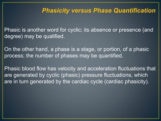
This document discusses hepatic Doppler ultrasound waveforms. It defines key terms like antegrade and retrograde flow. The normal hepatic artery waveform is pulsatile with low resistance. The normal hepatic vein waveform is biphasic or tetraphasic. Abnormal portal vein waveforms can be pulsatile, demonstrate slow flow, show retrograde flow, or have absent flow. The document provides detailed descriptions of hepatic artery, hepatic vein, and portal vein waveforms both normal and abnormal.
In this presentation we will discuss normal doppler parameters in portal and hepatic veins and hepatic artery. We will discuss the pathologies regarding hepatic, and portal veins and hepatic artery.
we will discuss Role of sonography in TIPS evaluation.
we will discuss the role of Doppler in post op follow up of hepatic transplant.
Doppler ultrasound of visceral arteriesSamir Haffar
Doppler ultrasound of different diseases of visceral arteries including arterial stenosis and occlusion, arterial aneurysm, artrial pseudoaneurysm, arterio-venous fistula, artrial dissection, and abdominal vascular compression syndromes
Role of contrast enhanced ultrasonography in characterization of hepatobiliar...Dr. Muhammad Bin Zulfiqar
Role of contrast enhanced ultrasonography in characterization of hepatobiliary disease Dr. Muhammad Bin Zulfiqar
Here we will discuss the state of the art technique of CEUS imaging in Charecterization of benign and malignant pathologies of liver.
The venous system contains about 70–80% of the circulating blood volume which is non-pulsatile. However, changes in flow and pressure caused by the right atrial and right ventricular filling produce pulsations in the central veins that are transmitted to the peripheral veins (e.g. jugular veins) and are opposite to the direction of the blood flow.
●
The arterial pulse and blood pressure reflects the dynamics of the left side of the heart, while the jugular veins provide the information about the hemodynamic events from the right side of the heart-right atrial pressure during systole and right ventricular filling pressure during diastole.
●
Hence, an accurate assessment of the venous pulse, the jugular venous pulse (JVP) reflects the dynamics of the right side of the heart.1
History ●
Lancis (1728) first described the cervical venous pulse of the external jugular vein in a patient with tricuspid regurgitation (see Table 16.1).
●
However, the classic graphic recordings of the JVP were done by Chauvea and Marey (1863).
●
But it was Potain (1869) who accurately described the wave pattern in the internal jugular vein.
Giant a Waves or Cannon Waves
These occur whenever the RA contracts against the closed TV during RV systole. Paul Wood described the giant a wave as ‘venous Corrigan’. Cannon waves may occur either regularly or irregularly and are most common in the presence of arrhythmias. ●
Regular cannon waves occur in – Junctional rhythm – Ventricular tachycardia (VT) 1:1 retrograde conduction – Isorhythmic AV dissociation
●
Irregular cannon waves occur in – Complete heart block (see Fig. 16.6) – Classic AV dissociation –VT – Ventricular pacing – Ventricular ectopics
NVBDCP.pptx Nation vector borne disease control programSapna Thakur
NVBDCP was launched in 2003-2004 . Vector-Borne Disease: Disease that results from an infection transmitted to humans and other animals by blood-feeding arthropods, such as mosquitoes, ticks, and fleas. Examples of vector-borne diseases include Dengue fever, West Nile Virus, Lyme disease, and malaria.
The prostate is an exocrine gland of the male mammalian reproductive system
It is a walnut-sized gland that forms part of the male reproductive system and is located in front of the rectum and just below the urinary bladder
Function is to store and secrete a clear, slightly alkaline fluid that constitutes 10-30% of the volume of the seminal fluid that along with the spermatozoa, constitutes semen
A healthy human prostate measures (4cm-vertical, by 3cm-horizontal, 2cm ant-post ).
It surrounds the urethra just below the urinary bladder. It has anterior, median, posterior and two lateral lobes
It’s work is regulated by androgens which are responsible for male sex characteristics
Generalised disease of the prostate due to hormonal derangement which leads to non malignant enlargement of the gland (increase in the number of epithelial cells and stromal tissue)to cause compression of the urethra leading to symptoms (LUTS
Report Back from SGO 2024: What’s the Latest in Cervical Cancer?bkling
Are you curious about what’s new in cervical cancer research or unsure what the findings mean? Join Dr. Emily Ko, a gynecologic oncologist at Penn Medicine, to learn about the latest updates from the Society of Gynecologic Oncology (SGO) 2024 Annual Meeting on Women’s Cancer. Dr. Ko will discuss what the research presented at the conference means for you and answer your questions about the new developments.
Acute scrotum is a general term referring to an emergency condition affecting the contents or the wall of the scrotum.
There are a number of conditions that present acutely, predominantly with pain and/or swelling
A careful and detailed history and examination, and in some cases, investigations allow differentiation between these diagnoses. A prompt diagnosis is essential as the patient may require urgent surgical intervention
Testicular torsion refers to twisting of the spermatic cord, causing ischaemia of the testicle.
Testicular torsion results from inadequate fixation of the testis to the tunica vaginalis producing ischemia from reduced arterial inflow and venous outflow obstruction.
The prevalence of testicular torsion in adult patients hospitalized with acute scrotal pain is approximately 25 to 50 percent
New Directions in Targeted Therapeutic Approaches for Older Adults With Mantl...i3 Health
i3 Health is pleased to make the speaker slides from this activity available for use as a non-accredited self-study or teaching resource.
This slide deck presented by Dr. Kami Maddocks, Professor-Clinical in the Division of Hematology and
Associate Division Director for Ambulatory Operations
The Ohio State University Comprehensive Cancer Center, will provide insight into new directions in targeted therapeutic approaches for older adults with mantle cell lymphoma.
STATEMENT OF NEED
Mantle cell lymphoma (MCL) is a rare, aggressive B-cell non-Hodgkin lymphoma (NHL) accounting for 5% to 7% of all lymphomas. Its prognosis ranges from indolent disease that does not require treatment for years to very aggressive disease, which is associated with poor survival (Silkenstedt et al, 2021). Typically, MCL is diagnosed at advanced stage and in older patients who cannot tolerate intensive therapy (NCCN, 2022). Although recent advances have slightly increased remission rates, recurrence and relapse remain very common, leading to a median overall survival between 3 and 6 years (LLS, 2021). Though there are several effective options, progress is still needed towards establishing an accepted frontline approach for MCL (Castellino et al, 2022). Treatment selection and management of MCL are complicated by the heterogeneity of prognosis, advanced age and comorbidities of patients, and lack of an established standard approach for treatment, making it vital that clinicians be familiar with the latest research and advances in this area. In this activity chaired by Michael Wang, MD, Professor in the Department of Lymphoma & Myeloma at MD Anderson Cancer Center, expert faculty will discuss prognostic factors informing treatment, the promising results of recent trials in new therapeutic approaches, and the implications of treatment resistance in therapeutic selection for MCL.
Target Audience
Hematology/oncology fellows, attending faculty, and other health care professionals involved in the treatment of patients with mantle cell lymphoma (MCL).
Learning Objectives
1.) Identify clinical and biological prognostic factors that can guide treatment decision making for older adults with MCL
2.) Evaluate emerging data on targeted therapeutic approaches for treatment-naive and relapsed/refractory MCL and their applicability to older adults
3.) Assess mechanisms of resistance to targeted therapies for MCL and their implications for treatment selection
Title: Sense of Taste
Presenter: Dr. Faiza, Assistant Professor of Physiology
Qualifications:
MBBS (Best Graduate, AIMC Lahore)
FCPS Physiology
ICMT, CHPE, DHPE (STMU)
MPH (GC University, Faisalabad)
MBA (Virtual University of Pakistan)
Learning Objectives:
Describe the structure and function of taste buds.
Describe the relationship between the taste threshold and taste index of common substances.
Explain the chemical basis and signal transduction of taste perception for each type of primary taste sensation.
Recognize different abnormalities of taste perception and their causes.
Key Topics:
Significance of Taste Sensation:
Differentiation between pleasant and harmful food
Influence on behavior
Selection of food based on metabolic needs
Receptors of Taste:
Taste buds on the tongue
Influence of sense of smell, texture of food, and pain stimulation (e.g., by pepper)
Primary and Secondary Taste Sensations:
Primary taste sensations: Sweet, Sour, Salty, Bitter, Umami
Chemical basis and signal transduction mechanisms for each taste
Taste Threshold and Index:
Taste threshold values for Sweet (sucrose), Salty (NaCl), Sour (HCl), and Bitter (Quinine)
Taste index relationship: Inversely proportional to taste threshold
Taste Blindness:
Inability to taste certain substances, particularly thiourea compounds
Example: Phenylthiocarbamide
Structure and Function of Taste Buds:
Composition: Epithelial cells, Sustentacular/Supporting cells, Taste cells, Basal cells
Features: Taste pores, Taste hairs/microvilli, and Taste nerve fibers
Location of Taste Buds:
Found in papillae of the tongue (Fungiform, Circumvallate, Foliate)
Also present on the palate, tonsillar pillars, epiglottis, and proximal esophagus
Mechanism of Taste Stimulation:
Interaction of taste substances with receptors on microvilli
Signal transduction pathways for Umami, Sweet, Bitter, Sour, and Salty tastes
Taste Sensitivity and Adaptation:
Decrease in sensitivity with age
Rapid adaptation of taste sensation
Role of Saliva in Taste:
Dissolution of tastants to reach receptors
Washing away the stimulus
Taste Preferences and Aversions:
Mechanisms behind taste preference and aversion
Influence of receptors and neural pathways
Impact of Sensory Nerve Damage:
Degeneration of taste buds if the sensory nerve fiber is cut
Abnormalities of Taste Detection:
Conditions: Ageusia, Hypogeusia, Dysgeusia (parageusia)
Causes: Nerve damage, neurological disorders, infections, poor oral hygiene, adverse drug effects, deficiencies, aging, tobacco use, altered neurotransmitter levels
Neurotransmitters and Taste Threshold:
Effects of serotonin (5-HT) and norepinephrine (NE) on taste sensitivity
Supertasters:
25% of the population with heightened sensitivity to taste, especially bitterness
Increased number of fungiform papillae
Tom Selleck Health: A Comprehensive Look at the Iconic Actor’s Wellness Journeygreendigital
Tom Selleck, an enduring figure in Hollywood. has captivated audiences for decades with his rugged charm, iconic moustache. and memorable roles in television and film. From his breakout role as Thomas Magnum in Magnum P.I. to his current portrayal of Frank Reagan in Blue Bloods. Selleck's career has spanned over 50 years. But beyond his professional achievements. fans have often been curious about Tom Selleck Health. especially as he has aged in the public eye.
Follow us on: Pinterest
Introduction
Many have been interested in Tom Selleck health. not only because of his enduring presence on screen but also because of the challenges. and lifestyle choices he has faced and made over the years. This article delves into the various aspects of Tom Selleck health. exploring his fitness regimen, diet, mental health. and the challenges he has encountered as he ages. We'll look at how he maintains his well-being. the health issues he has faced, and his approach to ageing .
Early Life and Career
Childhood and Athletic Beginnings
Tom Selleck was born on January 29, 1945, in Detroit, Michigan, and grew up in Sherman Oaks, California. From an early age, he was involved in sports, particularly basketball. which played a significant role in his physical development. His athletic pursuits continued into college. where he attended the University of Southern California (USC) on a basketball scholarship. This early involvement in sports laid a strong foundation for his physical health and disciplined lifestyle.
Transition to Acting
Selleck's transition from an athlete to an actor came with its physical demands. His first significant role in "Magnum P.I." required him to perform various stunts and maintain a fit appearance. This role, which he played from 1980 to 1988. necessitated a rigorous fitness routine to meet the show's demands. setting the stage for his long-term commitment to health and wellness.
Fitness Regimen
Workout Routine
Tom Selleck health and fitness regimen has evolved. adapting to his changing roles and age. During his "Magnum, P.I." days. Selleck's workouts were intense and focused on building and maintaining muscle mass. His routine included weightlifting, cardiovascular exercises. and specific training for the stunts he performed on the show.
Selleck adjusted his fitness routine as he aged to suit his body's needs. Today, his workouts focus on maintaining flexibility, strength, and cardiovascular health. He incorporates low-impact exercises such as swimming, walking, and light weightlifting. This balanced approach helps him stay fit without putting undue strain on his joints and muscles.
Importance of Flexibility and Mobility
In recent years, Selleck has emphasized the importance of flexibility and mobility in his fitness regimen. Understanding the natural decline in muscle mass and joint flexibility with age. he includes stretching and yoga in his routine. These practices help prevent injuries, improve posture, and maintain mobilit
2. Flow Direction
Antegrade versus Retrograde
The term antegrade refers to flow in the forward direction with
respect to its expected direction in the circulatory system. For
example, antegrade flow moves away from the heart in the
systemic arteries and toward the heart in the systemic veins.
Antegrade flow may be either toward or away from the
transducer, depending on the spatial relationship of the
transducer to the vessel; therefore, antegrade flow may be
displayed above or below the baseline, depending on the
vessel being interrogated.
An example of antegrade flow away from the transducer
(displayed below the baseline) is seen in the systolic wave (S
wave) and diastolic wave (D wave) of the normal hepatic
venous waveform.
Waveform Nomenclature
3.
4. The term retrograde refers to flow in the reverse direction with
respect to its expected direction in the circulatory system. For
example, retrograde flow may be seen in severe portal
hypertension, in which portal venous flow reverses direction
(hepatofugal flow).
5. Phasicity versus Phase Quantification
Phasic is another word for cyclic; its absence or presence (and
degree) may be qualified.
On the other hand, a phase is a stage, or portion, of a phasic
process; the number of phases may be quantified.
Phasic blood flow has velocity and acceleration fluctuations that
are generated by cyclic (phasic) pressure fluctuations, which
are in turn generated by the cardiac cycle (cardiac phasicity).
6.
7. Unidirectional versus Bidirectional
Vessels with flow in only one direction (whether antegrade or
retrograde) can be said to have unidirectional flow, which can
only be monophasic.
Vessels that have flow in two directions are said to have
bidirectional flow, which may be biphasic, triphasic, or
tetraphasic, depending on how many times blood flows in each
direction.
8. Inflection Quantification.—
Waveform can be characterized according to the number of
inflections in each cycle.
Inflections must occur in pairs; otherwise, what goes up doesn't
come down.
Waveforms without inflection are aninflectional; those with two
inflections are di-inflectional; and those with four inflections—
the maximum number of inflections per cardiac cycle—are
tetrainflectional.
9. Arterial Resistance.—In the physiologic state, arteries have the
capacity to change their resistance to divert flow toward the
organs that need it most.
Arteries that normally have low resistance in resting (ie,
nonexercising) patients include:
Arteries that normally have high resistance in resting patients
include:
10. High-resistance artery (left) allows less blood flow during end
diastole (the trough is lower) than does a low-resistance artery
(right).
These visual findings are confirmed by calculating an RI. High-
resistance arteries normally have RIs over 0.7, whereas low-
resistance arteries have RIs ranging from 0.55 to 0.7.
The hepatic artery is a low-resistance artery.
11. Waveform Nomenclature
Systematic characterization of all waveforms includes :
1. Predominant flow direction (antegrade versus retrograde),
2. Phasicity (pulsatile, phasic, nonphasic, or aphasic),
3. Phase quantification (monophasic, biphasic, triphasic, or
tetraphasic),
4. Inflection quantification (aninflectional, di-inflectional, or
tetrainflectional).
Additional features include the presence or absence of spectral
broadening and, in arteries, the level of resistance (high versus
low)
15. Tetrainflectional = a, S, v and D inflection points.
Normal hepatic venous flow has historically been called
triphasic; in reality, however, it is biphasic with predominantly
antegrade flow and four inflection points.
Normal waveforms
17. Liver Doppler Waveforms
Hepatic Arteries
The flow is antegrade throughout the entire cardiac cycle and
is displayed above the baseline. Because the liver requires
continuous blood flow, the hepatic artery is a low-resistance
vessel, with an expected RI ranging from 0.55 to 0.7. In
summary, the hepatic arterial waveform is normally pulsatile
with low resistance.
18. Liver disease may manifest in the hepatic artery as abnormally
elevated (RI >0.7) or decreased (RI <0.55) resistance.
High resistance is a nonspecific finding that may be seen :
19. Spectrum of increasing hepatic
arterial resistance (bottom to
top).
The hepatic artery normally has
low resistance (RI = 0.55–0.7)
(middle).
Resistance below this range
(bottom) is abnormal. Similarly,
any resistance above this range
(top) may also be abnormal.
High resistance is less specific
20. Low hepatic arterial resistance is more specific for disease and
has a more limited differential diagnosis. It includes :
21. The effect of cirrhosis on hepatic arterial microcirculation is
complex and variable.
Arterial resistance has been shown to be decreased, normal,
or increased in cirrhotic patients.
Some aspects of the disease process, such as inflammatory
edema, arterial compression by regenerative nodules, and
arterial compression by stiff noncompliant (fibrotic)
parenchyma, have been thought to increase resistance.
Other aspects, such as the “hepatic arterial buffer response”
(compensatory small artery proliferation and increased
numbers of arteriolar beds) and arteriovenous shunting, are
thought to decrease resistance).
The overall balance of these factors presumably dictates the
observed resistance, and it has been shown that hepatic
arterial RI is not useful for diagnosing cirrhosis or predicting its
severity.
22. Hepatic Veins
The bulk of hepatic venous flow is antegrade. Although there are
moments of retrograde flow, the majority of blood flow must be
antegrade to get back to the heart. Antegrade flow is away from
the liver and toward the heart; thus, it will also be away from the
transducer and, therefore, displayed below the baseline.
Pressure changes in the right atrium is transmitted directly to the
hepatic veins.
23. The term triphasic, which refers to the a, S, and D inflection
points, is commonly used to describe the shape of this
waveform; according to some authors, however, this term is a
misnomer, and the term tetrainflectional is more accurate, since
it includes the v wave and avoids inaccurate phase
quantification. Normal hepatic venous waveforms may be
24. • The peak of the retrograde a wave corresponds with atrial contraction, which
occurs at end diastole.
• The trough of the antegrade S wave correlates with peak negative pressure
created by the downward motion of the atrioventricular septum during early to
midsystole.
• The peak of the upward-facing v wave correlates with opening of the tricuspid
valve, which marks the transition from systole to diastole.
• The trough of the antegrade D wave correlates with rapid early diastolic right
ventricular filling.
25. • It is generated by increased right atrial pressure resulting
from atrial contraction, which occurs toward end diastole.
• The a wave is an upward-pointing wave with a peak that
corresponds to maximal retrograde hepatic venous flow.
• In physiologic states, the peak of the a wave is above the
baseline, and the a wave is wider and taller than the v wave.
• The only time this rule breaks down is in cases of severe
tricuspid regurgitation, when the S wave becomes retrograde
and merges with the a and v waves to form one large
retrograde a-S-v complex.
a wave
26. S wave
• Its initial downward-sloping portion is generated by
decreasing right atrial pressure, as a result of the “sucking”
effect created by the downward motion of the atrioventricular
septum.
• The S wave corresponds to antegrade hepatic venous flow.
• The lowest point occurs in mid-systole and is the point at
which negative pressure is minimally opposed and
antegrade velocity is maximal. After this low point, the wave
27. v wave
• The upward-sloping portion is generated by increasing right
atrial pressure resulting from continued systemic venous
return against the still-closed tricuspid valve, all of which
occurs toward the end of systole.
• The peak of the wave marks the opening of the tricuspid
valve and the transition from systole to diastole.
• Thereafter, the wave slopes downward because right atrial
pressure is relieved during rapid early diastolic right
ventricular filling.
• The position of the peak of the v wave varies from above to
below the baseline in normal states.
28. D wave
• Its initial downward-sloping portion is generated by
decreasing right atrial pressure resulting from rapid early
diastolic right ventricular filling.
• The D wave corresponds to antegrade hepatic venous flow
and is the smaller of the two downward-pointing waves.
29. Normal variant C wave
(a)As ventricular systole begins, retrograde velocity begins to decrease (small
yellow arrow in the IVC), causing the retrograde velocity on the spectral
tracing to decrease, as in a regular A wave.
(b)With the pulmonic valve closed, the ventricular pressure increases before
ejection
of blood into the pulmonary artery. The tricuspid valve may bulge into the right
atrium,
30. c)When the pulmonic valve opens, blood is ejected from the
ventricle (large yellow arrow), and the retrograde velocity
progressively decreases (small yellow arrow in the IVC) as part
of the normal conclusion of the A wave.
31. Abnormal (pathologic) portal venous flow usually manifests in
one of four ways.
1. Increased pulsatility (pulsatile waveform)
The hepatic sinusoids connect the portal veins with the hepatic
arteries and veins.
Hepatic sinusoids connect the portal veins with the hepatic
arteries and veins. In the normal state, the arteries do not
contribute significantly to pulsatility, whereas the hepatic veins
contribute as described earlier.
Anything that abnormally transmits pressure to the sinusoids
32. On the hepatic venous side, tricuspid regurgitation and right-
sided CHF transmit pressure and increase pulsatility.
On the arterial side, arteriovenous shunting (as seen in severe
cirrhosis) or arteriovenous fistulas (as seen in hereditary
hemorrhagic telangiectasia) may have this effect.
Causes of Pulsatile Portal Venous Waveform
33. Spectral Doppler US image shows a pulsatile waveform with
flow reversal in the right portal vein. The waveform may be
systematically characterized as predominantly antegrade,
pulsatile, biphasic-bidirectional, and di-inflectional.
34. 2. Slow portal venous flow.
Abnormally slow flow occurs when back pressure limits forward
velocity.
Slow flow is diagnostic for portal hypertension, which is
diagnosed when peak velocity is less than 16 cm/sec.
Findings That Are Diagnostic for Portal Hypertension
36. 3. Hepatofugal (retrograde) flow
Hepatofugal flow occurs when back pressure exceeds forward
pressure, with flow subsequently reversing direction.
This results in a waveform that is below the baseline.
37. 4. Absent (aphasic) portal venous flow.
Absent flow in the portal vein may be due to stagnant flow
(portal hypertension) or occlusive disease, usually caused by
thrombosis.
38. Causes of Absent Portal Venous Flow
In severe portal hypertension, there is a period of time during
the disease course when flow is neither hepatopetal nor
hepatofugal, but stagnant.
This results in absent portal venous flow and puts the patient at
increased risk for portal vein thrombosis.
Drawings (top) show predominantly antegrade flow from the hepatic veins (blue) to the heart and in the hepatic arteries (red) toward the liver. Retrograde flow would be in the opposite direction.
Diagrams (bottom) illustrate typical spectral Doppler waveforms in these vessels. Note that antegrade flow in the hepatic veins is displayed below the baseline, whereas antegrade flow in the hepatic arteries is displayed above the baseline.
Antegrade flow may be either toward the transducer (hepatic artery) or away from the transducer (hepatic vein). Similarly, retrograde flow may be either toward the transducer (displayed above the baseline) or away from the transducer (displayed below the baseline).
Pulsatile flow is exaggerated phasicity, which is normally seen in arteries but can also be seen in diseased veins. Nonphasic flow does in fact have a phase; however, the phase has no velocity variation (nonphasic could be thought of as meaning “nonvariation”). The term aphasic literally means “without phase,” which is the case when there is no flow.
Because some sonologists have traditionally considered each inflection point in a cycle (rather than components on each side of the waveform in a cycle) to constitute a phase, there is considerable ambiguity in waveform nomenclature.
The median arcuate ligament is a fibrous arch that unites the diaphragmatic crura on either side of the aortic hiatus
*The ligament usually passes superior to the origin of the celiac axis
** in Arcuate ligament syndrome compression of the celiac axis, compromising blood flow and causing symptoms
The peak of the v wave may cross above the baseline (retrograde flow) or may stay below the baseline (ie, remain antegrade).
Note the overall W shape of the hepatic venous waveform, which can be remembered by using the word “waveform” as a mnemonic device.