Downloaded 811 times

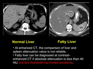
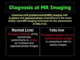
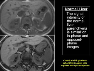
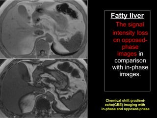
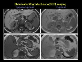
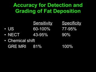
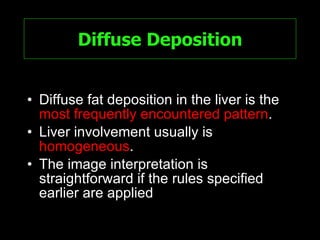
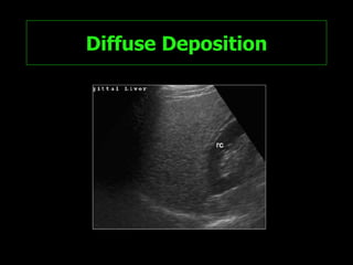
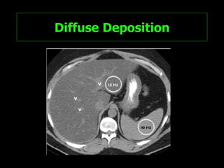
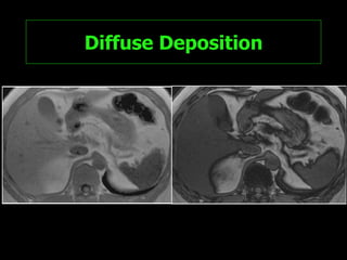
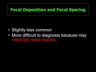
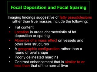
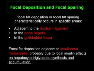
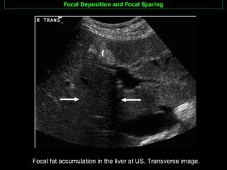
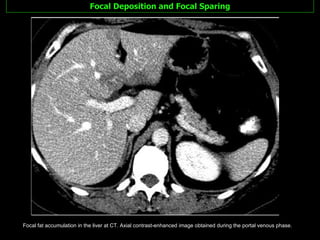
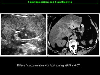
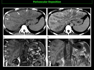
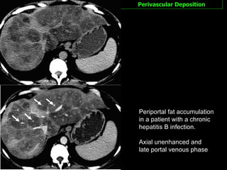
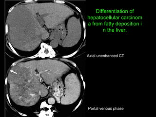
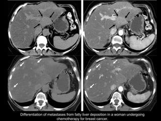
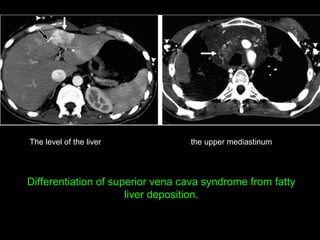
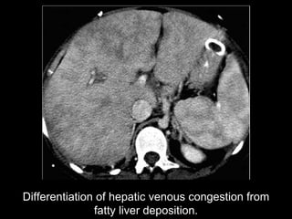
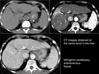
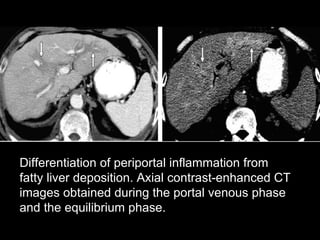
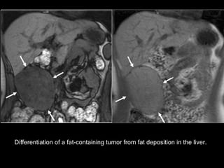


Fatty liver has various imaging patterns that can mimic other conditions, leading to unnecessary testing. The most common pattern is diffuse and homogeneous fat deposition, but other patterns include focal deposits, diffuse with focal sparing, and perivascular or subcapsular deposits. A diagnosis of fatty liver can be made with ultrasound, CT or MRI by applying established criteria regarding liver echogenicity and attenuation compared to the spleen. Chemical shift MRI is the most reliable method for assessing fat content when findings are equivocal. Considering features like location, morphology, enhancement and mass effect usually allows differentiation of fat deposits from tumors or other abnormalities.


