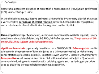
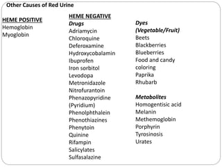
Dr Jitendra verma
Assistant prof.
Dept of pediatric medicine
RVRS Medical collage,Bhilwara
Clinical Evaluation of the Child
With
Hematuria
Defination
Hematuria, persistent presence of more than 5 red blood cells (RBCs)/high power field
(HPF) in uncentrifuged urine.
In the clinical setting, qualitative estimates are provided by a urinary dipstick that uses
a very sensitive peroxidase chemical reaction between hemoglobin (or myoglobin)
and a colorimetric chemical indicator impregnated on the dipstick.
Chemstrip (Boehringer Mannheim), a common commercially available dipstick, is very
sensitive and capable of detecting 3-5 RBCs/HPF of unspun urine. The presence of 10-
50 RBCs/μL may suggest underlying pathology,
significant hematuria is generally considered as > 50 RBCs/HPF. False-negative results
can occur in the presence of formalin (used as a urine preservative) or high urinary
concentrations of ascorbic acid (i.e., in patients with vitamin C intake > 2,000 mg/day).
False-positive results may be seen in a child with an alkaline urine (pH > 8), or more
commonly following contamination with oxidizing agents such as hydrogen peroxide
used to clean the perineum before obtaining a specimen.
Other Causes of Red Urine
HEME POSITIVE
Hemoglobin
Myoglobin
HEME NEGATIVE
Drugs
Adriamycin
Chloroquine
Deferoxamine
Hydroxycobalamin
Ibuprofen
Iron sorbitol
Levodopa
Metronidazole
Nitrofurantoin
Phenazopyridine
(Pyridium)
Phenolphthalein
Phenothiazines
Phenytoin
Quinine
Rifampin
Salicylates
Sulfasalazine
Dyes
(Vegetable/Fruit)
Beets
Blackberries
Blueberries
Food and candy
coloring
Paprika
Rhubarb
Metabolites
Homogentisic acid
Melanin
Methemoglobin
Porphyrin
Tyrosinosis
Urates
Evaluation of the child with hematuria begins with a careful history, physical
examination, and microscopic urinalysis. This information is used to determine the
level of hematuria (upper vs lower urinary tract) and to determine the urgency of the
evaluation based on symptomatology.
→Upper urinary tract sources of hematuria originate within the nephron (glomerulus,
tubular system, or interstitium).
•Color-brown, cola- or tea-colored, or burgundy urine.
•Proteinuria- > 100mg/dL via dipstick.
•microscopic findings - RBC casts, and deformed urinary RBCs (particularly
acanthocytes).
→Lower urinary tract sources of hematuria originate from the pelvocaliceal system,
ureter, bladder, or urethra.
•Color-bright red or pink.
•microscopic findings -blood clots, normal urinary RBC.
•proteinuria on dipstick is minimal (<100 mg/dL).
Causes of Hematuria in
Children
Isolated Renal Disease
1.IgA nephropathy (Berger
disease)
2.Alport syndrome (hereditary
nephritis)
3.Thin glomerular basement
membrane nephropathy
4.Postinfectious GN (PSGN)*
5.Membranous
nephropathy(MGN)
6.Membranoproliferative
GN(MPGN)
7.Rapidly progressive
GN(RPGN)
8.Focal segmental
glomerulosclerosis
(FSGS)
9.Anti–glomerular basement
membrane disease
Multisystem
Disease
1.SLE-nephritis*
2.HSP-nephritis
3.Granulomatosis
with polyangiitis
(formerly
4.Wegener
granulomatosis)
5.PAN
6.Goodpasture
syndrome
7.HUS
8.Sickle cell
glomerulopathy
9.HIV
nephropathy
Tubulointerstitial
Disease
1.Pyelonephritis
2.Interstitial nephritis
3.Papillary necrosis
4.Acute tubular necrosis
Vascular Disorders
1.Arterial or venous
thrombosis
Malformations
(aneurysms,
hemangiomas)
2.Nutcracker syndrome
3.Hemoglobinopathy
(sickle cell trait/disease)
4.Crystalluria
Anatomic Disorders
1.Hydronephrosis
2,Cystic-syndromic kidney disease
3.Polycystic kidney disease
4.Multicystic dysplasia
5.Tumor (Wilms tumor, rhabdomyosarcoma, angiomyolipoma, medullary
carcinoma)
6.Trauma
LOWER URINARY TRACT DISEASE
1. Inflammation (infectious and noninfectious)
2. Cystitis
3. Urethritis
4. Urolithiasis
5. Trauma
6. Coagulopathy
7. Heavy exercise
8. Bladder tumor
9. Factitious syndrome, factitious syndrome by proxy
Specific symptoms
1.Tea- or cola-colored urine, facial or body edema, hypertension, and oliguria classic
symptoms of glomerulonephritis.
2.A history of recent upper respiratory, skin, or gastrointestinal infection suggests
postinfectious glomerulonephritis, hemolytic-uremic syndrome, or HSP.
3.Rash and joint complaints suggest HSP or SLE nephritis.
4. Hematuria associated with glomerulonephritis is typically painless, but can be
associated with flank pain when acute or unusually severe.
5. Frequency, dysuria, and unexplained fevers suggest a urinary tract infection,
whereas renal colic suggests nephrolithiasis
,
6. A flank mass can suggest hydronephrosis, renal cystic diseases, renal vein
thrombosis, or tumor.
7. Hematuria associated with headache, mental status changes, visual changes
(diplopia), epistaxis, or heart failure suggests associated severe hypertension.
8. Child abuse must always be suspected in the child presenting with unexplained
perineal bruising and hematuria.
9. family history is critical in the initial assessment of isolated Alport syndrome, thin
glomerular basement membrane disease; SLE nephritis, HANAC, IgA
nephropathy,PCKD.
10. Physical examination may also suggest possible causes of hematuria.
11.Several malformation syndromes are associated with renal disease, including VATER
(vertebral-body anomalies, anal-atresia, trache-oesophageal fistula, and renal
dysplasia) syndrome.
12.Abdominal masses may be caused by bladder distention in posterior urethral
valves, hydronephrosis in ureteropelvic junction obstruction, polycystic kidney disease,
or Wilms tumor.
13. Hematuria seen in patients with neurologic or cutaneous abnormalities may be the
result of a number of syndromic renal disorders, including tuberous sclerosis, von
Hippel-Lindau syndrome, and Zellweger (cerebrohepatorenal) syndrome.
14. Anatomic abnormalities of the external genitalia may be associated with hematuria
and/or renal disease.
15.Urethrorrhagia --which is urethral bleeding in the absence of urine, is associated
with dysuria and blood spots on underwear after voiding. in pre-pubertal boys ,benign
self-limited course.
16.Recurrent episodes of gross hematuria suggest IgA nephropathy, Alport syndrome,
or thin glomerular basement membrane disease.
17.Dysuria and abdominal or flank pain are symptoms of idiopathic hypercalciuria, or
urolithiasis.
Asymptomatic patients with isolated microscopic
hematuria should not undergo extensive diagnostic
evaluation, because such hematuria is often
transient and benign.
Common Causes of Gross Hematuria--
1. Urinary tract infection--MCC
2. Meatal stenosis with ulcer
3. Perineal irritation
4. Trauma
5. Urolithiasis
6. Hypercalciuria
7. Obstruction
8. Coagulopathy
9. Tumor
10.Glomerular disease
11.Postinfectious glomerulonephritis
12.Henoch-Schönlein purpura nephritis
13.IgA nephropathy
14.Alport syndrome (hereditary nephritis)
15.Thin glomerular basement membrane disease
16.Systemic lupus erythematosus nephritis
CAUSE OF ANAEMIA
1.acute kidney injury --Anemia in this setting may be caused by hypervolemia with
dilution
2. chronic kidney disease --decreased RBC production(REDUCED ERYTHROPOITIN)
3.hemolytic-uremic syndrome- hemolysis
4. SLE – chronic hemolytic anemia,.
5. Goodpasture syndrome --blood loss from pulmonary hemorrhage.
6. HSP or hemolytic-uremic Syndrome—melena.
Diagnostic
algorithm
INVESTIGATIONS
1.CBC-- A. Thrombocytopenia
decreased platelet production--
(malignancies)
increased platelet consumption--
(SLE, idiopathic thrombocytopenic
purpura,
hemolytic-uremic syndrome, renal vein
thrombosis, or congenital hepatic
fibrosis with portal hypertension
secondary to autosomal recessive
polycystic
kidney disease)
B. Low Hb
C.Reduced RBCs
2. PBF-- A. microangiopathic process consistent with the hemolytic-uremic
syndrome
B. Shape of RBCs-upper and lower urinary tract.
3. Chemistry
- Electrolytes, BUN/ Creatinine /Creatinine clearance, Serum protein/Albumin
/Cholesterol
‘’ A bleeding diathesis is an unusual cause of hematuria,
and coagulation studies are not routinely obtained unless a
personal or family history suggests a bleeding tendency.’’
4. Level of C3---Low in PIGN in 90% cases (due to activation of compliment
pathway.
return to normal within 6 weeks.
C4 level remains normal.
Reduced C3 in - Post infectious glomerulonephritis
- Systemic lupus nephritis (and low C4)
- Nephritis with chronic infection
- Membrano proliferative
glomerulonephritis
5. ANA
6.ASO/Anti-DNase B
5.voiding cystourethrogram-- only required in patients with a urinary tract
infection, renal scarring, hydroureter, or pyelocaliectasis.
6 Cystoscopy --an unnecessary and costly procedure in most pediatric
patients with hematuria, and carries the associated risks of anesthesia. This
procedure should be reserved for
evaluating the rare child with a bladder mass noted on ultrasound, urethral
abnormalities caused by trauma, posterior urethral
valves, or tumor.
7. Children with persistent asymptomatic isolated hematuria and a completel
normal evaluation should have their blood pressure and urine checked every
3 mo until the hematuria resolves.
8. Renal biopsy is indicated for some children with persistent microscopic
hematuria and for most children with recurrent gross hematuria associated with
decreased renal function, proteinuria, or hypertension.
Source. 1.Nelson 21st edition
2. Kishor pediatric
nephrology
THANK YOU
Dr Jitendra verma
Assistant prof.
Dept of pediatric medicine
RVRS Medical collage,Bhilwara

Hematuria.pptx

  • 1.
    Dr Jitendra verma Assistantprof. Dept of pediatric medicine RVRS Medical collage,Bhilwara Clinical Evaluation of the Child With Hematuria
  • 2.
    Defination Hematuria, persistent presenceof more than 5 red blood cells (RBCs)/high power field (HPF) in uncentrifuged urine. In the clinical setting, qualitative estimates are provided by a urinary dipstick that uses a very sensitive peroxidase chemical reaction between hemoglobin (or myoglobin) and a colorimetric chemical indicator impregnated on the dipstick. Chemstrip (Boehringer Mannheim), a common commercially available dipstick, is very sensitive and capable of detecting 3-5 RBCs/HPF of unspun urine. The presence of 10- 50 RBCs/μL may suggest underlying pathology, significant hematuria is generally considered as > 50 RBCs/HPF. False-negative results can occur in the presence of formalin (used as a urine preservative) or high urinary concentrations of ascorbic acid (i.e., in patients with vitamin C intake > 2,000 mg/day). False-positive results may be seen in a child with an alkaline urine (pH > 8), or more commonly following contamination with oxidizing agents such as hydrogen peroxide used to clean the perineum before obtaining a specimen.
  • 3.
    Other Causes ofRed Urine HEME POSITIVE Hemoglobin Myoglobin HEME NEGATIVE Drugs Adriamycin Chloroquine Deferoxamine Hydroxycobalamin Ibuprofen Iron sorbitol Levodopa Metronidazole Nitrofurantoin Phenazopyridine (Pyridium) Phenolphthalein Phenothiazines Phenytoin Quinine Rifampin Salicylates Sulfasalazine Dyes (Vegetable/Fruit) Beets Blackberries Blueberries Food and candy coloring Paprika Rhubarb Metabolites Homogentisic acid Melanin Methemoglobin Porphyrin Tyrosinosis Urates
  • 4.
    Evaluation of thechild with hematuria begins with a careful history, physical examination, and microscopic urinalysis. This information is used to determine the level of hematuria (upper vs lower urinary tract) and to determine the urgency of the evaluation based on symptomatology.
  • 5.
    →Upper urinary tractsources of hematuria originate within the nephron (glomerulus, tubular system, or interstitium). •Color-brown, cola- or tea-colored, or burgundy urine. •Proteinuria- > 100mg/dL via dipstick. •microscopic findings - RBC casts, and deformed urinary RBCs (particularly acanthocytes). →Lower urinary tract sources of hematuria originate from the pelvocaliceal system, ureter, bladder, or urethra. •Color-bright red or pink. •microscopic findings -blood clots, normal urinary RBC. •proteinuria on dipstick is minimal (<100 mg/dL).
  • 6.
    Causes of Hematuriain Children Isolated Renal Disease 1.IgA nephropathy (Berger disease) 2.Alport syndrome (hereditary nephritis) 3.Thin glomerular basement membrane nephropathy 4.Postinfectious GN (PSGN)* 5.Membranous nephropathy(MGN) 6.Membranoproliferative GN(MPGN) 7.Rapidly progressive GN(RPGN) 8.Focal segmental glomerulosclerosis (FSGS) 9.Anti–glomerular basement membrane disease Multisystem Disease 1.SLE-nephritis* 2.HSP-nephritis 3.Granulomatosis with polyangiitis (formerly 4.Wegener granulomatosis) 5.PAN 6.Goodpasture syndrome 7.HUS 8.Sickle cell glomerulopathy 9.HIV nephropathy Tubulointerstitial Disease 1.Pyelonephritis 2.Interstitial nephritis 3.Papillary necrosis 4.Acute tubular necrosis Vascular Disorders 1.Arterial or venous thrombosis Malformations (aneurysms, hemangiomas) 2.Nutcracker syndrome 3.Hemoglobinopathy (sickle cell trait/disease) 4.Crystalluria
  • 7.
    Anatomic Disorders 1.Hydronephrosis 2,Cystic-syndromic kidneydisease 3.Polycystic kidney disease 4.Multicystic dysplasia 5.Tumor (Wilms tumor, rhabdomyosarcoma, angiomyolipoma, medullary carcinoma) 6.Trauma LOWER URINARY TRACT DISEASE 1. Inflammation (infectious and noninfectious) 2. Cystitis 3. Urethritis 4. Urolithiasis 5. Trauma 6. Coagulopathy 7. Heavy exercise 8. Bladder tumor 9. Factitious syndrome, factitious syndrome by proxy
  • 8.
    Specific symptoms 1.Tea- orcola-colored urine, facial or body edema, hypertension, and oliguria classic symptoms of glomerulonephritis. 2.A history of recent upper respiratory, skin, or gastrointestinal infection suggests postinfectious glomerulonephritis, hemolytic-uremic syndrome, or HSP. 3.Rash and joint complaints suggest HSP or SLE nephritis. 4. Hematuria associated with glomerulonephritis is typically painless, but can be associated with flank pain when acute or unusually severe. 5. Frequency, dysuria, and unexplained fevers suggest a urinary tract infection, whereas renal colic suggests nephrolithiasis ,
  • 9.
    6. A flankmass can suggest hydronephrosis, renal cystic diseases, renal vein thrombosis, or tumor. 7. Hematuria associated with headache, mental status changes, visual changes (diplopia), epistaxis, or heart failure suggests associated severe hypertension. 8. Child abuse must always be suspected in the child presenting with unexplained perineal bruising and hematuria. 9. family history is critical in the initial assessment of isolated Alport syndrome, thin glomerular basement membrane disease; SLE nephritis, HANAC, IgA nephropathy,PCKD. 10. Physical examination may also suggest possible causes of hematuria. 11.Several malformation syndromes are associated with renal disease, including VATER (vertebral-body anomalies, anal-atresia, trache-oesophageal fistula, and renal dysplasia) syndrome.
  • 10.
    12.Abdominal masses maybe caused by bladder distention in posterior urethral valves, hydronephrosis in ureteropelvic junction obstruction, polycystic kidney disease, or Wilms tumor. 13. Hematuria seen in patients with neurologic or cutaneous abnormalities may be the result of a number of syndromic renal disorders, including tuberous sclerosis, von Hippel-Lindau syndrome, and Zellweger (cerebrohepatorenal) syndrome. 14. Anatomic abnormalities of the external genitalia may be associated with hematuria and/or renal disease. 15.Urethrorrhagia --which is urethral bleeding in the absence of urine, is associated with dysuria and blood spots on underwear after voiding. in pre-pubertal boys ,benign self-limited course. 16.Recurrent episodes of gross hematuria suggest IgA nephropathy, Alport syndrome, or thin glomerular basement membrane disease. 17.Dysuria and abdominal or flank pain are symptoms of idiopathic hypercalciuria, or urolithiasis.
  • 11.
    Asymptomatic patients withisolated microscopic hematuria should not undergo extensive diagnostic evaluation, because such hematuria is often transient and benign.
  • 12.
    Common Causes ofGross Hematuria-- 1. Urinary tract infection--MCC 2. Meatal stenosis with ulcer 3. Perineal irritation 4. Trauma 5. Urolithiasis 6. Hypercalciuria 7. Obstruction 8. Coagulopathy 9. Tumor 10.Glomerular disease 11.Postinfectious glomerulonephritis 12.Henoch-Schönlein purpura nephritis 13.IgA nephropathy 14.Alport syndrome (hereditary nephritis) 15.Thin glomerular basement membrane disease 16.Systemic lupus erythematosus nephritis
  • 13.
    CAUSE OF ANAEMIA 1.acutekidney injury --Anemia in this setting may be caused by hypervolemia with dilution 2. chronic kidney disease --decreased RBC production(REDUCED ERYTHROPOITIN) 3.hemolytic-uremic syndrome- hemolysis 4. SLE – chronic hemolytic anemia,. 5. Goodpasture syndrome --blood loss from pulmonary hemorrhage. 6. HSP or hemolytic-uremic Syndrome—melena.
  • 14.
  • 15.
    INVESTIGATIONS 1.CBC-- A. Thrombocytopenia decreasedplatelet production-- (malignancies) increased platelet consumption-- (SLE, idiopathic thrombocytopenic purpura, hemolytic-uremic syndrome, renal vein thrombosis, or congenital hepatic fibrosis with portal hypertension secondary to autosomal recessive polycystic kidney disease) B. Low Hb C.Reduced RBCs 2. PBF-- A. microangiopathic process consistent with the hemolytic-uremic syndrome B. Shape of RBCs-upper and lower urinary tract. 3. Chemistry - Electrolytes, BUN/ Creatinine /Creatinine clearance, Serum protein/Albumin /Cholesterol
  • 16.
    ‘’ A bleedingdiathesis is an unusual cause of hematuria, and coagulation studies are not routinely obtained unless a personal or family history suggests a bleeding tendency.’’ 4. Level of C3---Low in PIGN in 90% cases (due to activation of compliment pathway. return to normal within 6 weeks. C4 level remains normal. Reduced C3 in - Post infectious glomerulonephritis - Systemic lupus nephritis (and low C4) - Nephritis with chronic infection - Membrano proliferative glomerulonephritis 5. ANA 6.ASO/Anti-DNase B
  • 17.
    5.voiding cystourethrogram-- onlyrequired in patients with a urinary tract infection, renal scarring, hydroureter, or pyelocaliectasis. 6 Cystoscopy --an unnecessary and costly procedure in most pediatric patients with hematuria, and carries the associated risks of anesthesia. This procedure should be reserved for evaluating the rare child with a bladder mass noted on ultrasound, urethral abnormalities caused by trauma, posterior urethral valves, or tumor. 7. Children with persistent asymptomatic isolated hematuria and a completel normal evaluation should have their blood pressure and urine checked every 3 mo until the hematuria resolves. 8. Renal biopsy is indicated for some children with persistent microscopic hematuria and for most children with recurrent gross hematuria associated with decreased renal function, proteinuria, or hypertension.
  • 18.
    Source. 1.Nelson 21stedition 2. Kishor pediatric nephrology
  • 19.
    THANK YOU Dr Jitendraverma Assistant prof. Dept of pediatric medicine RVRS Medical collage,Bhilwara