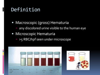
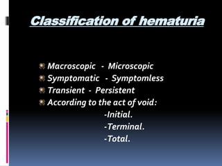
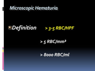
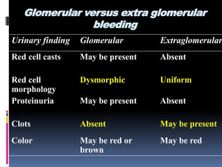
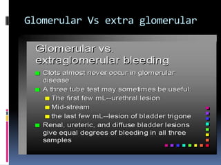
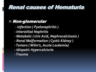
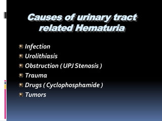
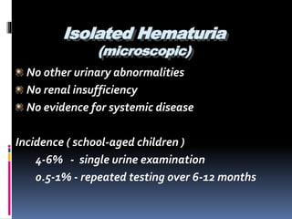
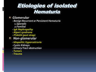
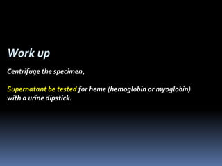
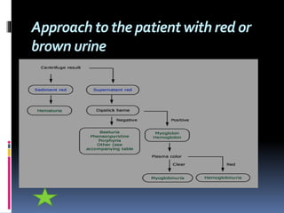
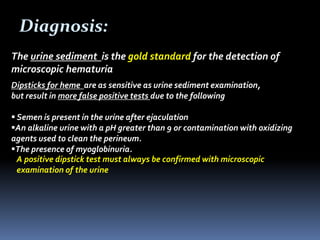
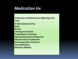
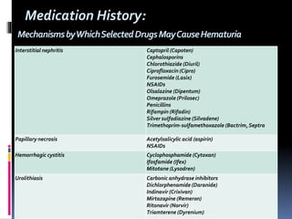
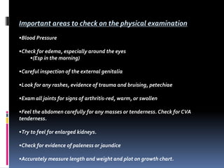
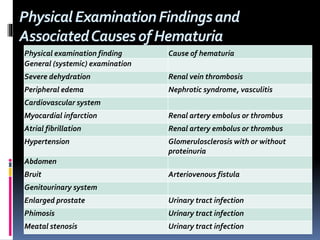
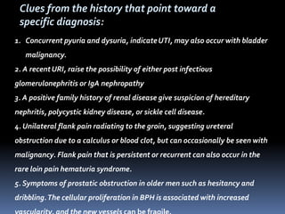
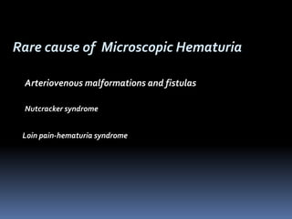
The document discusses the evaluation of hematuria. It defines macroscopic and microscopic hematuria and provides a classification. Important clues from the history and physical exam are outlined that can point to specific diagnoses. Distinguishing between glomerular and extraglomerular bleeding is important. Common causes of hematuria are reviewed including those related to the kidney, urinary tract, and unrelated conditions. Transient versus persistent hematuria is an important distinction to make in guiding evaluation and workup. Recommended initial laboratory tests and potential radiologic tests are presented.
The document provides guidelines for evaluating a patient presenting with hematuria. It begins with terminology and background on gross versus microscopic hematuria. Common causes are discussed including infection, stones, trauma, and tumors. The evaluation involves taking a thorough history, physical exam, urinalysis, and based on risk factors, further tests may include renal imaging, cystoscopy, or nephrology referral. The goal is to identify any underlying renal disease or urologic malignancy as the cause of the hematuria.
Hematuria, or blood in the urine, can be caused by diseases of the urinary system or other systemic disorders. It is classified as microscopic or gross based on visibility, and as early, terminal, or diffuse based on timing during urination. Common causes include glomerular diseases, infections, cancers, trauma, and stones. Diagnosis involves urinalysis, microscopy, imaging, and sometimes kidney biopsy. Treatment focuses on the underlying condition causing the hematuria. Prognosis depends on associated clinical or laboratory abnormalities, with isolated microscopic hematuria generally having a good prognosis.
Hematuria is defined as the presence of red blood cells in urine. It can be classified as gross or microscopic based on visibility. Hematuria requires investigation to identify underlying causes like malignancy, stones, or renal disease. Evaluation involves history, exam, urinalysis, imaging, and potentially biopsy. Treatment depends on the identified cause, which may include surgery, chemotherapy, or monitoring for progressive disease. Differential diagnosis considers non-blood substances that can appear red in urine. The epidemiology of hematuria depends on demographics and the primary disease process causing red urine.
1. The document provides guidance on evaluating a patient presenting with red or brown colored urine (hematuria). An initial workup includes a urine analysis and microscopy to determine the source and characteristics of the red blood cells.
2. Further evaluation depends on whether the hematuria is glomerular or extraglomerular in origin, which can be suggested by factors like RBC morphology and presence of proteinuria. Potential causes include infections, stones, tumors, and glomerular diseases.
3. If hematuria persists after initial workup, referral to nephrology or urology may be warranted for cystoscopy, imaging tests, or renal biopsy to identify the underlying cause. Thorough history and physical
This document discusses hematuria, or the presence of red blood cells in urine. It defines gross hematuria as visible red or brown urine, and microscopic hematuria as 3 or more red blood cells per high-powered field in urine samples. The causes, patterns, and workup of hematuria are described. Transient microscopic hematuria is common and often does not require extensive evaluation unless the patient is over 50 or has other risk factors. Evaluation of persistent hematuria may involve urine analysis, cystoscopy, imaging tests, and renal biopsy depending on findings.
This document provides information on hematuria (blood in the urine) including causes, evaluation, diagnosis, and management. It discusses glomerular and non-glomerular causes of hematuria and lists various renal diseases that can cause hematuria such as IgA nephropathy, Alport syndrome, thin basement membrane disease, and membranoproliferative glomerulonephritis. The evaluation of hematuria involves urine analysis, urine culture, renal function tests, imaging studies, and sometimes renal biopsy. Management depends on the underlying cause and may include antibiotics, diuretics, immunosuppressive therapy, dialysis, or surgery.
The document discusses hematuria (blood in the urine) including its definition, classification, causes, evaluation, and management. It addresses topics such as the difference between glomerular and extraglomerular hematuria, post-streptococcal glomerulonephritis (PSGN), and lupus nephritis. The key points are:
1) Hematuria can be either microscopic or macroscopic and can have many potential causes including infections, tumors, trauma, inflammation, and structural abnormalities.
2) Evaluation of hematuria involves urine analysis, blood tests, imaging, and sometimes renal biopsy to determine the cause and guide treatment.
3) Glomerular hematuria is suggested by
What is Urine
Indication of UA
Methods of collection of urine sample
Types of urine sample
Macroscopic examination of urine
Chemical examination of urine
Microscopic examination of urine
Hematuria, or blood in the urine, is a common reason children are referred to pediatricians and nephrologists. While sometimes transient and insignificant, hematuria can also be a sign of serious renal disease. The document outlines the evaluation and workup of hematuria in children, noting that urinalysis, history, and physical exam usually guide the cause. For isolated microscopic hematuria, repeat testing and minimal workup can rule out significant disease in most cases. However, if hematuria presents with proteinuria, rising creatinine, failure to thrive, or hypertension, it increases the likelihood of an underlying serious renal condition requiring further evaluation.
1. The patient, a 48-year-old otherwise healthy woman, presented with microscopic hematuria on a routine urinalysis.
2. Microscopic hematuria is defined as more than 3 red blood cells per high-power field on urinalysis.
3. Evaluation of the patient with hematuria includes urine analysis, urine culture if indicated by presence of white blood cells, renal ultrasound or CT urogram, cystoscopy if lower urinary tract is suspected, and further testing depending on initial findings.
Approch to Hematuria in pediatric age groupMohammed Saadi
The document defines hematuria and its types, identifies common causes like UTIs and glomerulonephritis, and outlines the approach to evaluating and managing a patient with hematuria. This involves taking a thorough history, conducting a physical exam, ordering urine and blood tests to identify the cause, and treating any underlying conditions found like cystitis, pyelonephritis, or acute glomerulonephritis. Imaging tests may also be used to identify issues like stones, tumors, or structural abnormalities.
This document defines hematuria as the presence of red blood cells in the urine and discusses its types, causes, evaluation, and management. It outlines the main objectives as understanding the definition and types of hematuria, identifying the causes, and how to investigate and treat patients with hematuria. Key points include that hematuria can be grossly visible or microscopic, and its causes can be structural disruptions in the kidneys, infections, tumors, bleeding disorders, or toxic insults to the kidneys. The evaluation of hematuria involves taking a medical history, physical exam, urine analysis, blood tests, imaging, and sometimes renal biopsy. Treatment depends on the underlying cause but may include antibiotics for infections, supportive care, immunosuppress
Approach to Hematuria including:
Definition of Hematuria.
Pathophysiology of Hematuria.
Differential Diagnosis of Red Urine.
Causes of Hematuria.
Approach to a patient with Hematuria.
Diagnostic Algorithms.
Management and Disposition.
Hematuria refers to the presence of blood in the urine. A diagnosis requires red blood cells to be present in urine samples obtained at least a week apart. Hematuria can be classified as microscopic or macroscopic, intermittent or persistent, and by its location in the urinary tract. Potential causes include glomerular disease, tumors, infections, vascular abnormalities, stones and trauma. Evaluation involves urinalysis, urine culture, imaging tests like ultrasound and CT urography, and cystoscopy depending on risk factors. Treatment focuses on the underlying cause if identified, while asymptomatic microscopic hematuria often requires monitoring without intervention.
This document discusses hematuria, or the presence of blood in the urine. Evaluation is warranted when hematuria is present, as it can be a sign of medical or urological issues. An initial workup includes a medical history, physical exam, urine analysis, and urine microscopy. Further imaging with ultrasound, CT scan, or cystoscopy may be used to investigate the urinary tract for causes like cancer, stones, infections, or structural abnormalities. For high risk patients, especially those older than 40 or with a history of smoking, a cystoscopy and urine cytology are recommended to screen for bladder cancer. Most cases of asymptomatic microscopic hematuria remain unexplained despite a full urological evaluation.
1. Hematuria is defined as more than 5 red blood cells per high power field in urine. It can be transient and microscopic or gross.
2. Causes of hematuria include glomerular and non-glomerular diseases of the kidneys, as well as conditions of the ureters, bladder, and urethra.
3. Evaluation of hematuria involves microscopic examination to confirm red blood cells and exclude other causes that can cause a positive dipstick. Further tests are aimed at identifying the specific underlying condition and its location.
1. Hematuria is defined as more than 5 red blood cells per high power field in urine. It can be transient and microscopic or gross.
2. Causes of hematuria include glomerular and non-glomerular diseases of the kidneys, ureters, bladder, and urethra as well as coagulation disorders and malignancies.
3. Evaluation of hematuria involves urine microscopy to exclude myoglobinuria or hemoglobinuria, urine culture, renal ultrasound, and possibly renal biopsy depending on the suspected cause and severity.
Hematuria in children can be detected by urinary dipstick or microscopic examination. Common causes include glomerular disease, hypercalciuria, and nutcracker syndrome. Evaluation depends on whether hematuria is isolated, accompanied by proteinuria, or symptomatic. For isolated hematuria, follow-up includes urine culture and screening for hypercalciuria if persistent. Hematuria with proteinuria warrants further testing including serum creatinine. Symptomatic hematuria evaluation includes history, physical exam, and urinalysis to determine glomerular vs extraglomerular cause. Renal biopsy is considered if hematuria is substantial or progressive.
This document discusses upper gastrointestinal bleeding (UGIB). It defines UGIB as bleeding from the gastrointestinal tract proximal to the ligament of Trietz, which usually manifests as hematemesis or melena and sometimes hematochezia. Risk stratification systems like the Blatchford and Rockall scores are used to predict outcomes like rebleeding and mortality. Endoscopy is important for diagnosis, prognosis, and potential therapy. Management involves fluid resuscitation, identifying the bleeding source, stopping active bleeding, treating underlying causes, and preventing rebleed. Proton pump inhibitors are the standard medical treatment. Endoscopic modalities like injection, thermal, and mechanical methods are used for non-variceal bleeding.
Chronic liver disease in children22.pptxAmmaraHameed6
This document discusses chronic liver disease and portal hypertension in children. It defines chronic liver disease as ongoing liver injury for at least 6 months that can progress to cirrhosis and liver failure. Cirrhosis is the end result of liver cell damage and destruction. The etiology of chronic liver disease in children varies according to age and includes infectious, metabolic, autoimmune, anatomical, and toxic/drug-induced causes. Some common chronic liver diseases in children discussed in detail include biliary atresia, alpha-1 antitrypsin deficiency, cystic fibrosis, and Wilson's disease.
The document discusses nephrolithiasis (kidney stones) and pyelonephritis (kidney infection). It covers the types, risk factors, pathogenesis, clinical manifestations, diagnosis and treatment of both conditions. Calcium oxalate stones are the most common type of kidney stones. Risk factors for stone formation include dietary factors like calcium intake as well as urinary abnormalities. Pyelonephritis is commonly caused by gram-negative bacteria ascending from the bladder. It can cause kidney swelling and damage if left untreated.
This document defines hematuria and describes its classification and various etiologies. It can be caused by diseases of the urinary system, glomerular issues, interstitial diseases, uroepithelium malignancy, systemic disorders, adjacent organ diseases, drugs, and unknown sources. Clinical features depend on red blood cell amount and urine pH. Differential diagnosis considers contaminated urine, drugs, porphyria, and hemoglobinuria. Evaluation includes a three-glass urine test, accompanying symptoms, laboratory tests, renal biopsy if indicated, and cystoscopy or imaging tests. Glomerular bleeding shows red cell casts, dysmorphic cells, and proteinuria while extraglomerular bleeding lacks casts and shows uniform cells.
Hematuria can be caused by various upper and lower urinary tract diseases. Common causes of glomerular hematuria include IgA nephropathy, Alport syndrome, and thin basement membrane disease. Post-streptococcal glomerulonephritis is associated with a preceding streptococcal infection. Membranous nephropathy presents with nephrotic syndrome. Goodpasture disease involves anti-GBM antibodies attacking the lungs and kidneys. Diagnosis involves urinalysis, renal biopsy, and identifying underlying causes or associations. Treatment depends on the specific condition but may include antibiotics, steroids, immunosuppressants, blood pressure control, and addressing complications.
This document discusses hematuria (blood in the urine) and obstructive uropathy (blockage of urine flow in the urinary tract). It covers evaluating hematuria through urinalysis, imaging tests, and cystoscopy. Common causes of hematuria include infections, stones, tumors, and glomerulonephritis. Obstructive uropathy can be congenital or acquired and cause changes to the urethra, bladder, ureters, and kidneys over time. Relieving the obstruction through surgery, stents, or nephrostomy is the main treatment approach.
These lecture slides, by Dr Sidra Arshad, offer a quick overview of the physiological basis of a normal electrocardiogram.
Learning objectives:
1. Define an electrocardiogram (ECG) and electrocardiography
2. Describe how dipoles generated by the heart produce the waveforms of the ECG
3. Describe the components of a normal electrocardiogram of a typical bipolar lead (limb II)
4. Differentiate between intervals and segments
5. Enlist some common indications for obtaining an ECG
6. Describe the flow of current around the heart during the cardiac cycle
7. Discuss the placement and polarity of the leads of electrocardiograph
8. Describe the normal electrocardiograms recorded from the limb leads and explain the physiological basis of the different records that are obtained
9. Define mean electrical vector (axis) of the heart and give the normal range
10. Define the mean QRS vector
11. Describe the axes of leads (hexagonal reference system)
12. Comprehend the vectorial analysis of the normal ECG
13. Determine the mean electrical axis of the ventricular QRS and appreciate the mean axis deviation
14. Explain the concepts of current of injury, J point, and their significance
Study Resources:
1. Chapter 11, Guyton and Hall Textbook of Medical Physiology, 14th edition
2. Chapter 9, Human Physiology - From Cells to Systems, Lauralee Sherwood, 9th edition
3. Chapter 29, Ganong’s Review of Medical Physiology, 26th edition
4. Electrocardiogram, StatPearls - https://www.ncbi.nlm.nih.gov/books/NBK549803/
5. ECG in Medical Practice by ABM Abdullah, 4th edition
6. Chapter 3, Cardiology Explained, https://www.ncbi.nlm.nih.gov/books/NBK2214/
7. ECG Basics, http://www.nataliescasebook.com/tag/e-c-g-basics
Cell Therapy Expansion and Challenges in Autoimmune DiseaseHealth Advances
There is increasing confidence that cell therapies will soon play a role in the treatment of autoimmune disorders, but the extent of this impact remains to be seen. Early readouts on autologous CAR-Ts in lupus are encouraging, but manufacturing and cost limitations are likely to restrict access to highly refractory patients. Allogeneic CAR-Ts have the potential to broaden access to earlier lines of treatment due to their inherent cost benefits, however they will need to demonstrate comparable or improved efficacy to established modalities.
In addition to infrastructure and capacity constraints, CAR-Ts face a very different risk-benefit dynamic in autoimmune compared to oncology, highlighting the need for tolerable therapies with low adverse event risk. CAR-NK and Treg-based therapies are also being developed in certain autoimmune disorders and may demonstrate favorable safety profiles. Several novel non-cell therapies such as bispecific antibodies, nanobodies, and RNAi drugs, may also offer future alternative competitive solutions with variable value propositions.
Widespread adoption of cell therapies will not only require strong efficacy and safety data, but also adapted pricing and access strategies. At oncology-based price points, CAR-Ts are unlikely to achieve broad market access in autoimmune disorders, with eligible patient populations that are potentially orders of magnitude greater than the number of currently addressable cancer patients. Developers have made strides towards reducing cell therapy COGS while improving manufacturing efficiency, but payors will inevitably restrict access until more sustainable pricing is achieved.
Despite these headwinds, industry leaders and investors remain confident that cell therapies are poised to address significant unmet need in patients suffering from autoimmune disorders. However, the extent of this impact on the treatment landscape remains to be seen, as the industry rapidly approaches an inflection point.
8 Surprising Reasons To Meditate 40 Minutes A Day That Can Change Your Life.pptxHolistified Wellness
We’re talking about Vedic Meditation, a form of meditation that has been around for at least 5,000 years. Back then, the people who lived in the Indus Valley, now known as India and Pakistan, practised meditation as a fundamental part of daily life. This knowledge that has given us yoga and Ayurveda, was known as Veda, hence the name Vedic. And though there are some written records, the practice has been passed down verbally from generation to generation.
Adhd Medication Shortage Uk - trinexpharmacy.comreignlana06
The UK is currently facing a Adhd Medication Shortage Uk, which has left many patients and their families grappling with uncertainty and frustration. ADHD, or Attention Deficit Hyperactivity Disorder, is a chronic condition that requires consistent medication to manage effectively. This shortage has highlighted the critical role these medications play in the daily lives of those affected by ADHD. Contact : +1 (747) 209 – 3649 E-mail : sales@trinexpharmacy.com
Promoting Wellbeing - Applied Social Psychology - Psychology SuperNotesPsychoTech Services
A proprietary approach developed by bringing together the best of learning theories from Psychology, design principles from the world of visualization, and pedagogical methods from over a decade of training experience, that enables you to: Learn better, faster!
Integrating Ayurveda into Parkinson’s Management: A Holistic ApproachAyurveda ForAll
Explore the benefits of combining Ayurveda with conventional Parkinson's treatments. Learn how a holistic approach can manage symptoms, enhance well-being, and balance body energies. Discover the steps to safely integrate Ayurvedic practices into your Parkinson’s care plan, including expert guidance on diet, herbal remedies, and lifestyle modifications.
Rasamanikya is a excellent preparation in the field of Rasashastra, it is used in various Kushtha Roga, Shwasa, Vicharchika, Bhagandara, Vatarakta, and Phiranga Roga. In this article Preparation& Comparative analytical profile for both Formulationon i.e Rasamanikya prepared by Kushmanda swarasa & Churnodhaka Shodita Haratala. The study aims to provide insights into the comparative efficacy and analytical aspects of these formulations for enhanced therapeutic outcomes.
Histololgy of Female Reproductive System.pptxAyeshaZaid1
Dive into an in-depth exploration of the histological structure of female reproductive system with this comprehensive lecture. Presented by Dr. Ayesha Irfan, Assistant Professor of Anatomy, this presentation covers the Gross anatomy and functional histology of the female reproductive organs. Ideal for students, educators, and anyone interested in medical science, this lecture provides clear explanations, detailed diagrams, and valuable insights into female reproductive system. Enhance your knowledge and understanding of this essential aspect of human biology.
Local Advanced Lung Cancer: Artificial Intelligence, Synergetics, Complex Sys...Oleg Kshivets
Overall life span (LS) was 1671.7±1721.6 days and cumulative 5YS reached 62.4%, 10 years – 50.4%, 20 years – 44.6%. 94 LCP lived more than 5 years without cancer (LS=2958.6±1723.6 days), 22 – more than 10 years (LS=5571±1841.8 days). 67 LCP died because of LC (LS=471.9±344 days). AT significantly improved 5YS (68% vs. 53.7%) (P=0.028 by log-rank test). Cox modeling displayed that 5YS of LCP significantly depended on: N0-N12, T3-4, blood cell circuit, cell ratio factors (ratio between cancer cells-CC and blood cells subpopulations), LC cell dynamics, recalcification time, heparin tolerance, prothrombin index, protein, AT, procedure type (P=0.000-0.031). Neural networks, genetic algorithm selection and bootstrap simulation revealed relationships between 5YS and N0-12 (rank=1), thrombocytes/CC (rank=2), segmented neutrophils/CC (3), eosinophils/CC (4), erythrocytes/CC (5), healthy cells/CC (6), lymphocytes/CC (7), stick neutrophils/CC (8), leucocytes/CC (9), monocytes/CC (10). Correct prediction of 5YS was 100% by neural networks computing (error=0.000; area under ROC curve=1.0).
- Video recording of this lecture in English language: https://youtu.be/kqbnxVAZs-0
- Video recording of this lecture in Arabic language: https://youtu.be/SINlygW1Mpc
- Link to download the book free: https://nephrotube.blogspot.com/p/nephrotube-nephrology-books.html
- Link to NephroTube website: www.NephroTube.com
- Link to NephroTube social media accounts: https://nephrotube.blogspot.com/p/join-nephrotube-on-social-media.html
share - Lions, tigers, AI and health misinformation, oh my!.pptxTina Purnat
• Pitfalls and pivots needed to use AI effectively in public health
• Evidence-based strategies to address health misinformation effectively
• Building trust with communities online and offline
• Equipping health professionals to address questions, concerns and health misinformation
• Assessing risk and mitigating harm from adverse health narratives in communities, health workforce and health system
share - Lions, tigers, AI and health misinformation, oh my!.pptx
10714657.ppt
1. Evaluation of the Hematuria,
MUHAMMAD Y EBRAHIM, MD.
Consultant Nephrologists SRMC & MBH
Director of inpatient dialysis at SRMC, SRCH & MBH.
Co-medical director at out patient dialysis unit at
Dialysis & Clinics Inc DCI.
Chief Department of Nephrology at SRMC.
4. Definition
Macroscopic (gross) Hematuria
any discolored urine visible to the human eye
Microscopic Hematuria
>5 RBC/hpf seen under microscope
5. Classification of hematuria
Macroscopic - Microscopic
Symptomatic - Symptomless
Transient - Persistent
According to the act of void:
-Initial.
-Terminal.
-Total.
8. Glomerular versus extra glomerular
bleeding
Urinary finding Glomerular Extraglomerular
Red cell casts May be present Absent
Red cell
morphology
Dysmorphic Uniform
Proteinuria May be present Absent
Clots Absent May be present
Color May be red or
brown
May be red
10. Causes of Hematuria
Kidney disease
Lesions along the urinary tract
Conditions unrelated to kidney and
urinary tract
11. Hematuria not representing
kidney or urinary tract disorder
Following exercise
Febrile disorders
Gastroenteritis with dehydration
Contamination from external
genitalia
16. Causes of urinary tract
related Hematuria
Infection
Urolithiasis
Obstruction ( UPJ Stenosis )
Trauma
Drugs ( Cyclophosphamide )
Tumors
17. Isolated Hematuria
(microscopic)
No other urinary abnormalities
No renal insufficiency
No evidence for systemic disease
Incidence ( school-aged children )
4-6% - single urine examination
0.5-1% - repeated testing over 6-12 months
20. Gross hematuria:
Suspected if a red or brown color change of urine
Intermittent red or brown color urine a/w variety of clinical setting
Medications (phenazopyridine, microbid, NSAID)
Ingestion of beets or certain dyes
Metabolities
Myoglobinuria or hemoglobinuria
If pass clot, indicate lower urinary source
21. Work up
Centrifuge the specimen,
Supernatant be tested for heme (hemoglobin or myoglobin)
with a urine dipstick.
22. Causes of heme-negative red urine
Medications Food dyes Metabolities
Doxorubicin
Beets (in selected
patients)
Bile pigments
Chloroquine Blackberries Homogentisic acid
Deferoxamine Food coloring Melanin
Ibuprofen Methemoglobin
Iron sorbitol Porphyrin
Nitrofurantoin Tyrosinosis
Phenazopyridine Urates
Phenolphthalein
Rifampin
25. Microscopic hematuria:
Accidental finding from UA or urine dipstick
3 or more RBC/hpf in spun urine sediment.
No "safe" lower limit below which significant disease can be excluded
Often asymptomatic
The degree of hematuria does not correlate with the seriousness of the
underlying cause of the bleeding.
26. Diagnosis:
The urine sediment is the gold standard for the detection of
microscopic hematuria
Dipsticks for heme are as sensitive as urine sediment examination,
but result in more false positive tests due to the following
Semen is present in the urine after ejaculation
An alkaline urine with a pH greater than 9 or contamination with oxidizing
agents used to clean the perineum.
The presence of myoglobinuria.
A positive dipstick test must always be confirmed with microscopic
examination of the urine
27. The evaluation should address the following
three questions
1. Are there any clues from the history or physical
examination that suggest a particular diagnosis?
2. Does the hematuria represent glomerular or
extraglomerular bleeding?
3. Is the hematuria transient or persistent?
28. Urethral: First 10-15 mL
Bladder: Final 10-30 mL
Upper urinary tract:Throughout
a three-tube test may also help to locate
the source of bleeding in selected cases.
30. Important questions to ask in patients History
•Has there been any signs of a UTI as dysuria & frequency? Any suprapubic pain?
•Has there been any recent URI symptoms or sore throat?
•Has there been any type of skin rashes or sores?
•Any abdominal pain or colicky pain?
•Are the stools loose or bloody?
•Has there been any recent trauma?
•Has there been any joint pains or swellings?
•Is there any history of sickle cell disease or trait?
•Is there any family history of renal disease, transplants, or dialysis? Is there a
family history of hearing deficits?
•What medications does the child take?
34. Important areas to check on the physical examination
•Blood Pressure
•Check for edema, especially around the eyes
•(Esp in the morning)
•Careful inspection of the external genitalia
•Look for any rashes, evidence of trauma and bruising, petechiae
•Exam all joints for signs of arthritis-red, warm, or swollen
•Feel the abdomen carefully for any masses or tenderness. Check for CVA
tenderness.
•Try to feel for enlarged kidneys.
•Check for evidence of paleness or jaundice
•Accurately measure length and weight and plot on growth chart.
35. PhysicalExaminationFindingsand
AssociatedCausesofHematuria
Physical examination finding Cause of hematuria
General (systemic) examination
Severe dehydration Renal vein thrombosis
Peripheral edema Nephrotic syndrome, vasculitis
Cardiovascular system
Myocardial infarction Renal artery embolus or thrombus
Atrial fibrillation Renal artery embolus or thrombus
Hypertension Glomerulosclerosis with or without
proteinuria
Abdomen
Bruit Arteriovenous fistula
Genitourinary system
Enlarged prostate Urinary tract infection
Phimosis Urinary tract infection
Meatal stenosis Urinary tract infection
36. 1. Concurrent pyuria and dysuria, indicateUTI, may also occur with bladder
malignancy.
2. A recentURI, raise the possibility of either post infectious
glomerulonephritis or IgA nephropathy
3. A positive family history of renal disease give suspicion of hereditary
nephritis, polycystic kidney disease, or sickle cell disease.
4. Unilateral flank pain radiating to the groin, suggesting ureteral
obstruction due to a calculus or blood clot, but can occasionally be seen with
malignancy. Flank pain that is persistent or recurrent can also occur in the
rare loin pain hematuria syndrome.
5. Symptoms of prostatic obstruction in older men such as hesitancy and
dribbling.The cellular proliferation in BPH is associated with increased
Clues from the history that point toward a
specific diagnosis:
37. 6. Recent vigorous exercise or trauma
7. History of a bleeding disorder or bleeding from multiple sites due to
uncontrolled anticoagulant therapy.
8. Cyclic hematuria in women that is most prominent during and shortly after
menstruation, suggesting endometriosis of the urinary tract .
9. Medications that might cause nephritis (usually with other findings,
typically with renal insufficiency).
10. AA should be screened for sickle cell trait or disease, which can lead to
papillary necrosis and hematuria.
11.Travel or residence in areas endemic forSchistosoma hematobium .
12.Sterile pyuria with hematuria, which may occur with renal tuberculosis,
analgesic nephropathy and other interstitial diseases.
Clues from the history that point toward a specific diagnosis:
40. Rare cause of Microscopic Hematuria
Arteriovenous malformations and fistulas
Nutcracker syndrome
Loin pain-hematuria syndrome
41. Arteriovenous malformations and fistulas — An AV malformation (AVM) or
fistula of the urologic tract may be either congenital or acquired.The primary
presenting sign is gross hematuria, but high-output heart failure and
hypertension also may be seen .The latter is presumably due to activation of
the renin-angiotensin system resulting from ischemia distal to the AVM
Nutcracker syndrome —The nutcracker syndrome refers to compression of the
left renal vein between the aorta and proximal superior mesenteric artery.
Nutcracker syndrome can cause both microscopic and gross hematuria, primarily
in children (but also adults) in Asia .The hematuria is usually asymptomatic but
may be associated with left flank pain. Nutcracker syndrome has also been
associated with orthostatic proteinuria.
Loin pain-hematuria syndrome —The loin pain-hematuria syndrome is a poorly
defined disorder characterized by loin or flank pain that is often severe and
unrelenting, and hematuria with dysmorphic red cell features suggesting a
glomerular origin. Affected patients usually have normal kidney function.
43. Exception:
Malignancy risk in older patients with transient
hematuria
In older patients, even transient hematuria carries an appreciable
risk of malignancy (assuming no evidence of glomerular bleeding)
The risks includes : age >50, smoker and Hx of analgesic abuse.
Transient hematuria
Transient microscopic hematuria is a common problem in adults
Fever, infection, trauma, and exercise are potential causes
It is reasonable to repeat an abnormal urinalysis in a few days
44. When persistent hematuria is essentially the only
manifestation of glomerular disease, one of three
disorders is most likely
IgA nephropathy, in which there is often gross hematuria, and
sometimes a positive family history but without any clear
pattern of autosomal inheritance
Alport syndrome (hereditary nephritis), in which gross
hematuria can occur in association with a positive family
history of renal failure, and sometimes deafness or corneal
abnormalities.
Thin basement membrane nephropathy (also called thin
basement membrane disease or benign familial hematuria), in
which gross hematuria is unusual and the family history may
be positive (with an autonomic dominant pattern of
inheritance) for microscopic hematuria but not for renal failure
.
45. LaboratoryTests (initial work up)
• Repeat UA in a few days
•UA and microscopy to determine the number and morphology of RBC, crystal and
casts,Consider urine Cx
• CBC, PT, INR, electrolytes, kidney function
•Further urologic evaluation is warranted if more than three RBC/phf are found on at
least 2 of 3 properly collected urine specimens or if high-grade microscopic hematuria
(>than 100 red blood cells per high-power field) is found on a single urinalysis.17
• Serum chemistries and serologic studies for glomerular causes of hematuria as
directed by the medical history ANA, C3 , C4, Hepatitis B and C, HIV, ESR, Anti DNA
and other lupus studies, ASO, ANCA, AntiGBM antibodies,SPEP, UPEP, M spike.
• Imaging stuidies like US kidney and Bladder . CT Scan, MRI , MRA, Renal angiogram ,
IVP, Cystoscopy, Retrograde Pyelogram, kidney biopsy
• Consultation to Nephrologist or Urologist
46. Radiologicandothertestsfortheevaluationofhematuria
Test Advantages Disadvantages
Intravenous pyelogram (IVP)
Excellent visualization of the
kidney, collecting system, and
ureter
May miss bladder lesions; can
cause nephrotoxicity,
idiosyncratic reactions (1/10,000)
Cystoscopy
Best way to examine the bladder,
which is not as well visualized by
IVP or ultrasound
Invasive, uncomfortable and
expensive
Ultrasound
If of good quality, as sensitive as
IVP for renal lesions, with less
morbidity and cost
Less sensitive than IVP for ureter
and bladder
Retrograde pyelography
The best test for examing the
ureters, can be combined with
cystoscopy
Invasive, not useful for examining
other parts of the urinary
collecting system
Urinary cytology
Sensitivity 67 percent, specificity
96 percent for uroepithelial
cancer
Useful only for cancer, mainly of
the bladder
CT scan
Excellent for examining the renal
parenchyma
Expensive
Angiography
Useful for gross hematuria when
other tests have not revealed the
cause; the only good test for
vascular malformations
Invasive, expensive
47. A biopsy is not usually performed for isolated
glomerular hematuria (i.e., no proteinuria or renal
insufficiency,) since there is no specific therapy for
these conditions, unless the patient is considering
becoming a kidney donor
However, biopsy should be considered if there
is evidence of progressive disease as
manifested by an elevation in the plasma
creatinine concentration, increasing protein
excretion, or an otherwise unexplained rise in
blood pressure, even when the values remain
within the normal range.
Renal Biopsy: