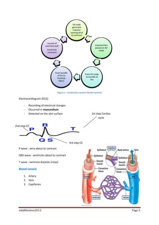
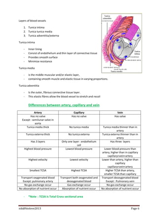
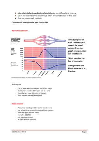
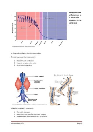
HEART
Heart structures (human)

   -     Enclosed by pericardium (double sac of serous
         membrane)
   -     Pericardium lined by squamous serous membrane
   -     Filled with serous fluid
   -     Serous fluid produced by serous pericardial membrane
   -     Serous fluid function to eliminate friction during beating

Layers :

   -     Epicardium
   -     Myocardium
              Act as barrier from spread of infection and inflammation from adjacent structures
   -     Endocardium

Valves

   -     Artrioventricular valve (AV)
              Between atria and ventricles
              Right side referred as tricuspid valve
              Left side referred as bicuspid valve or
                 mitral valve
   -     Semilunar valve
              Between ventricle and artery
              At Pulmonary artery (pulmonary
                 semilunar valve)
              At aorta known as Aortic semilunar valve


Why pulmonary circuit is a short loop?

   -     Because start at heart right half and go to the lung and into heart
         left half


Systemic circuit is a longer loop

   -     Because start at heart left half and end at the heart right half


nik@biolove2013                                                                             Page 1
Cardiac cycle

Referred as one complete heart beat

Systole – contract

Diastole – relax


    1 - atrial   - Blood flow into atria                                 2- atrial    - remaining blood is pushed out from
       and       and ventricle through                                    systole     atria to ventricle
   ventricular   AV valve                                               ventricular   - blood rushes out from ventricle due to
    diastole     - AV valve open,                                        diastole     high pressure
                 semilunar valve closed                                               -AV open
                                                                                      - semilunar valve closed
                                             3- atria
                                            diastole
                                           ventricular   - blood pushes out
                                                         from ventricle
                                             systole     completely
                                                         -semilunar valve open
                                                         -atrioventricular
                                                         closed


‘lup’ – closing of atroventricular valve

‘dup’ – closing of semilunar valve

Cardiac output

    -    Amount of blood pumped by each ventricle in one minute
    -    Product of heart rate and stroke volume
    -    Normal : 5liter/min


nik@biolove2013                                                                                                           Page 2
Heart rate

   -   Pulse/number of heart beat per minute
   -   Normal : 75 beats/min

Stroke volume

   -   Amount of blood pumped out by each ventricle in each heart beat
   -   70ml/beat
   -   Regulation depend on venous return

Venous return

   -   Amount of blood entering the heart

Question : Why our heart beat become fast after exercise?

   -   Skeletal muscle contract and relax causing blood flow to be faster
   -   Speed up venous return
   -   Venous return increase, stroke volume increase
   -   Causing more contraction

Regulation of heart beat

                                                                  - signal passes to AV node
                                                                     - ventricle contract
  - sinoatrial node (SA node) generate impulse
                      to atria
                - atria contract


   -   During stress or physical activities nerves of sympathetic division triggers AV and SA node to
       increase heart beat.
   -   Parasympathetic nerves slow down heart rate.
   -   Hormone :
             Epinephrine and thyroxine increase heart rate
   -   Ions :
             Low ion, low heart rate
   -   Other factors affecting heart rate :
             Age
             Gender
             Body Temp.
             Activities


nik@biolove2013                                                                                Page 3
Conduction system of the heart

Two systems :

        Autonomic nervous system
    -   Slow down or speeds heart rate
    -   Depend on which division it activated

        Nodal system or intrinsic condustion system
    -   A specialized tissue
    -   Function as it is a combination of muscle and nervous tissue

Nodal system

    1) Depend on AV node and SA node
    2) SA node located at right atrium
    3) Also called as pacemaker because it starts the heart beat
            From SA node, impulse spread to the atria
            Atria contract
            Then spread to AV node
    4) AV node located at the junction of atria and ventricle
    5) Then impulse send to bundle of His
    6) Then spread to Purkinje fibers
    7) From purkinje spread to muscle of ventricle walls

At AV node, impulse is delayed because to wait for atria to finish contract


nik@biolove2013                                                               Page 4
SA node
                                                      generate
                                                       impulse
                                                    causing atria
                                                     to contract


                           muscle of
                         ventricle wall                                      impulse then
                                                                             passes to AV
                           (ventricle                                           node
                           contract)


                                   from bundle
                                                                    from AV node
                                     of his to
                                                                     to bundle of
                                     Purkinje
                                                                         His
                                      fibers


                                        Figure 1 : Conduction system (Nodal System)

  Electrocardiogram (ECG)

      -   Recording of electrical changes
      -   Occurred in myocardium
      -   Detected on the skin surface                                    1st step Cardiac
                                                                                cycle

2nd step CC


                                                   3rd step CC
  P wave : atria about to contract

  QRS wave : ventricles about to contract

  T wave : ventricle diastole (relax)

  Blood vessels

      1. Artery
      2. Vein
      3. Capillaries


  nik@biolove2013                                                                            Page 5
Layers of blood vessels

    1. Tunica intima
    2. Tunica tunica media
    3. Tunica adventitia/externa

Tunica intima

    -     Inner lining
    -     Consist of endothelium and thin layer of connective tissue
    -     Provides smooth surface
    -     Minimize resistance

Tunica media

    -     is the middle muscular and/or elastic layer,
    -     containing smooth muscle and elastic tissue in varying proportions.

Tunica adventitia

    -     Is the outer, fibrous connective tissue layer.
    -     This elastic fibres allow the blood vessel to stretch and recoil


          Differences between artery, capillary and vein
             Artery                              Capillary                                Vein
         Has no valve                           Has no valve                            Has valve
  Except : semilunar valve in
             aorta
     Tunica media thick                       No tunica media                 Tunica media thinner than in
                                                                                         artery
        Tunica externa thick                 No tunica externa               Tunica externa thinner than in
                                                                                         artery
            Has 3 layers               Only one layer : endothelium                 Has three layers
                                                   cell
    Highest blood pressure                Lowest blood pressure                 Lower blood pressure than
                                                                              artery, higher than in capillary
                                                                                   capillary<vein<artery
          Highest velocity                    Lowest velocity                Lower than artery, higher than
                                                                                          capillary
                                                                                   capillary<vein<artery
           Smallest TCSA                        Highest TCSA                     Higher TCSA than artery,
                                                                               smaller TCSA than capillary
 Transport oxygenated blood           Transport both oxygenated and          Transport deoxygenated blood
   Except: pulmonary artery                deoxygenated blood                    Except : Pulmonary vein
    No gas exchange occur                  Gas exchange occur                     No gas exchange occur
No absorption of nutrient occur        Absorption of nutrient occur          No absorption of nutrient occur

    -     *Note : TCSA is Total Cross sectional area


nik@biolove2013                                                                                        Page 6
Internal elastic lamina and external elastic lamina can be found only in artery
         Gases and nutrient cannot pass through artery and veins because of thick wall
         Only can pass through capillaries

Capillaries only have endothelial layer. One cell thick


Blood flow velocity


                                                                             velocity depend on
                                                                             total cross sectional
                                                                             area of the blood
                                                                             vessels. From the
                                                                             graph all information
                                                                             can be obtained.

                                                                             This is based on the
                                                                             law of continuity.

                                                                             !! Imagine that the
                                                                             blood is the water in
                                                                             the pipe.


Arterial pulse

    -    Can be detected in radial artery and carotid artery
    -    Radial artery : border of the palm side of a wrist
    -    Carotid artery : side of trachea of the neck
    -    Pulse indicate the rate of heart beat


Blood pressure

    -    Pressure of blood against the wall of blood vessels
    -    Use sphygmomanometer to measure blood pressure
    -    Detected at the brachial artery
    -    Example : (120/80)
    -    120 is systolic pressure
    -    80 is the diastolic pressure


nik@biolove2013                                                                               Page 7
Blood pressure
                                                             will decrease as
                                                             it move from
                                                             the aorta to the
                                                             vena cava


In the venules and veins, blood pressure is low.

Therefore, venous return depends on :

    1.   Skeletal muscle contraction
    2.   Presence of valves in the veins
    3.   Respiratory movements


Inhalation (respiratory movement) :

    1.   Thoracic P decreases
    2.   Abdominal P increases because chest expands
    3.   Allows blood in veins to return back to the heart


nik@biolove2013                                                        Page 8

Heart structures

  • 1.
    HEART Heart structures (human) - Enclosed by pericardium (double sac of serous membrane) - Pericardium lined by squamous serous membrane - Filled with serous fluid - Serous fluid produced by serous pericardial membrane - Serous fluid function to eliminate friction during beating Layers : - Epicardium - Myocardium  Act as barrier from spread of infection and inflammation from adjacent structures - Endocardium Valves - Artrioventricular valve (AV)  Between atria and ventricles  Right side referred as tricuspid valve  Left side referred as bicuspid valve or mitral valve - Semilunar valve  Between ventricle and artery  At Pulmonary artery (pulmonary semilunar valve)  At aorta known as Aortic semilunar valve Why pulmonary circuit is a short loop? - Because start at heart right half and go to the lung and into heart left half Systemic circuit is a longer loop - Because start at heart left half and end at the heart right half nik@biolove2013 Page 1
  • 2.
    Cardiac cycle Referred asone complete heart beat Systole – contract Diastole – relax 1 - atrial - Blood flow into atria 2- atrial - remaining blood is pushed out from and and ventricle through systole atria to ventricle ventricular AV valve ventricular - blood rushes out from ventricle due to diastole - AV valve open, diastole high pressure semilunar valve closed -AV open - semilunar valve closed 3- atria diastole ventricular - blood pushes out from ventricle systole completely -semilunar valve open -atrioventricular closed ‘lup’ – closing of atroventricular valve ‘dup’ – closing of semilunar valve Cardiac output - Amount of blood pumped by each ventricle in one minute - Product of heart rate and stroke volume - Normal : 5liter/min nik@biolove2013 Page 2
  • 3.
    Heart rate - Pulse/number of heart beat per minute - Normal : 75 beats/min Stroke volume - Amount of blood pumped out by each ventricle in each heart beat - 70ml/beat - Regulation depend on venous return Venous return - Amount of blood entering the heart Question : Why our heart beat become fast after exercise? - Skeletal muscle contract and relax causing blood flow to be faster - Speed up venous return - Venous return increase, stroke volume increase - Causing more contraction Regulation of heart beat - signal passes to AV node - ventricle contract - sinoatrial node (SA node) generate impulse to atria - atria contract - During stress or physical activities nerves of sympathetic division triggers AV and SA node to increase heart beat. - Parasympathetic nerves slow down heart rate. - Hormone :  Epinephrine and thyroxine increase heart rate - Ions :  Low ion, low heart rate - Other factors affecting heart rate :  Age  Gender  Body Temp.  Activities nik@biolove2013 Page 3
  • 4.
    Conduction system ofthe heart Two systems : Autonomic nervous system - Slow down or speeds heart rate - Depend on which division it activated Nodal system or intrinsic condustion system - A specialized tissue - Function as it is a combination of muscle and nervous tissue Nodal system 1) Depend on AV node and SA node 2) SA node located at right atrium 3) Also called as pacemaker because it starts the heart beat  From SA node, impulse spread to the atria  Atria contract  Then spread to AV node 4) AV node located at the junction of atria and ventricle 5) Then impulse send to bundle of His 6) Then spread to Purkinje fibers 7) From purkinje spread to muscle of ventricle walls At AV node, impulse is delayed because to wait for atria to finish contract nik@biolove2013 Page 4
  • 5.
    SA node generate impulse causing atria to contract muscle of ventricle wall impulse then passes to AV (ventricle node contract) from bundle from AV node of his to to bundle of Purkinje His fibers Figure 1 : Conduction system (Nodal System) Electrocardiogram (ECG) - Recording of electrical changes - Occurred in myocardium - Detected on the skin surface 1st step Cardiac cycle 2nd step CC 3rd step CC P wave : atria about to contract QRS wave : ventricles about to contract T wave : ventricle diastole (relax) Blood vessels 1. Artery 2. Vein 3. Capillaries nik@biolove2013 Page 5
  • 6.
    Layers of bloodvessels 1. Tunica intima 2. Tunica tunica media 3. Tunica adventitia/externa Tunica intima - Inner lining - Consist of endothelium and thin layer of connective tissue - Provides smooth surface - Minimize resistance Tunica media - is the middle muscular and/or elastic layer, - containing smooth muscle and elastic tissue in varying proportions. Tunica adventitia - Is the outer, fibrous connective tissue layer. - This elastic fibres allow the blood vessel to stretch and recoil Differences between artery, capillary and vein Artery Capillary Vein Has no valve Has no valve Has valve Except : semilunar valve in aorta Tunica media thick No tunica media Tunica media thinner than in artery Tunica externa thick No tunica externa Tunica externa thinner than in artery Has 3 layers Only one layer : endothelium Has three layers cell Highest blood pressure Lowest blood pressure Lower blood pressure than artery, higher than in capillary capillary<vein<artery Highest velocity Lowest velocity Lower than artery, higher than capillary capillary<vein<artery Smallest TCSA Highest TCSA Higher TCSA than artery, smaller TCSA than capillary Transport oxygenated blood Transport both oxygenated and Transport deoxygenated blood Except: pulmonary artery deoxygenated blood Except : Pulmonary vein No gas exchange occur Gas exchange occur No gas exchange occur No absorption of nutrient occur Absorption of nutrient occur No absorption of nutrient occur - *Note : TCSA is Total Cross sectional area nik@biolove2013 Page 6
  • 7.
    Internal elastic laminaand external elastic lamina can be found only in artery Gases and nutrient cannot pass through artery and veins because of thick wall Only can pass through capillaries Capillaries only have endothelial layer. One cell thick Blood flow velocity velocity depend on total cross sectional area of the blood vessels. From the graph all information can be obtained. This is based on the law of continuity. !! Imagine that the blood is the water in the pipe. Arterial pulse - Can be detected in radial artery and carotid artery - Radial artery : border of the palm side of a wrist - Carotid artery : side of trachea of the neck - Pulse indicate the rate of heart beat Blood pressure - Pressure of blood against the wall of blood vessels - Use sphygmomanometer to measure blood pressure - Detected at the brachial artery - Example : (120/80) - 120 is systolic pressure - 80 is the diastolic pressure nik@biolove2013 Page 7
  • 8.
    Blood pressure will decrease as it move from the aorta to the vena cava In the venules and veins, blood pressure is low. Therefore, venous return depends on : 1. Skeletal muscle contraction 2. Presence of valves in the veins 3. Respiratory movements Inhalation (respiratory movement) : 1. Thoracic P decreases 2. Abdominal P increases because chest expands 3. Allows blood in veins to return back to the heart nik@biolove2013 Page 8