1. Heart structures (human)
- Enclosed by pericardium (double sac of serous membrane)
- Pericardium lined by squamous serous membrane
- Filled with serous fluid
- Serous fluid produced by serous pericardial membrane
- Serous fluid function to eliminate friction during beating
Layers :
- Epicardium
- Myocardium
Act as barrier from spread of infection and inflammation from adjacent structures
- Endocardium
Valves
- Artrioventricular valve (AV)
Between atria and ventricles
Right side referred as tricuspid valve
Left side referred as bicuspid valve or mitral valve
- Semilunar valve
Between ventricle and artery
At Pulmonary artery (pulmonary semilunar valve)
At aorta known as Aortic semilunar valve
Why pulmonary circuit is a short loop?
- Because start at heart right half and go to the lung and into heart left half
Systemic circuit is a longer loop
- Because start at heart left half and end at the heart right half
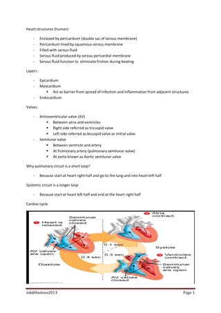
Cardiac cycle
nik@biolove2013 Page 1
2. 1 - atrial 2- atrial
- Blood flow into atria and - remaining blood is pushed out from atria to
and ventricle through AV valve systole ventricle
ventricular - AV valve open, ventricular - blood rushes out from ventricle due to high
diastole semilunar valve closed
diastole pressure
-AV open
- semilunar closed
3- atria
diastole
ventricular
systole - blood pushes out from
ventricle completely
-semilunar valve open
-atrioventricular closed
‘lup’ – closing of atroventricular valve
‘dup’ – closing of semilunar valve
Cardiac output
- Amount of blood pumped by each ventricle in one minute
- Product of heart rate and stroke volume
- Normal : 5liter/min
Heart rate
- Pulse/number of heart beat per minute
- Normal : 75 beats/min
Stroke volume
- Amount of blood pumped out by each ventricle in each heart beat
- 70ml/beat
- Regulation depend on venous return
Venous return
- Amount of blood entering the heart
Why our heart beat become fast after exercise?
- Skeletal muscle contract and relax causing blood flow to be faster
- Speed up venous return
- Venous return increase, stroke volume increase
- Causing more contraction
nik@biolove2013 Page 2
3. Regulation of heart beat
- signal passes to AV node
- ventricle contract
- sinoatrial node (SA node) generate impulse to atria
- atria contract
- During stress or physical activities nerves of sympathetic division triggers AV and SA node to
increase heart beat.
- Parasympathetic nerves slow down heart rate.
- Hormone :
Epinephrine and thyroxine increase heart rate
- Ions :
Low ion, low heart rate
- Other factors affecting heart rate :
Age
Gender
Body Temp.
Activities
Conduction system of the heart
Two systems :
Autonomic nervous system
- Slow down or speeds heart rate
- Depend on which division it activated
Nodal system or intrinsic condustion system
- A specialized tissue
- Function as it is a combination of muscle and nervous tissue
Nodal system
1) Depend on AV node and SA node
2) SA node located at right atrium
3) Also called as pacemaker because it starts the heart beat
From SA node, impulse spread to the atria
Atria contract
Then spread to AV node
4) AV node located at the junction of atria and ventricle
5) Then impulse send to bundle of His
nik@biolove2013 Page 3
4. 6) Then spread to Purkinje fibers
7) From purkinje spread to muscle of ventricle walls
At AV node, impulse is delayed because to wait for atria to finish contract
SA node
generate
impulse
causing atria
to contract
muscle of
ventricle wall impulse then
passes to AV
(ventricle node
contract)
from bundle
from AV node
of his to
to bundle of
Purkinje
His
fibers
Figure 1 : Conduction system (Nodal System)
nik@biolove2013 Page 4