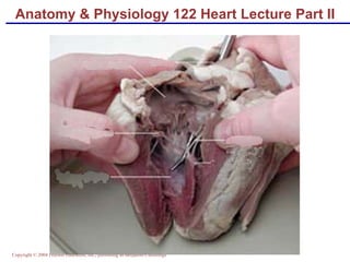
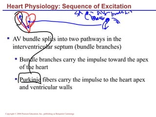
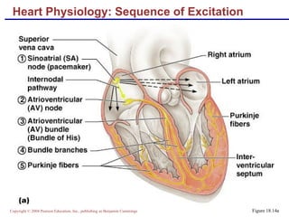
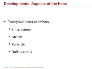
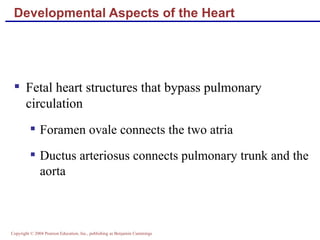
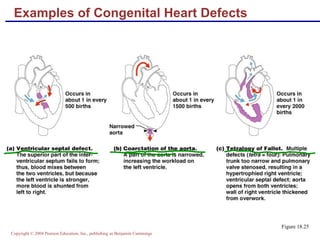
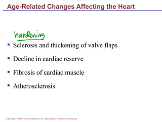
Anatomy & Physiology 122 Heart Lecture Part II
Cardiac Muscle Contraction Heart muscle: Is stimulated by nerves and is self-excitable (automaticity) Contracts as a unit Has a long (250 ms) absolute refractory period Cardiac muscle contraction is similar to skeletal muscle contraction
Heart Cells
Heart Physiology: Sequence of Excitation Sinoatrial (SA) node generates impulses about 75 times/minute Atrioventricular (AV) node delays the impulse approximately 0.1 second Impulse passes from atria to ventricles via the atrioventricular bundle (bundle of His)
Heart Physiology: Sequence of Excitation AV bundle splits into two pathways in the interventricular septum (bundle branches) Bundle branches carry the impulse toward the apex of the heart Purkinje fibers carry the impulse to the heart apex and ventricular walls
Heart Physiology: Sequence of Excitation Figure 18.14a
Sequence of Excitation - 2
Heart Excitation Related to ECG Figure 18.17
Electrocardiography Electrical activity is recorded by electrocardiogram (ECG) P wave corresponds to depolarization of SA node QRS complex corresponds to ventricular depolarization T wave corresponds to ventricular repolarization Atrial repolarization record is masked by the larger QRS complex
Electrocardiograms The electocardiogram or ECG is a major diagnostic  tool for the assessment of the health of the heart. It is a measurement taken at the surface of the skin which reflects the  electrical phenomena  in the heart when the  SA node  triggers the  electrical sequence  that controls heart action .
Electrocardiography Figure 18.16
Heart Sounds Heart sounds (lub-dup) are associated with closing of heart valves First sound occurs as AV valves close and signifies beginning of systole Second sound occurs when SL valves close at the beginning of ventricular diastole
Cardiac Cycle Cardiac cycle refers to all events associated with blood flow through the heart Systole – contraction of heart muscle Diastole – relaxation of heart muscle
Phases of the Cardiac Cycle Ventricular filling – mid-to-late diastole Heart blood pressure is low as blood enters atria and flows into ventricles AV valves are open, then atrial systole occurs
Phases of the Cardiac Cycle Ventricular systole Atria relax  Rising ventricular pressure results in closing of AV valves Isovolumetric contraction phase Ventricular ejection phase opens semilunar valves
Phases of the Cardiac Cycle Isovolumetric relaxation – early diastole Ventricles relax Backflow of blood in aorta and pulmonary trunk closes semilunar valves Dicrotic notch – brief rise in aortic pressure caused by backflow of blood rebounding off semilunar valves
Sympathetic nervous system (SNS) stimulation is activated by stress, anxiety, excitement, or exercise  Parasympathetic nervous system (PNS) stimulation is mediated by acetylcholine and opposes the SNS  PNS dominates the autonomic stimulation, slowing heart rate and causing vagal tone Regulation of Heart Rate: Autonomic Nervous System
Atrial (Bainbridge) Reflex Atrial (Bainbridge) reflex – a sympathetic reflex initiated by increased blood in the atria Causes stimulation of the SA node Stimulates baroreceptors in the atria, causing increased SNS stimulation
Chemical Regulation of the Heart The hormones epinephrine and thyroxine increase heart rate Intra- and extracellular ion concentrations must be maintained for normal heart function InterActive Physiology ® :  Cardiovascular System: Cardiac Output PLAY
Congestive Heart Failure (CHF) Congestive heart failure (CHF) is caused by: Coronary atherosclerosis Persistent high blood pressure Multiple myocardial infarcts Dilated cardiomyopathy (DCM)
Developmental Aspects of the Heart Embryonic heart chambers Sinus venous Atrium Ventricle Bulbus cordis
Developmental Aspects of the Heart Figure 18.24
Developmental Aspects of the Heart Fetal heart structures that bypass pulmonary circulation Foramen ovale connects the two atria  Ductus arteriosus connects pulmonary trunk and the aorta
Examples of Congenital Heart Defects Figure 18.25
Age-Related Changes Affecting the Heart Sclerosis and thickening of valve flaps Decline in cardiac reserve Fibrosis of cardiac muscle Atherosclerosis

Ap heart chp18part2

  • 1.
    Anatomy & Physiology122 Heart Lecture Part II
  • 2.
    Cardiac Muscle ContractionHeart muscle: Is stimulated by nerves and is self-excitable (automaticity) Contracts as a unit Has a long (250 ms) absolute refractory period Cardiac muscle contraction is similar to skeletal muscle contraction
  • 3.
  • 4.
    Heart Physiology: Sequenceof Excitation Sinoatrial (SA) node generates impulses about 75 times/minute Atrioventricular (AV) node delays the impulse approximately 0.1 second Impulse passes from atria to ventricles via the atrioventricular bundle (bundle of His)
  • 5.
    Heart Physiology: Sequenceof Excitation AV bundle splits into two pathways in the interventricular septum (bundle branches) Bundle branches carry the impulse toward the apex of the heart Purkinje fibers carry the impulse to the heart apex and ventricular walls
  • 6.
    Heart Physiology: Sequenceof Excitation Figure 18.14a
  • 7.
  • 8.
    Heart Excitation Relatedto ECG Figure 18.17
  • 9.
    Electrocardiography Electrical activityis recorded by electrocardiogram (ECG) P wave corresponds to depolarization of SA node QRS complex corresponds to ventricular depolarization T wave corresponds to ventricular repolarization Atrial repolarization record is masked by the larger QRS complex
  • 10.
    Electrocardiograms The electocardiogramor ECG is a major diagnostic tool for the assessment of the health of the heart. It is a measurement taken at the surface of the skin which reflects the electrical phenomena in the heart when the SA node triggers the electrical sequence that controls heart action .
  • 11.
  • 12.
    Heart Sounds Heartsounds (lub-dup) are associated with closing of heart valves First sound occurs as AV valves close and signifies beginning of systole Second sound occurs when SL valves close at the beginning of ventricular diastole
  • 13.
    Cardiac Cycle Cardiaccycle refers to all events associated with blood flow through the heart Systole – contraction of heart muscle Diastole – relaxation of heart muscle
  • 14.
    Phases of theCardiac Cycle Ventricular filling – mid-to-late diastole Heart blood pressure is low as blood enters atria and flows into ventricles AV valves are open, then atrial systole occurs
  • 15.
    Phases of theCardiac Cycle Ventricular systole Atria relax Rising ventricular pressure results in closing of AV valves Isovolumetric contraction phase Ventricular ejection phase opens semilunar valves
  • 16.
    Phases of theCardiac Cycle Isovolumetric relaxation – early diastole Ventricles relax Backflow of blood in aorta and pulmonary trunk closes semilunar valves Dicrotic notch – brief rise in aortic pressure caused by backflow of blood rebounding off semilunar valves
  • 17.
    Sympathetic nervous system(SNS) stimulation is activated by stress, anxiety, excitement, or exercise Parasympathetic nervous system (PNS) stimulation is mediated by acetylcholine and opposes the SNS PNS dominates the autonomic stimulation, slowing heart rate and causing vagal tone Regulation of Heart Rate: Autonomic Nervous System
  • 18.
    Atrial (Bainbridge) ReflexAtrial (Bainbridge) reflex – a sympathetic reflex initiated by increased blood in the atria Causes stimulation of the SA node Stimulates baroreceptors in the atria, causing increased SNS stimulation
  • 19.
    Chemical Regulation ofthe Heart The hormones epinephrine and thyroxine increase heart rate Intra- and extracellular ion concentrations must be maintained for normal heart function InterActive Physiology ® : Cardiovascular System: Cardiac Output PLAY
  • 20.
    Congestive Heart Failure(CHF) Congestive heart failure (CHF) is caused by: Coronary atherosclerosis Persistent high blood pressure Multiple myocardial infarcts Dilated cardiomyopathy (DCM)
  • 21.
    Developmental Aspects ofthe Heart Embryonic heart chambers Sinus venous Atrium Ventricle Bulbus cordis
  • 22.
    Developmental Aspects ofthe Heart Figure 18.24
  • 23.
    Developmental Aspects ofthe Heart Fetal heart structures that bypass pulmonary circulation Foramen ovale connects the two atria Ductus arteriosus connects pulmonary trunk and the aorta
  • 24.
    Examples of CongenitalHeart Defects Figure 18.25
  • 25.
    Age-Related Changes Affectingthe Heart Sclerosis and thickening of valve flaps Decline in cardiac reserve Fibrosis of cardiac muscle Atherosclerosis