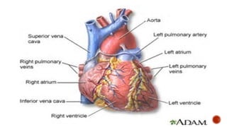
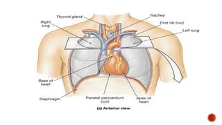
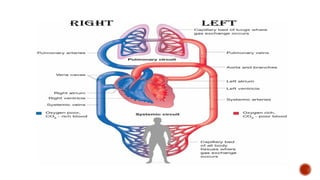
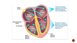
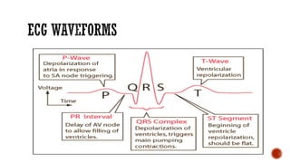
Nor Masita Esa
Universiti Kebangsaan Malaysia
At the end of the session,the students should be able to:
▪ Identify the major structures and functions of the cardiovascular
system.
▪ explain the circulation of blood through the heart and coronary
vessels.
▪ define the cardiac output and cardiac cycle.
▪ name and locate the elements of heart’s conduction system.
▪ The heart is a muscular pump; it beats an average of 70
times per minute.
▪ This continues pumping moves blood through the body
nourishing tissue cell and body, removing wastes. Deficit in
the structure or function of the heart affect all body tissue
and may limit almost all human functions.
Shape
▪ The heart is a hollow, cone-shape organ approximately the
▪ Size of an adult’s fist.
Weight
▪ 200 to 425 gram
Location
▪ Mediasternum of the thoracic cavity
▪ Between the vertebral column and the sternum
▪ Flanked laterally by the lungs
▪ Two- thirds of the heart mass lies to the left of the sternum
▪ The upper base lies beneath the second rib
▪ The pointed apex approximately with the 5th intercostals space, midpoint to the clavicle.
▪ Covered by a double layer of fibroserous membrane (pericardium).
The pericardium:
▪ encases the heart and anchors it to surrounding structures, forming the pericardial
sac.
▪ prevents the heart from overfilling with blood.
▪ the parietal pericardium is the outermost layer.
▪ the visceral pericardium or epicardium adheres to the heart surface.
▪ the small space between visceral and parietal layers - pericardialcavity.
▪ serous lubricating fluid produced in this space cushions the heart as it beats.
▪ Three layers:
1. Epicardium
• the outermost epicardium
• same structure as the visceral pericardium.
2. Myocardium
• the middle layer of the heart
• consists of specialized cardiac muscle cell (myofibrils) – provide the
bulk of contractile heart muscle.
3. Endocardium
• the innermost layer
• sheath of endothelium - lines the inside of the heart’s chamber and
great vessels
▪ Four hollow chamber
- two upper chamber called atrium
- two lower chamber called ventricles
• Separated by septum.
• Each of the heart’s chambers is separated by a valve.
• The atriums are separated from the ventricles by two atrioventricular (AV) valve.
- The tricuspid valve - right side
- The bicuspid or mitral valve – left side.
- The flaps of each of these valves are anchored to the papillary muscles of the ventricles by
the chordae tendineae.
• The ventricles are connected to their great vessels by semilunar valves.
- On the right the pulmonary valve joins the right ventricle with the pulmonary artery.
- On the left, the aortic valve joins the left ventricle to the aorta.
▪ Systemic Circulation consists of:
– Left side of the heart
– Aorta and its branches
– Capillaries – supply the brain & peripheral tissues
– Systemic venous system
– Vena cava
* The systemic system – move blood to peripheral areas of the body
high pressure system
▪ Pulmonary Circulation consists of:
– Right side of the heart
– Pulmonary artery
– Pulmonary capillaries
– Pulmonary vein
* Located in the thorax near the heart, the pulmonary circulation is a
low-pressure system.
▪ Coronary Circulation
– The heart muscle itself is supplied by its own network of vessels.
The left & right coronary arteries originate at the base of the aorta and
branch out to encircle the myocardium, supplying blood, O2 and
nutrients to the myocardium.
- The left main coronary artery divides to form
• anterior descending arteries
• circumflex arteries
• The anterior descending artery:
– supplies the anterior interventricular septum and the left ventricle.
• The circumflex branch supplies:
– Left lateral wall of the left ventricle
• The right coronary artery (RCA) supplies:
– Right ventricle and forms the posterior descending artery (PDA)
– PDA supplies – posterior portion of the heart.
▪ While ventricular contraction delivers blood through the pulmonary and systemic
circulation, during ventricular relaxation the coronary arteries fill with O2-rich
blood
• After the blood perfuses the heart muscle, the cardiac veins drain the blood into the
coronary sinus, which empties into the right atrium of the heart.
• Blood flow through the coronary arteries is regulated by:
▪ – Aortic pressure
▪ – Heart rate
▪ – Metabolic activity of the heart
▪ – Blood vessel tone (constriction)
▪ Cardiac cycle
– The contraction and relaxation of the heart constitutes one heartbeat.
- Thus cardiac cycle consists of systole and diastole of the atria plus systole and diastole
of the ventricles
▪ Atrial Systole (lasts about 0.1sec)
- The atria are contracting at the same time ventricles are relaxed
- Depolarization of SA node causes atrial depolarization and marked by PWave in the
ECG
- Atrial depolarization causes atrial systole,they exert pressure on the blood within ,
which forces blood through the open AV valves into the ventricles
- End DiastoleVolume (EDV) : the blood volume contains in the ventricles at the end of its
relaxation period.
▪ Ventricular Systole (Last about 0.3sec)
- marked as QRS complex in the ECG onset of ventricular
depolarization
- The ventricles are contracting, at the same time, atria are relaxed in
atrial diastole
- Ventricular depolarization causes ventricular systole
- Pressure rises inside the ventricles and pushes the blood up against
the atrioventricular (AV) valves, forcing them shut. Both AV and
Semilunar Valves are closed about 0.05sec. This is period of
Isovolumetric contraction. Cardiac muscle are contracting and
exerting force but are not yet shortening
- Continued pressure in the ventricles causes pressure inside the
chamber to rise sharply and both Semilunar valves open and
ejection of the blood from the heart begins. This period is called
Ventricular Ejection
- Both ventricles ejects about 70mls blood into the aorta and
pulmonary trunk. The volume remaining in each ventricle at the end
of systole about 60mls called End-systolicVolume (ESV)
- Stroke Volume, the volume ejected per beat from each ventricle
- SV = EDV – ESV (about 130mls – 60mls = 70mls)
▪ Relaxation Period (lasts about 0.4sec)
- T wave in the ECG marks the onset of ventricle repolarization
- Both atria and ventricles are relaxed
- Ventricular relaxation causes ventricular diastole. As the ventricles
relaxed the pressure in the chamber falls and blood in the aorta and
pulmonary trunk begins to flow backward toward the regions of lower
pressure in the ventricle
- The SL valves closes as the valve cusps catches the backflowing of the
blood
- Isovolumetric relaxation, period when the four valves are closed and
the ventricle blood volume does not change.
- Ventricular filling begins as the ventricle continue to relax and the
pressure quickly falls and drop below atrial pressure and the AV
valves open. At the end of relaxation period, the ventricles are about
three-quarter full.
▪ There are 4 heart sounds:
- The normal heart sound are only the first and second heart sound (S1
and S2)
- S1 described as Lubb louder and bit longer than S2. Caused by
blood turbulence associated with closure of the AV Valve soon after
the ventricular systole begins
- S2 can be described as dub shorter and not loud as first heart sound
caused by blood turbulence associated with closure of SL valves.
- S3 is due to blood turbulence during rapid ventricular filling
- S4 is due to blood turbulence during atrial systole
▪ Cardiac output (CO)
– the amount of blood pumped by the ventricles into the pulmonary and systemic
circulation in 1 minute
– The average adult CO - 4 to 8 l/min
CO = SV X HR
▪ Ejection fraction (EF)
– the percentage of total blood in the ventricle at the end of the diastole ejected from
the heart with each beat.
– The normal EF - 50 to 70%.
– CO is an indicator of how well the heart is functioning as a pump.
▪ Preload
– the amount of cardiac muscle fiber tension or stretch that exists at the end of
diastole, just before contraction of the ventricles.
▪ Afterload
– the force the ventricles must overcome to eject their blood volume.
– pressure in the arterial system ahead of the ventricles.
▪ Contractility
– the inherent (natural) capability of the cardiac muscle fibers to shorten.
▪ Sinoatrial node (SA node)
– ‘pacemaker’ of the heart
– generating an impulse 60 to 100 times/min
– this impulse travel across the atria via internodal pathways to the atrioventricular
(AV) node, in the floor of the interatrial septum
▪ Atrioventricular (AV) node
– the very small junction fiber of the AV node slows the impulse, slightly delaying its
transmission to the ventricles.
▪ Bundle of His
– It then passes though the bundle of His at the atrioventricular junction and
continues down the interventricular septum through the right and left bundle
branches and out to the Perkinje fibers in the ventricular muscle walls.
THANK YOU

Anatomy & PHYSIOLOGY Cardiovascular System

  • 1.
    Nor Masita Esa UniversitiKebangsaan Malaysia
  • 2.
    At the endof the session,the students should be able to: ▪ Identify the major structures and functions of the cardiovascular system. ▪ explain the circulation of blood through the heart and coronary vessels. ▪ define the cardiac output and cardiac cycle. ▪ name and locate the elements of heart’s conduction system.
  • 3.
    ▪ The heartis a muscular pump; it beats an average of 70 times per minute. ▪ This continues pumping moves blood through the body nourishing tissue cell and body, removing wastes. Deficit in the structure or function of the heart affect all body tissue and may limit almost all human functions.
  • 5.
    Shape ▪ The heartis a hollow, cone-shape organ approximately the ▪ Size of an adult’s fist. Weight ▪ 200 to 425 gram Location ▪ Mediasternum of the thoracic cavity ▪ Between the vertebral column and the sternum ▪ Flanked laterally by the lungs ▪ Two- thirds of the heart mass lies to the left of the sternum ▪ The upper base lies beneath the second rib ▪ The pointed apex approximately with the 5th intercostals space, midpoint to the clavicle.
  • 7.
    ▪ Covered bya double layer of fibroserous membrane (pericardium). The pericardium: ▪ encases the heart and anchors it to surrounding structures, forming the pericardial sac. ▪ prevents the heart from overfilling with blood. ▪ the parietal pericardium is the outermost layer. ▪ the visceral pericardium or epicardium adheres to the heart surface. ▪ the small space between visceral and parietal layers - pericardialcavity. ▪ serous lubricating fluid produced in this space cushions the heart as it beats.
  • 8.
    ▪ Three layers: 1.Epicardium • the outermost epicardium • same structure as the visceral pericardium. 2. Myocardium • the middle layer of the heart • consists of specialized cardiac muscle cell (myofibrils) – provide the bulk of contractile heart muscle. 3. Endocardium • the innermost layer • sheath of endothelium - lines the inside of the heart’s chamber and great vessels
  • 10.
    ▪ Four hollowchamber - two upper chamber called atrium - two lower chamber called ventricles • Separated by septum. • Each of the heart’s chambers is separated by a valve. • The atriums are separated from the ventricles by two atrioventricular (AV) valve. - The tricuspid valve - right side - The bicuspid or mitral valve – left side. - The flaps of each of these valves are anchored to the papillary muscles of the ventricles by the chordae tendineae. • The ventricles are connected to their great vessels by semilunar valves. - On the right the pulmonary valve joins the right ventricle with the pulmonary artery. - On the left, the aortic valve joins the left ventricle to the aorta.
  • 12.
    ▪ Systemic Circulationconsists of: – Left side of the heart – Aorta and its branches – Capillaries – supply the brain & peripheral tissues – Systemic venous system – Vena cava * The systemic system – move blood to peripheral areas of the body high pressure system
  • 13.
    ▪ Pulmonary Circulationconsists of: – Right side of the heart – Pulmonary artery – Pulmonary capillaries – Pulmonary vein * Located in the thorax near the heart, the pulmonary circulation is a low-pressure system.
  • 15.
    ▪ Coronary Circulation –The heart muscle itself is supplied by its own network of vessels. The left & right coronary arteries originate at the base of the aorta and branch out to encircle the myocardium, supplying blood, O2 and nutrients to the myocardium. - The left main coronary artery divides to form • anterior descending arteries • circumflex arteries
  • 16.
    • The anteriordescending artery: – supplies the anterior interventricular septum and the left ventricle. • The circumflex branch supplies: – Left lateral wall of the left ventricle • The right coronary artery (RCA) supplies: – Right ventricle and forms the posterior descending artery (PDA) – PDA supplies – posterior portion of the heart.
  • 18.
    ▪ While ventricularcontraction delivers blood through the pulmonary and systemic circulation, during ventricular relaxation the coronary arteries fill with O2-rich blood • After the blood perfuses the heart muscle, the cardiac veins drain the blood into the coronary sinus, which empties into the right atrium of the heart. • Blood flow through the coronary arteries is regulated by: ▪ – Aortic pressure ▪ – Heart rate ▪ – Metabolic activity of the heart ▪ – Blood vessel tone (constriction)
  • 19.
    ▪ Cardiac cycle –The contraction and relaxation of the heart constitutes one heartbeat. - Thus cardiac cycle consists of systole and diastole of the atria plus systole and diastole of the ventricles ▪ Atrial Systole (lasts about 0.1sec) - The atria are contracting at the same time ventricles are relaxed - Depolarization of SA node causes atrial depolarization and marked by PWave in the ECG - Atrial depolarization causes atrial systole,they exert pressure on the blood within , which forces blood through the open AV valves into the ventricles - End DiastoleVolume (EDV) : the blood volume contains in the ventricles at the end of its relaxation period.
  • 20.
    ▪ Ventricular Systole(Last about 0.3sec) - marked as QRS complex in the ECG onset of ventricular depolarization - The ventricles are contracting, at the same time, atria are relaxed in atrial diastole - Ventricular depolarization causes ventricular systole - Pressure rises inside the ventricles and pushes the blood up against the atrioventricular (AV) valves, forcing them shut. Both AV and Semilunar Valves are closed about 0.05sec. This is period of Isovolumetric contraction. Cardiac muscle are contracting and exerting force but are not yet shortening
  • 21.
    - Continued pressurein the ventricles causes pressure inside the chamber to rise sharply and both Semilunar valves open and ejection of the blood from the heart begins. This period is called Ventricular Ejection - Both ventricles ejects about 70mls blood into the aorta and pulmonary trunk. The volume remaining in each ventricle at the end of systole about 60mls called End-systolicVolume (ESV)
  • 22.
    - Stroke Volume,the volume ejected per beat from each ventricle - SV = EDV – ESV (about 130mls – 60mls = 70mls) ▪ Relaxation Period (lasts about 0.4sec) - T wave in the ECG marks the onset of ventricle repolarization - Both atria and ventricles are relaxed - Ventricular relaxation causes ventricular diastole. As the ventricles relaxed the pressure in the chamber falls and blood in the aorta and pulmonary trunk begins to flow backward toward the regions of lower pressure in the ventricle - The SL valves closes as the valve cusps catches the backflowing of the blood
  • 23.
    - Isovolumetric relaxation,period when the four valves are closed and the ventricle blood volume does not change. - Ventricular filling begins as the ventricle continue to relax and the pressure quickly falls and drop below atrial pressure and the AV valves open. At the end of relaxation period, the ventricles are about three-quarter full.
  • 26.
    ▪ There are4 heart sounds: - The normal heart sound are only the first and second heart sound (S1 and S2) - S1 described as Lubb louder and bit longer than S2. Caused by blood turbulence associated with closure of the AV Valve soon after the ventricular systole begins - S2 can be described as dub shorter and not loud as first heart sound caused by blood turbulence associated with closure of SL valves. - S3 is due to blood turbulence during rapid ventricular filling - S4 is due to blood turbulence during atrial systole
  • 27.
    ▪ Cardiac output(CO) – the amount of blood pumped by the ventricles into the pulmonary and systemic circulation in 1 minute – The average adult CO - 4 to 8 l/min CO = SV X HR ▪ Ejection fraction (EF) – the percentage of total blood in the ventricle at the end of the diastole ejected from the heart with each beat. – The normal EF - 50 to 70%. – CO is an indicator of how well the heart is functioning as a pump.
  • 28.
    ▪ Preload – theamount of cardiac muscle fiber tension or stretch that exists at the end of diastole, just before contraction of the ventricles. ▪ Afterload – the force the ventricles must overcome to eject their blood volume. – pressure in the arterial system ahead of the ventricles. ▪ Contractility – the inherent (natural) capability of the cardiac muscle fibers to shorten.
  • 29.
    ▪ Sinoatrial node(SA node) – ‘pacemaker’ of the heart – generating an impulse 60 to 100 times/min – this impulse travel across the atria via internodal pathways to the atrioventricular (AV) node, in the floor of the interatrial septum ▪ Atrioventricular (AV) node – the very small junction fiber of the AV node slows the impulse, slightly delaying its transmission to the ventricles. ▪ Bundle of His – It then passes though the bundle of His at the atrioventricular junction and continues down the interventricular septum through the right and left bundle branches and out to the Perkinje fibers in the ventricular muscle walls.
  • 32.