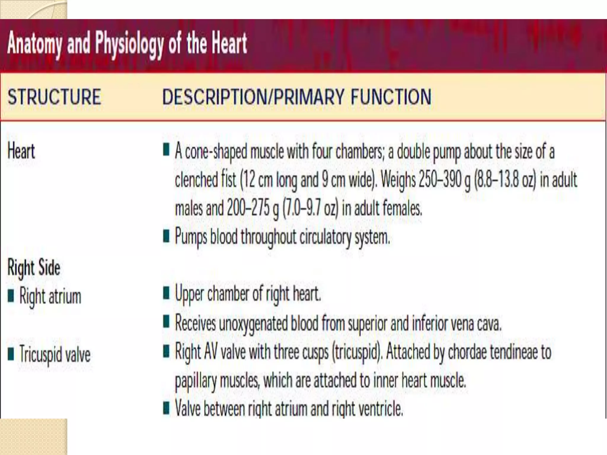
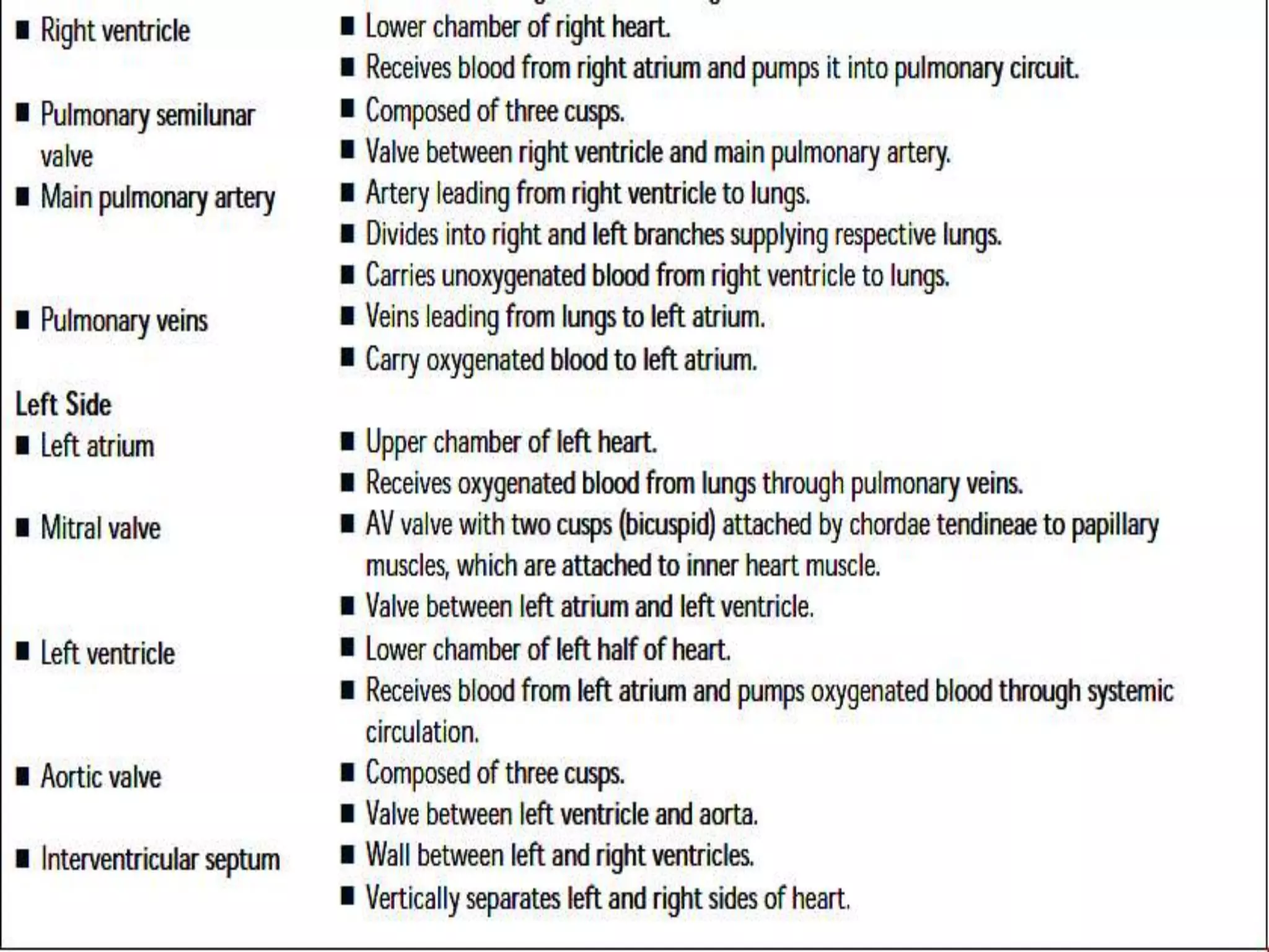
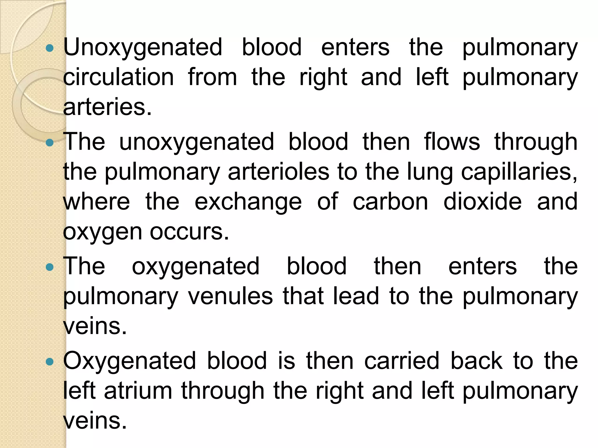
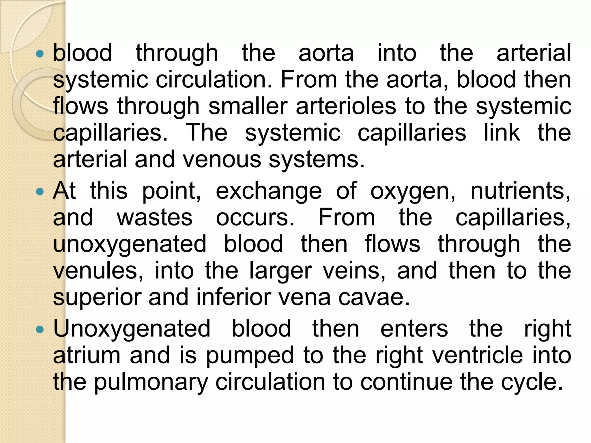
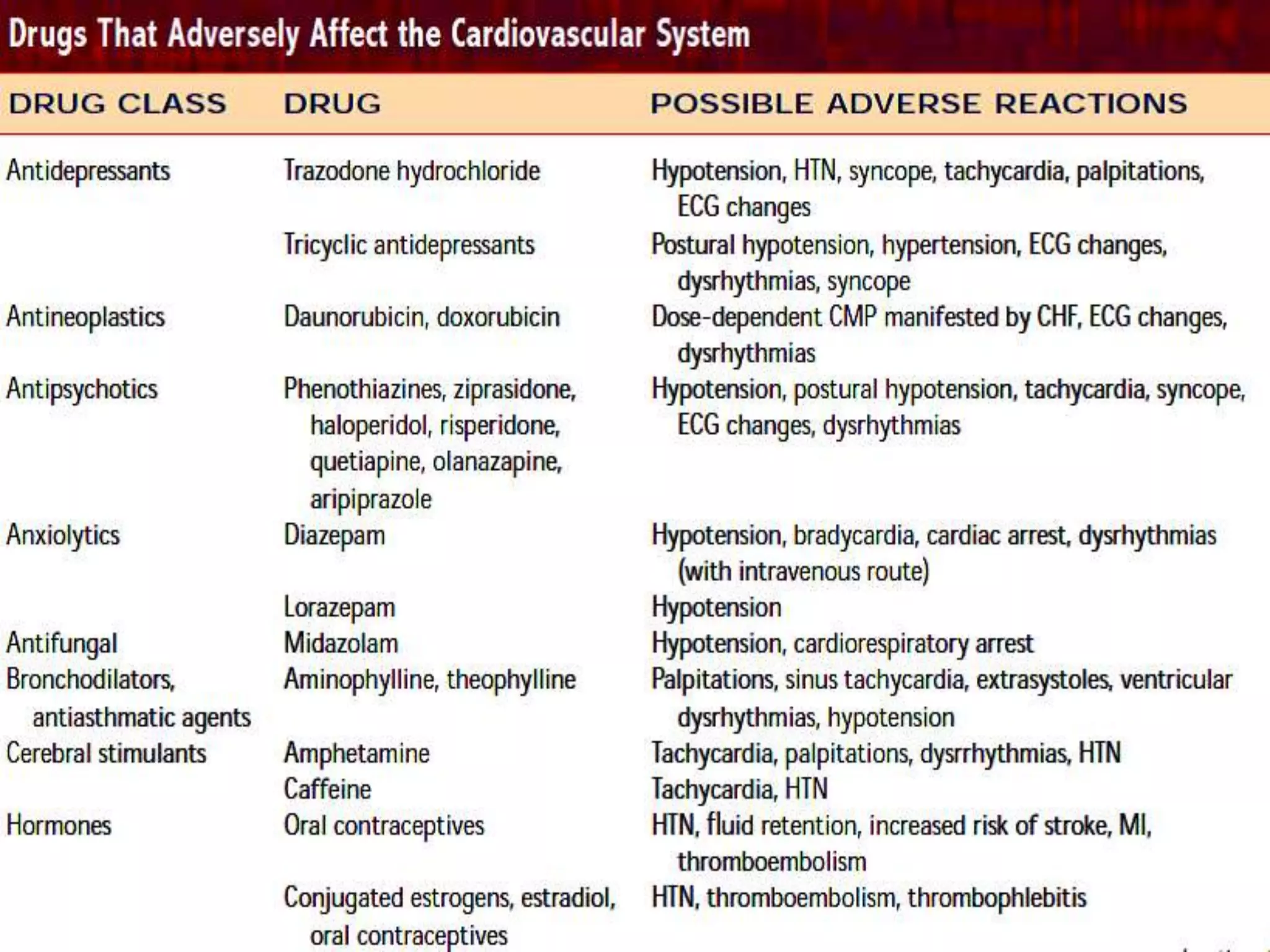
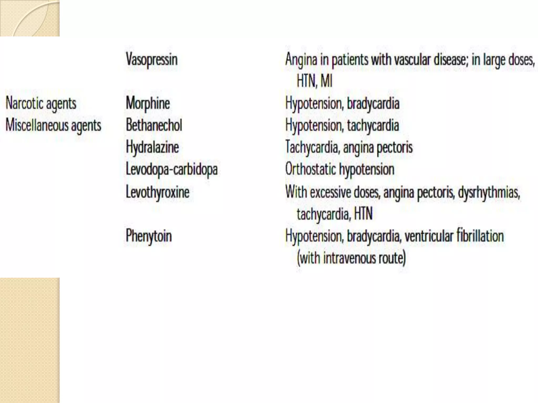
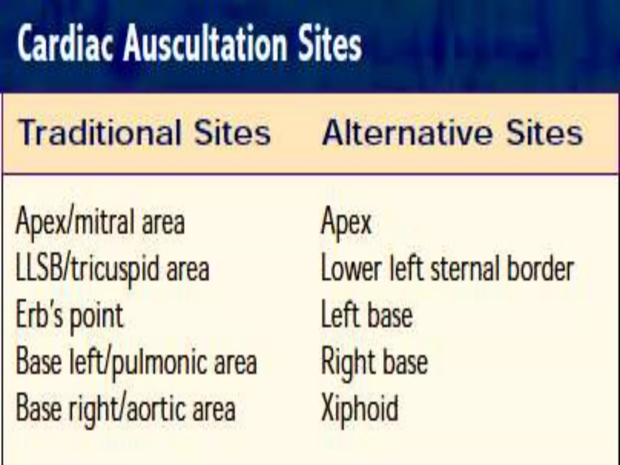
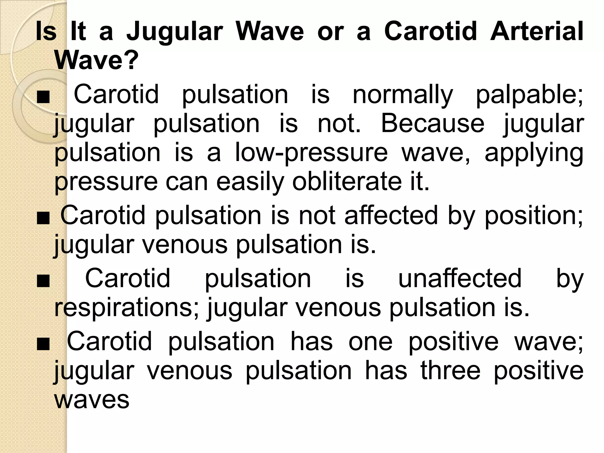
Assessing the cardiovascular
System


Dr/Magda Bayoumi
Layers of the Heart
 Endocardium: Inner layer of heart; a smooth,
  thin layer of endothelium and connective
  tissue. smooth inner lining of the heart.
 Myocardium:
 Middle and thickest layer of heart;       heart
  muscle.
 Responsible for cardiac contraction.
 Epicardium: The layer of serous pericardium
  on heart’s surface. Contains main coronary
  blood vessels.
 Pericardium: Sac that surrounds the heart and roots
  of the great vessels.
 Composed of two layers: fibrous pericardium
  (outer layer of fibrous connective tissue) and serous
  pericardium.
 Serous pericardium also has two layers: outer or
  parietal layer
 That lines the fibrous layer and visceral or inner layer
  that lines the heart and is also called the
   Epicardium.
   Serous pericardium contains pericardial fluid
   (10–20 mL of serous fluid).
   Pericardial fluid moistens the pericardial sac
    between the two layers and prevents friction
    during systole and diastole.
The Circulatory System
 The circulatory system has two main
  networks, the pulmonary circulation and
  the systemic circulation.
 coronary circulation is part of the systemic
  circulation and supplies the heart itself.
 The pulmonary circulation involves blood
  vessels that circulate blood through the
  pulmonary arteries, the lungs, and the
  pulmonary veins.
 Unoxygenated blood enters the pulmonary
  circulation from the right and left pulmonary
  arteries.
 The unoxygenated blood then flows through
  the pulmonary arterioles to the lung capillaries,
  where the exchange of carbon dioxide and
  oxygen occurs.
 The oxygenated blood then enters the
  pulmonary venules that lead to the pulmonary
  veins.
 Oxygenated blood is then carried back to the
  left atrium through the right and left pulmonary
  veins.
Systemic Circulation
 The systemic circulation is responsible for
  supplying oxygen to every cell in the body
  through the arterial system and then
  returning unoxygenated blood to the heart
  through the venous system.
 Oxygenated blood flows into the left atrium
  from the pulmonary circulation.
 The left atrium then pumps the oxygenated
  blood into the left ventricle, which in turn
  expels the oxygenated
Coronary Circulation
 The coronary circulation consists of the
  right and left coronary arteries and the
  coronary sinus and cardiac veins. The
  coronary arteries are the first branches
  off the aorta.
 The cardiac veins drain into the coronary
  sinus, which in turn drains directly into
  the right atrium.
   blood through the aorta into the arterial
    systemic circulation. From the aorta, blood then
    flows through smaller arterioles to the systemic
    capillaries. The systemic capillaries link the
    arterial and venous systems.
   At this point, exchange of oxygen, nutrients,
    and wastes occurs. From the capillaries,
    unoxygenated blood then flows through the
    venules, into the larger veins, and then to the
    superior and inferior vena cavae.
   Unoxygenated blood then enters the right
    atrium and is pumped to the right ventricle into
    the pulmonary circulation to continue the cycle.
Mechanisms of Heart Sounds

   Heart sounds are the result of events
    within the heart. The movement and
    pressure       of       the     blood
    (hemodynamics), the activity of the
    electrical conduction system, and the
    movement of the valves affect the
    sounds that you hear
The Cardiac Cycle
 The cardiac cycle comprises systolic and
  diastolic phases. The systolic phase is
  the      contraction      or    emptying
  phase, and the diastolic phase is the
  resting or filling phase.
 The atria and ventricles alternate through
  the systolic and Diastolic phases; while
  the atria are contracting, the ventricles
  are relaxing, and vice versa.
Ventricular Diastole
   Ventricular diastole marks the beginning
    of the filling phase. Very shortly after the
    onset of ventricular diastole, the pressure
    in the ventricles is less than that of the
    atria, and the mitral and tricuspid
    valves open to allow filling of the
    ventricles (the rapid-filling phase of
    diastole).
Ventricular Systole
 With ventricular contraction, the pressure
  in the ventricles will exceed that in the
  atria.
 This results in closure of the mitral and
  tricuspid valves and marks the
  beginning of ventricular systole.
 While the ventricles are contracting and
  blood is being propelled from them, the
  atria are relaxed (atrial diastole) and are
  filling.
 The amount of blood ejected from the
  heartwith each contraction is referred to
  as the stroke volume.
 The cardiac output is the amount of
  blood ejected per minute.
 Cardiac output equals stroke volume
  multiplied by heart rate.
The Stroke Volume
 Preload refers to the volume of blood in the
  ventricles at t he end of diastole.
 An increase in venous return to the heart
  will, in turn, increase preload. Factors that
  affect venous return include:
 Venous blood reservoirs
 Skeletal muscle pump:
 Venous tone:
 Respiratory pump:
After load is simply the work that the
heart has to do to push blood into the
aorta and around the body.
   Afterload reflects the end-systolic volume. It is
    affected by the amount of resistance the
    ventricles have to contract against. An increase
    in after-load results in a decrease in stroke
    volume. After-load may be affected by:
   ■ Arterial elasticity.
   ■ Peripheral vascular resistance.
   ■ Aortic valve resistance.
   ■ Viscosity and volume of blood.
   ■ Contractility,which reflects the force of
    contraction.
   ■ Positive inotropes,which increase the force
    of contraction.
   ■ Negative inotropes, which decrease the
    force of contraction.
The Heart’s Electrical Conduction
    System
 The specialized cells of the conduction
  system                                      have
  automaticity,excitability, conductivity and
  refractoriness.
 Automaticity is the cell’s ability to initiate an
  impulse.
 Excitability is the cell’s ability to respond to
  an impulse and create an action potential.
  Conductivity is the cell’s ability to transmit
  an impulse.
 Refractoriness is the cell’s ability to respond
  to the transmitted impulse.
 The sinoatrial (SA) node is the normal
  pacemaker of the heart located in the right
  atrium near the superior vena cava
  entrance point.
 The SA node paces the normal adult heart
  at 60 to 100 BPM.
 The activation of the SA node passes
  through the atria and results in atrial
   electrical activity is then conducted to the
    atrioventricular (AV) node.This node is located at
    the base of the right atrium between the atria and the
    ventricles. It has the ability to pace the heart at a rate
    of 40 to 60 BPM.
   The electrical impulse is then transmitted.
   from the AV node to the bundle of His, which divides
    into two branches, the right and the left, which
    traverse the interventricular septum.
   Finally, the impulse is transmitted to small branches
    that eventuate into the Purkinje fibers, which
    stimulate the ventricles to contract.
   The pacer ability of the bundle of His is 20 to 40 BPM.
The Valves
   S1, the first heart sound, results from
    the closure of the mitral (M1) and
    tricuspid (T1) valves. M1 and T1
    normally close within approximately 0.02
    second or less. These valve sounds are
    often heard as a single sound. S1 is best
    heard at the apex or left lateral sternal
    border (LLSB) with the diaphragm of the
    stethoscope.
The Second Heart Sound (S2)
 When the systolic pressure in the
  ventricles decreases below that of the
  aorta and the pulmonary artery (toward
  the end of systole), the aortic (A2) and
  pulmonic (P2) valves close, producing
  the second heart sound.
 Clinically, this sound marks the end of
  systole and the beginning of diastole.
 A2 and P2 normally close about 0.02
  second from each other; consequently,
  they may occasionally be heard as a
  single sound.
Extra Heart Sounds
 Additional sounds that may be heard
  during auscultation ,        include early
  ejection click, mid systolic ejection click,
  opening snap, S3, and S4.These sounds
  do not always indicate pathology.
Interaction With Other Body Systems
 ENDOCRINE
  Distributes hormones throughout body via
   circulatory system.
  Cardiac muscle cells secrete atrial natriuretic
   peptide (ANP), which helps maintain fluid and
   electrolyte balance and lowers volume and
   blood pressure. Erythropoietin regulates RBC
   production. Epinephrine and norepinephrine
   increase heart rate and force of contraction.
 URINARY
  Helps regulate volume within vascular system.
   Renin/angiotensin system affects B/P.
  Erythropoietin affects RBC production.
LYMPHATIC
 Delivers WBCs and antibodies to fight
  pathogens.
 And heart from pathogens Protects vascular
  system.
REPRODUCTIVE
 Distributes reproductive hormones. Delivers
  nutrients to reproductive organs.
 Vascular system needed for changes that
  occur during sexual arousal.
 Premenopausal       women      have  lower
  incidence of heart disease.
RESPIRATORY

 RBCs  exchange oxygen and carbon
 dioxide in lungs and transport it to
 peripheral system. Provides oxygen to
 and      removes         wastes     from
 cardiovascular system. Lungs convert
 angiotensin I to II, which helps maintain
 blood pressure.
 INTEGUMENTARY
 Responds to skin injury or infection by
  delivering clotting factors and immune
  system response to affected area.
 Stimulation of mast cells in response to
  injury or infection.
 Produces      local changes in blood
  pressure and release of ADH, which
  helps      blood     flow and    capillary
  permeability.
SKELETAL
 Delivers calcium and minerals to bones for bone
  growth. Delivers parathormone and calcitonin.
 Provides calcium for normal heart muscle
  contraction.
 Produces blood cells in bone marrow.
 Skeletal framework protects heart.
MUSCULAR
 Delivers nutrients to muscles and throughout
  circulatory system.
 Removes carbon dioxide, lactic acid and heat
  produced by muscle activity.
 Muscles provide protection for neck vessels.
  Heart is muscle responsible for pumping blood.
 Muscle contraction of legs helps with venous
  return.
NEUROLOGICAL
 Endothelial cells of brain capillaries form a
  semi-permeable membrane that maintains
  blood-brain barrier.
 Controls peripheral circulation and heart
  rate and increases blood volume and
  pressure.
DIGESTIVE
 Delivers nutrients and hormones from site
  of absorption and transports nutrients and
  toxins to liver. Supplies cardiovascular
  system with nutrients and absorbs water
  and ions that help maintain blood volume.
Korotkoff’s Sounds:
 When you take your patient’s BP, you may hear five
  distinct phases called Korotkoff’s sounds.These
  phases occur because the BP cuff partially obstructs
  blood flow and disturbs the laminar flow pattern,
  causing turbulence. Korotkoff phases include the
  following:
■ Phase I: A faint, clear, rhythmic tapping noise that
  gradually increases in intensity. Intraluminal pressure
  and cuff pressure are equal.
■ Phase II: A swishing sound that is heard as the
  vessel distends with blood.
■ Phase III: Sounds become more intense. Vessel is
  open in systole but not in diastole.
■ Phase IV: Sounds begin to muffle, and pressure is
  closest to diastolic arterial pressure.
■ Phase V: Sounds disappear because vessel remains
  open.
Central Artery and Jugular
    Veins
 The sternocleidomastoid and trapezius
  muscles are helpful in locating these vessels.
 The carotid artery and the internal jugular run
  parallel   to   each     other    along     the
  sternocleidomastoid muscle toward the
  sternal notch.
 The external jugular crosses the internal
  jugular    and    lies   posterior    to    the
  sternocleidomastoid muscle.
Physical Assessment
Is It a Jugular Wave or a Carotid Arterial
  Wave?
■ Carotid pulsation is normally palpable;
  jugular pulsation is not. Because jugular
  pulsation is a low-pressure wave, applying
  pressure can easily obliterate it.
■ Carotid pulsation is not affected by position;
  jugular venous pulsation is.
■ Carotid pulsation is unaffected by
  respirations; jugular venous pulsation is.
■ Carotid pulsation has one positive wave;
  jugular venous pulsation has three positive
  waves
These graphics represent the normal cardiac pulsations and heart sounds. The jugular venous
pulsation normally has 3 positive waves—the a, c, and v waves and 2 negative troughs—x and y
troughs. The "a" wave is approximately synchronous with the first heart sound and just precedes
the carotid upstroke. The "v" wave coincides approximately with the second heart sound. The
normal carotid artery pulsation has a single positive wave during systole, followed by the
dicrotic notch (about the time of the second heart sound). The apex impulse represents the normal
brief, palpable systolic impulse occurring at the time of the first heart sound. In young normal
individuals there may be a palpable early diastolic filling wave representing the rapid filling
phase of ventricular diastole and corresponding to the normal third heart sound. Auscultation at
the aortic area reveals a normal first heart sound (S1) and second heart sound (S2). S2 is normally
louder than S1 in this area. At the pulmonary area there is normal inspiratory (physiologic)
splitting of the second sound due to asynchronous aortic and pulmonic closure. The aortic
component of the second heart sound (A2) normally precedes the pulmonic component (P2). At
the tricuspid area there is normal splitting of the first heart sound due to asynchronous mitral and
tricuspid closure. The mitral component of the first sound (M1) normally precedes the tricuspid
component (T1). At this area, physiologic splitting of the second sound may also be appreciated.
At the mitral area, the first and second heart sounds are normal. The first sound is normally
louder than the second heart sound and only the aortic component of the second heart sound is
normally appreciated. Occasionally a third heart sound is normal, reflecting deceleration of blood
into the left ventricle during the rapid filling phase of early diastole. Children and young adults
often have normal or physiologic third heart sounds.
Heart assessment
Heart assessment
Heart assessment
Heart assessment
Heart assessment
Heart assessment
Heart assessment
Heart assessment
Heart assessment
Heart assessment
Heart assessment
Heart assessment
Heart assessment
Heart assessment
Heart assessment
Heart assessment
Heart assessment
Heart assessment
Heart assessment
Heart assessment
Heart assessment
Heart assessment
Heart assessment
Heart assessment
Heart assessment
Heart assessment

Heart assessment

  • 1.
  • 7.
    Layers of theHeart  Endocardium: Inner layer of heart; a smooth, thin layer of endothelium and connective tissue. smooth inner lining of the heart.  Myocardium:  Middle and thickest layer of heart; heart muscle.  Responsible for cardiac contraction.  Epicardium: The layer of serous pericardium on heart’s surface. Contains main coronary blood vessels.
  • 8.
     Pericardium: Sacthat surrounds the heart and roots of the great vessels.  Composed of two layers: fibrous pericardium (outer layer of fibrous connective tissue) and serous pericardium.  Serous pericardium also has two layers: outer or parietal layer  That lines the fibrous layer and visceral or inner layer that lines the heart and is also called the  Epicardium.  Serous pericardium contains pericardial fluid  (10–20 mL of serous fluid).  Pericardial fluid moistens the pericardial sac between the two layers and prevents friction during systole and diastole.
  • 9.
    The Circulatory System The circulatory system has two main networks, the pulmonary circulation and the systemic circulation.  coronary circulation is part of the systemic circulation and supplies the heart itself.  The pulmonary circulation involves blood vessels that circulate blood through the pulmonary arteries, the lungs, and the pulmonary veins.
  • 10.
     Unoxygenated bloodenters the pulmonary circulation from the right and left pulmonary arteries.  The unoxygenated blood then flows through the pulmonary arterioles to the lung capillaries, where the exchange of carbon dioxide and oxygen occurs.  The oxygenated blood then enters the pulmonary venules that lead to the pulmonary veins.  Oxygenated blood is then carried back to the left atrium through the right and left pulmonary veins.
  • 11.
    Systemic Circulation  Thesystemic circulation is responsible for supplying oxygen to every cell in the body through the arterial system and then returning unoxygenated blood to the heart through the venous system.  Oxygenated blood flows into the left atrium from the pulmonary circulation.  The left atrium then pumps the oxygenated blood into the left ventricle, which in turn expels the oxygenated
  • 12.
    Coronary Circulation  Thecoronary circulation consists of the right and left coronary arteries and the coronary sinus and cardiac veins. The coronary arteries are the first branches off the aorta.  The cardiac veins drain into the coronary sinus, which in turn drains directly into the right atrium.
  • 15.
    blood through the aorta into the arterial systemic circulation. From the aorta, blood then flows through smaller arterioles to the systemic capillaries. The systemic capillaries link the arterial and venous systems.  At this point, exchange of oxygen, nutrients, and wastes occurs. From the capillaries, unoxygenated blood then flows through the venules, into the larger veins, and then to the superior and inferior vena cavae.  Unoxygenated blood then enters the right atrium and is pumped to the right ventricle into the pulmonary circulation to continue the cycle.
  • 17.
    Mechanisms of HeartSounds  Heart sounds are the result of events within the heart. The movement and pressure of the blood (hemodynamics), the activity of the electrical conduction system, and the movement of the valves affect the sounds that you hear
  • 18.
    The Cardiac Cycle The cardiac cycle comprises systolic and diastolic phases. The systolic phase is the contraction or emptying phase, and the diastolic phase is the resting or filling phase.  The atria and ventricles alternate through the systolic and Diastolic phases; while the atria are contracting, the ventricles are relaxing, and vice versa.
  • 19.
    Ventricular Diastole  Ventricular diastole marks the beginning of the filling phase. Very shortly after the onset of ventricular diastole, the pressure in the ventricles is less than that of the atria, and the mitral and tricuspid valves open to allow filling of the ventricles (the rapid-filling phase of diastole).
  • 20.
    Ventricular Systole  Withventricular contraction, the pressure in the ventricles will exceed that in the atria.  This results in closure of the mitral and tricuspid valves and marks the beginning of ventricular systole.  While the ventricles are contracting and blood is being propelled from them, the atria are relaxed (atrial diastole) and are filling.
  • 22.
     The amountof blood ejected from the heartwith each contraction is referred to as the stroke volume.  The cardiac output is the amount of blood ejected per minute.  Cardiac output equals stroke volume multiplied by heart rate.
  • 23.
    The Stroke Volume Preload refers to the volume of blood in the ventricles at t he end of diastole.  An increase in venous return to the heart will, in turn, increase preload. Factors that affect venous return include:  Venous blood reservoirs  Skeletal muscle pump:  Venous tone:  Respiratory pump:
  • 26.
    After load issimply the work that the heart has to do to push blood into the aorta and around the body.
  • 28.
    Afterload reflects the end-systolic volume. It is affected by the amount of resistance the ventricles have to contract against. An increase in after-load results in a decrease in stroke volume. After-load may be affected by:  ■ Arterial elasticity.  ■ Peripheral vascular resistance.  ■ Aortic valve resistance.  ■ Viscosity and volume of blood.  ■ Contractility,which reflects the force of contraction.  ■ Positive inotropes,which increase the force of contraction.  ■ Negative inotropes, which decrease the force of contraction.
  • 29.
    The Heart’s ElectricalConduction System  The specialized cells of the conduction system have automaticity,excitability, conductivity and refractoriness.  Automaticity is the cell’s ability to initiate an impulse.  Excitability is the cell’s ability to respond to an impulse and create an action potential. Conductivity is the cell’s ability to transmit an impulse.  Refractoriness is the cell’s ability to respond to the transmitted impulse.
  • 30.
     The sinoatrial(SA) node is the normal pacemaker of the heart located in the right atrium near the superior vena cava entrance point.  The SA node paces the normal adult heart at 60 to 100 BPM.  The activation of the SA node passes through the atria and results in atrial
  • 32.
    electrical activity is then conducted to the atrioventricular (AV) node.This node is located at the base of the right atrium between the atria and the ventricles. It has the ability to pace the heart at a rate of 40 to 60 BPM.  The electrical impulse is then transmitted.  from the AV node to the bundle of His, which divides into two branches, the right and the left, which traverse the interventricular septum.  Finally, the impulse is transmitted to small branches that eventuate into the Purkinje fibers, which stimulate the ventricles to contract.  The pacer ability of the bundle of His is 20 to 40 BPM.
  • 33.
    The Valves  S1, the first heart sound, results from the closure of the mitral (M1) and tricuspid (T1) valves. M1 and T1 normally close within approximately 0.02 second or less. These valve sounds are often heard as a single sound. S1 is best heard at the apex or left lateral sternal border (LLSB) with the diaphragm of the stethoscope.
  • 34.
    The Second HeartSound (S2)  When the systolic pressure in the ventricles decreases below that of the aorta and the pulmonary artery (toward the end of systole), the aortic (A2) and pulmonic (P2) valves close, producing the second heart sound.  Clinically, this sound marks the end of systole and the beginning of diastole.  A2 and P2 normally close about 0.02 second from each other; consequently, they may occasionally be heard as a single sound.
  • 35.
    Extra Heart Sounds Additional sounds that may be heard during auscultation , include early ejection click, mid systolic ejection click, opening snap, S3, and S4.These sounds do not always indicate pathology.
  • 36.
    Interaction With OtherBody Systems ENDOCRINE  Distributes hormones throughout body via circulatory system.  Cardiac muscle cells secrete atrial natriuretic peptide (ANP), which helps maintain fluid and electrolyte balance and lowers volume and blood pressure. Erythropoietin regulates RBC production. Epinephrine and norepinephrine increase heart rate and force of contraction. URINARY  Helps regulate volume within vascular system. Renin/angiotensin system affects B/P.  Erythropoietin affects RBC production.
  • 37.
    LYMPHATIC  Delivers WBCsand antibodies to fight pathogens.  And heart from pathogens Protects vascular system. REPRODUCTIVE  Distributes reproductive hormones. Delivers nutrients to reproductive organs.  Vascular system needed for changes that occur during sexual arousal.  Premenopausal women have lower incidence of heart disease.
  • 38.
    RESPIRATORY  RBCs exchange oxygen and carbon dioxide in lungs and transport it to peripheral system. Provides oxygen to and removes wastes from cardiovascular system. Lungs convert angiotensin I to II, which helps maintain blood pressure.
  • 39.
     INTEGUMENTARY  Respondsto skin injury or infection by delivering clotting factors and immune system response to affected area.  Stimulation of mast cells in response to injury or infection.  Produces local changes in blood pressure and release of ADH, which helps blood flow and capillary permeability.
  • 40.
    SKELETAL  Delivers calciumand minerals to bones for bone growth. Delivers parathormone and calcitonin.  Provides calcium for normal heart muscle contraction.  Produces blood cells in bone marrow.  Skeletal framework protects heart. MUSCULAR  Delivers nutrients to muscles and throughout circulatory system.  Removes carbon dioxide, lactic acid and heat produced by muscle activity.  Muscles provide protection for neck vessels. Heart is muscle responsible for pumping blood.  Muscle contraction of legs helps with venous return.
  • 41.
    NEUROLOGICAL  Endothelial cellsof brain capillaries form a semi-permeable membrane that maintains blood-brain barrier.  Controls peripheral circulation and heart rate and increases blood volume and pressure. DIGESTIVE  Delivers nutrients and hormones from site of absorption and transports nutrients and toxins to liver. Supplies cardiovascular system with nutrients and absorbs water and ions that help maintain blood volume.
  • 42.
    Korotkoff’s Sounds:  Whenyou take your patient’s BP, you may hear five distinct phases called Korotkoff’s sounds.These phases occur because the BP cuff partially obstructs blood flow and disturbs the laminar flow pattern, causing turbulence. Korotkoff phases include the following: ■ Phase I: A faint, clear, rhythmic tapping noise that gradually increases in intensity. Intraluminal pressure and cuff pressure are equal. ■ Phase II: A swishing sound that is heard as the vessel distends with blood. ■ Phase III: Sounds become more intense. Vessel is open in systole but not in diastole. ■ Phase IV: Sounds begin to muffle, and pressure is closest to diastolic arterial pressure. ■ Phase V: Sounds disappear because vessel remains open.
  • 48.
    Central Artery andJugular Veins  The sternocleidomastoid and trapezius muscles are helpful in locating these vessels.  The carotid artery and the internal jugular run parallel to each other along the sternocleidomastoid muscle toward the sternal notch.  The external jugular crosses the internal jugular and lies posterior to the sternocleidomastoid muscle.
  • 50.
  • 51.
    Is It aJugular Wave or a Carotid Arterial Wave? ■ Carotid pulsation is normally palpable; jugular pulsation is not. Because jugular pulsation is a low-pressure wave, applying pressure can easily obliterate it. ■ Carotid pulsation is not affected by position; jugular venous pulsation is. ■ Carotid pulsation is unaffected by respirations; jugular venous pulsation is. ■ Carotid pulsation has one positive wave; jugular venous pulsation has three positive waves
  • 53.
    These graphics representthe normal cardiac pulsations and heart sounds. The jugular venous pulsation normally has 3 positive waves—the a, c, and v waves and 2 negative troughs—x and y troughs. The "a" wave is approximately synchronous with the first heart sound and just precedes the carotid upstroke. The "v" wave coincides approximately with the second heart sound. The normal carotid artery pulsation has a single positive wave during systole, followed by the dicrotic notch (about the time of the second heart sound). The apex impulse represents the normal brief, palpable systolic impulse occurring at the time of the first heart sound. In young normal individuals there may be a palpable early diastolic filling wave representing the rapid filling phase of ventricular diastole and corresponding to the normal third heart sound. Auscultation at the aortic area reveals a normal first heart sound (S1) and second heart sound (S2). S2 is normally louder than S1 in this area. At the pulmonary area there is normal inspiratory (physiologic) splitting of the second sound due to asynchronous aortic and pulmonic closure. The aortic component of the second heart sound (A2) normally precedes the pulmonic component (P2). At the tricuspid area there is normal splitting of the first heart sound due to asynchronous mitral and tricuspid closure. The mitral component of the first sound (M1) normally precedes the tricuspid component (T1). At this area, physiologic splitting of the second sound may also be appreciated. At the mitral area, the first and second heart sounds are normal. The first sound is normally louder than the second heart sound and only the aortic component of the second heart sound is normally appreciated. Occasionally a third heart sound is normal, reflecting deceleration of blood into the left ventricle during the rapid filling phase of early diastole. Children and young adults often have normal or physiologic third heart sounds.