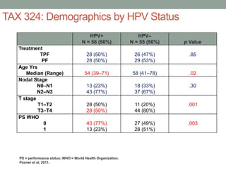
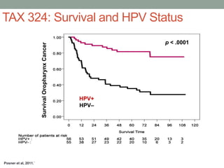
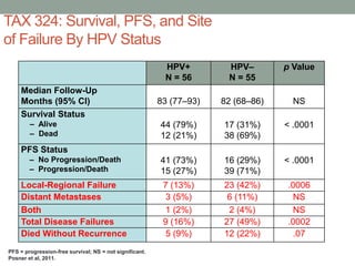
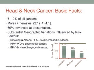
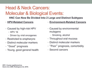
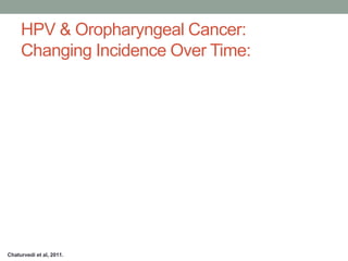
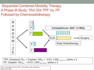
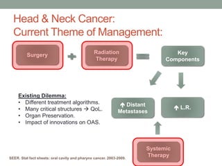
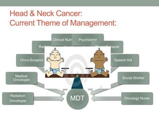
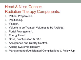
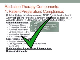
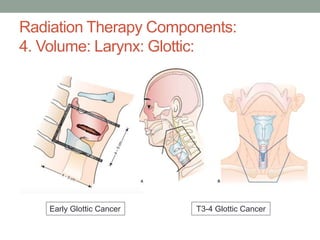
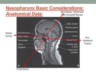
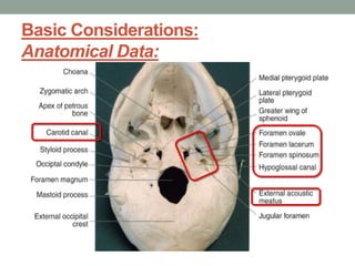
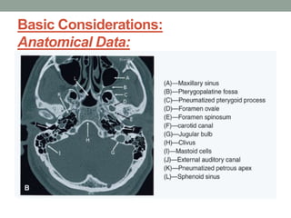
HEAD & NECK CANCER
HORIZONTAL
Mohamed Abdulla M.D.
Prof. of Clinical Oncology
Kasr Al-Aini School of Medicine
Cairo University
KASO – EXAM & RAP UP COURSE
THURSDAY 16/04/2015
GRAND NILE HOTEL & TOWER
FINDING
SIMILARITIES
IN COMMON
Disclosures:
• Amgen.
• Merck Serono.
• Sanofi.
• Astra Zeneca.
• Astellas.
• Roche.
• Pfizer.
• Novartis
Head & Neck Cancer: Basic Facts:
• 6 – 9% of all cancers.
• Males > Females; (2:1)  (4:1).
• 60% advanced at presentation.
• Substantial Geographic Variations Influenced by Risk
Factors:
• Smoking & Alcohol  5 – fold increased incidence.
• HPV  Oro-pharyngeal cancer.
• EPV  Nasopharyngeal cancer.
83%
59%
36%
0%
50%
100%
5-yearSurvival
Seminars in Oncology, Vol 41, No 6, December 2014, pp 798-806
Head & Neck Cancers:
Molecular & Biological Events:
• HPV-Related Cancers
• Caused by high-risk HPV
• HPV 16
• Driven by viral oncogenes
• Restricted to oropharynx
• Distinct molecular markers
• “Good” prognosis
• Young, good general health
• Environment-Related Cancers
• Caused by environmental
mutagens
• Smoking, alcohol
• Throughout oral mucosa
• Distinct molecular markers
• “Poor” prognosis, comorbidity
• Second cancers
HNC Can Now Be Divided Into 2 Large and Distinct Subtypes
HPV = human papillomavirus.
Goon et al, 2009; Rodriguez et al, 2010.
HPV & Oropharyngeal Cancer:
Changing Incidence Over Time:
Chaturvedi et al, 2011.
Sequential Combined Modality Therapy
APhase III Study: TAX 324 TPF Vs. PF
Followed by Chemoradiotherapy
R
A
N
D
O
M
I
Z
E
P
P
F
F
Carboplatinum: AUC 1.5 Wkly
Daily Radiotherapy
EUA
T
Surgery
TPF: Docetaxel 75D1 + Cisplatin 100D1 + 5-FU 1,000 CI: D1–4 q3wks x 3
PF: Cisplatin 100 D1 + 5-FU 1,000 CI: D1–5 q3wks x 3
AUC = area under the curve; EUA = examination under anesthesia.
Posner et al, 2007.
TAX 324: Demographics by HPV Status
HPV+
N = 56 (50%)
HPV–
N = 55 (50%) p Value
Treatment
TPF
PF
28 (50%)
28 (50%)
26 (47%)
29 (53%)
.85
Age Yrs
Median (Range) 54 (39–71) 58 (41–78) .02
Nodal Stage
N0–N1
N2–N3
13 (23%)
43 (77%)
18 (33%)
37 (67%)
.30
T stage
T1–T2
T3–T4
28 (50%)
28 (50%)
11 (20%)
44 (80%)
.001
PS WHO
0
1
43 (77%)
13 (23%)
27 (49%)
28 (51%)
.003
PS = performance status; WHO = World Health Organization.
Posner et al, 2011.
TAX 324: Survival and HPV Status
Posner et al, 2011.`
SurvivalOropharynxCancer
HPV+
HPV–
p < .0001
TAX 324: Survival, PFS, and Site
of Failure By HPV Status
HPV+
N = 56
HPV–
N = 55
p Value
Median Follow-Up
Months (95% CI) 83 (77–93) 82 (68–86) NS
Survival Status
– Alive
– Dead
44 (79%)
12 (21%)
17 (31%)
38 (69%)
< .0001
PFS Status
– No Progression/Death
– Progression/Death
41 (73%)
15 (27%)
16 (29%)
39 (71%)
< .0001
Local-Regional Failure 7 (13%) 23 (42%) .0006
Distant Metastases 3 (5%) 6 (11%) NS
Both 1 (2%) 2 (4%) NS
Total Disease Failures 9 (16%) 27 (49%) .0002
Died Without Recurrence 5 (9%) 12 (22%) .07
PFS = progression-free survival; NS = not significant.
Posner et al, 2011.
Head & Neck Cancer:
Current Theme of Management:
Surgery
Radiation
Therapy
Key
Components
 L.R.
 Distant
Metastases
Systemic
TherapySEER. Stat fact sheets: oral cavity and pharynx cancer. 2003-2009.
Existing Dilemma:
• Different treatment algorithms.
• Many critical structures  QoL.
• Organ Preservation.
• Impact of innovations on OAS.
MDT
Radiation
Oncologist
Medical
Oncologist
Onco-Surgeon
Radiologist
Clinical NutritionistPsychiatrist
Physiotherapist
Speech Aid
Social Worker
Oncology Nurse
Head & Neck Cancer:
Current Theme of Management:
Head & Neck Cancer:
Radiation Therapy Components:
1. Patient Preparation.
2. Positioning.
3. Fixation.
4. Volume to be Treated, Volumes to be Avoided.
5. Portal Arrangement.
6. Energy Used.
7. Dose, Fractionation & OAP
8. Acceptance and Quality Control.
9. Adding Systemic Therapy.
10. Management of Anticipated Complications & Follow Up
Radiation Therapy Components:
1. Patient Preparation: Compliance:
• Detailed history including previous HNSCC & radiation treatment.
• All investigations (Imaging, laboratory, pathologic, endoscopic) 
Accurate Staging  Clear Intention & Therapeutic Strategy.
• General Examination:
• Performance Status.
• Complexion: PALOR  ANEMIA., Depression.
• Nutritional Assessment.
• Co-morbid illness  DM
• Neurological Assessment.
• Pulmonary Assessment.
• Locoregional Examination:
• Palpable primary & lymph nodes.
• Oral Hygiene  Correction before treatment.
• Stoma Care.
• Understanding, Instructions, Interventions.
• Discuss with family.
• MDCT
• MRI
• PET/CT
• Others
• Hematological
• Organ Function
• Others
Poor Radiation Results From Non-compliance
in Radiation Technique
Critical Impact of Radiotherapy Protocol
Compliance and Quality in the Treatment of
Advanced Head and Neck Cancer: Results
From TROG 02.02
CO June 20, 2010 vol. 28 no. 18 2996-3001
Noncompliance, more relapses
Radiation Therapy Components:
2. Positioning: Comfortable & Reproducible
SUPINE
STRECHED
SHOULDERS
Snehal et al..Medical Dosimetry, Vol. 34, No. 3, pp. 225-227, 2009
• Acanthiomeatal Line is
perpendicular to table.
• Chin in neutral position.
Radiation Therapy Components:
3. Fixation:
• Thermoplastic Mask.
• Placement of Markings;
“Lead & Laser).
• Tongue Bite
(Depressor): In or Out.
Radiation Therapy Components:
4. Volume:
ICRU Reports 50 & 62:
• GTV: Gross Tumor Volume.
• CTV: Clinical Target Volume:
Extension of Subclinical
Disease.
• PTV: GTV + CTV + (3 – 5 mm).
• OAR or ORV.
ICRU Report 50 (1993) Prescribing, recording and
reporting photon beam therapy. International
Commission on Radiation Units and Measurements,
Bethesda, MD
• No Place for limited volumes except in Early glottic
cancers (T1-2).
• Neck nodes are usually included except in Early Glottic
Cancers and PNS.
• Bilaterality of neck nodes is mostly required.
Radiation Therapy Components:
4. Volume:
3D Techniques (Conformal, IMRT, IGRT)
 Spare Normal Tissues as much as we can
Accurate radiation dose delivery
No treatment volume Reduction.
Radiation Therapy Components:
4. Volume:
Radiation Therapy Components:
4. Volume: Larynx: Glottic:
Early Glottic Cancer T3-4 Glottic Cancer
Radiation Therapy Components:
4. Volume: Larynx: Supraglottic:
Nasopharynx Basic Considerations:
Anatomical Data:
Skull Base: Sphenoid
and Occipital Bones
Pre-
Vertebral
Fascia
Nasal
Cavity
Oropharynx
Basic Considerations:
Anatomical Data:
Basic Considerations:
Anatomical Data:
Basic Considerations:
Anatomical Data:
Basic Considerations:
Anatomical Data:Upwardextension
Basic Considerations:
Anatomical Data:
The Most
Common Site
Bounded by:
 Atlas vertebra
Axis vertebra
Sup. Constrictor ms
Buccopharyngeal
fascia
Retropharyngeal
space
Prevertebral fascia
Basic Considerations:
Anatomical Data:
Basic Considerations:
Anatomical Data:
Pharyngo-Basilar Fascia
Basic Considerations:
Anatomical Data:
Superior
Pharyngeal
Constrictor
Muscle
Skull Base
Pharyngobasilar fascia Muscle Deficiency Area
=
Sinus of Morgagni
Potential Route
for Intracranial
Spread
Radiation Therapy Components:
4. Volume: Nasopharynx:
Radiation Therapy Components:
4. Volume: Paranasal Sinuses:
Ipsilateral Tumor Bed + Ethmoid + Frontal + Openings on the
contralateral side. Nodes will be included if locally advanced or
high grade lesion.
Radiation Therapy Components:
4. Volume: Oropharynx:
Radiation Therapy Components:
4. Volume: Hypopharynx:
Radiation Therapy Components:
4. Volume: Oral Tongue and Floor of
Mouth:
• The best obtained is parallel opposed.
• Supplementations.
• IMRT.
• High Energy photon beams: 6-10 MV.
• Electron Beam: Energy according to desired depth as a
supplementation or sometimes for re-irradiation.
• Others.
Radiation Therapy Components:
5-6: Portal Arrangement & Energies:
Radiation Therapy Components:
7. Dose & Fractionation:
Radiation Therapy Components:
7. Dose & Fractionation:
Radiation Therapy Components:
8. Plan Acceptance:
Radiation Therapy Components:
9. Adding Systemic Therapy:
Seminars in Oncology, Vol 41, No 6, December 2014, pp 798-806
MACH-NC: 2009 Update:
93 Trials – 17346 Patients:
J.-P. Pignon et al. / Radiotherapy and Oncology 92 (2009) 4–14
CISPLATIN 100 mg/m2 (D1+22+43) +
RTH
• Early Reactions.
• Late Reactions.
• Follow up Strategy.
Radiation Therapy Components:
10. Management of Anticipated Complications:
Thank You

Head & neck cancer horizontal

  • 1.
    HEAD & NECKCANCER HORIZONTAL Mohamed Abdulla M.D. Prof. of Clinical Oncology Kasr Al-Aini School of Medicine Cairo University KASO – EXAM & RAP UP COURSE THURSDAY 16/04/2015 GRAND NILE HOTEL & TOWER FINDING SIMILARITIES IN COMMON
  • 2.
    Disclosures: • Amgen. • MerckSerono. • Sanofi. • Astra Zeneca. • Astellas. • Roche. • Pfizer. • Novartis
  • 3.
    Head & NeckCancer: Basic Facts: • 6 – 9% of all cancers. • Males > Females; (2:1)  (4:1). • 60% advanced at presentation. • Substantial Geographic Variations Influenced by Risk Factors: • Smoking & Alcohol  5 – fold increased incidence. • HPV  Oro-pharyngeal cancer. • EPV  Nasopharyngeal cancer. 83% 59% 36% 0% 50% 100% 5-yearSurvival Seminars in Oncology, Vol 41, No 6, December 2014, pp 798-806
  • 4.
    Head & NeckCancers: Molecular & Biological Events: • HPV-Related Cancers • Caused by high-risk HPV • HPV 16 • Driven by viral oncogenes • Restricted to oropharynx • Distinct molecular markers • “Good” prognosis • Young, good general health • Environment-Related Cancers • Caused by environmental mutagens • Smoking, alcohol • Throughout oral mucosa • Distinct molecular markers • “Poor” prognosis, comorbidity • Second cancers HNC Can Now Be Divided Into 2 Large and Distinct Subtypes HPV = human papillomavirus. Goon et al, 2009; Rodriguez et al, 2010.
  • 5.
    HPV & OropharyngealCancer: Changing Incidence Over Time: Chaturvedi et al, 2011.
  • 6.
    Sequential Combined ModalityTherapy APhase III Study: TAX 324 TPF Vs. PF Followed by Chemoradiotherapy R A N D O M I Z E P P F F Carboplatinum: AUC 1.5 Wkly Daily Radiotherapy EUA T Surgery TPF: Docetaxel 75D1 + Cisplatin 100D1 + 5-FU 1,000 CI: D1–4 q3wks x 3 PF: Cisplatin 100 D1 + 5-FU 1,000 CI: D1–5 q3wks x 3 AUC = area under the curve; EUA = examination under anesthesia. Posner et al, 2007.
  • 7.
    TAX 324: Demographicsby HPV Status HPV+ N = 56 (50%) HPV– N = 55 (50%) p Value Treatment TPF PF 28 (50%) 28 (50%) 26 (47%) 29 (53%) .85 Age Yrs Median (Range) 54 (39–71) 58 (41–78) .02 Nodal Stage N0–N1 N2–N3 13 (23%) 43 (77%) 18 (33%) 37 (67%) .30 T stage T1–T2 T3–T4 28 (50%) 28 (50%) 11 (20%) 44 (80%) .001 PS WHO 0 1 43 (77%) 13 (23%) 27 (49%) 28 (51%) .003 PS = performance status; WHO = World Health Organization. Posner et al, 2011.
  • 8.
    TAX 324: Survivaland HPV Status Posner et al, 2011.` SurvivalOropharynxCancer HPV+ HPV– p < .0001
  • 9.
    TAX 324: Survival,PFS, and Site of Failure By HPV Status HPV+ N = 56 HPV– N = 55 p Value Median Follow-Up Months (95% CI) 83 (77–93) 82 (68–86) NS Survival Status – Alive – Dead 44 (79%) 12 (21%) 17 (31%) 38 (69%) < .0001 PFS Status – No Progression/Death – Progression/Death 41 (73%) 15 (27%) 16 (29%) 39 (71%) < .0001 Local-Regional Failure 7 (13%) 23 (42%) .0006 Distant Metastases 3 (5%) 6 (11%) NS Both 1 (2%) 2 (4%) NS Total Disease Failures 9 (16%) 27 (49%) .0002 Died Without Recurrence 5 (9%) 12 (22%) .07 PFS = progression-free survival; NS = not significant. Posner et al, 2011.
  • 10.
    Head & NeckCancer: Current Theme of Management: Surgery Radiation Therapy Key Components  L.R.  Distant Metastases Systemic TherapySEER. Stat fact sheets: oral cavity and pharynx cancer. 2003-2009. Existing Dilemma: • Different treatment algorithms. • Many critical structures  QoL. • Organ Preservation. • Impact of innovations on OAS.
  • 11.
  • 12.
    Head & NeckCancer: Radiation Therapy Components: 1. Patient Preparation. 2. Positioning. 3. Fixation. 4. Volume to be Treated, Volumes to be Avoided. 5. Portal Arrangement. 6. Energy Used. 7. Dose, Fractionation & OAP 8. Acceptance and Quality Control. 9. Adding Systemic Therapy. 10. Management of Anticipated Complications & Follow Up
  • 13.
    Radiation Therapy Components: 1.Patient Preparation: Compliance: • Detailed history including previous HNSCC & radiation treatment. • All investigations (Imaging, laboratory, pathologic, endoscopic)  Accurate Staging  Clear Intention & Therapeutic Strategy. • General Examination: • Performance Status. • Complexion: PALOR  ANEMIA., Depression. • Nutritional Assessment. • Co-morbid illness  DM • Neurological Assessment. • Pulmonary Assessment. • Locoregional Examination: • Palpable primary & lymph nodes. • Oral Hygiene  Correction before treatment. • Stoma Care. • Understanding, Instructions, Interventions. • Discuss with family. • MDCT • MRI • PET/CT • Others • Hematological • Organ Function • Others
  • 14.
    Poor Radiation ResultsFrom Non-compliance in Radiation Technique Critical Impact of Radiotherapy Protocol Compliance and Quality in the Treatment of Advanced Head and Neck Cancer: Results From TROG 02.02 CO June 20, 2010 vol. 28 no. 18 2996-3001 Noncompliance, more relapses
  • 15.
    Radiation Therapy Components: 2.Positioning: Comfortable & Reproducible SUPINE STRECHED SHOULDERS Snehal et al..Medical Dosimetry, Vol. 34, No. 3, pp. 225-227, 2009 • Acanthiomeatal Line is perpendicular to table. • Chin in neutral position.
  • 16.
    Radiation Therapy Components: 3.Fixation: • Thermoplastic Mask. • Placement of Markings; “Lead & Laser). • Tongue Bite (Depressor): In or Out.
  • 17.
    Radiation Therapy Components: 4.Volume: ICRU Reports 50 & 62: • GTV: Gross Tumor Volume. • CTV: Clinical Target Volume: Extension of Subclinical Disease. • PTV: GTV + CTV + (3 – 5 mm). • OAR or ORV. ICRU Report 50 (1993) Prescribing, recording and reporting photon beam therapy. International Commission on Radiation Units and Measurements, Bethesda, MD
  • 18.
    • No Placefor limited volumes except in Early glottic cancers (T1-2). • Neck nodes are usually included except in Early Glottic Cancers and PNS. • Bilaterality of neck nodes is mostly required. Radiation Therapy Components: 4. Volume: 3D Techniques (Conformal, IMRT, IGRT)  Spare Normal Tissues as much as we can Accurate radiation dose delivery No treatment volume Reduction.
  • 19.
  • 20.
    Radiation Therapy Components: 4.Volume: Larynx: Glottic: Early Glottic Cancer T3-4 Glottic Cancer
  • 21.
    Radiation Therapy Components: 4.Volume: Larynx: Supraglottic:
  • 22.
    Nasopharynx Basic Considerations: AnatomicalData: Skull Base: Sphenoid and Occipital Bones Pre- Vertebral Fascia Nasal Cavity Oropharynx
  • 23.
  • 24.
  • 25.
  • 26.
  • 27.
  • 28.
    Bounded by:  Atlasvertebra Axis vertebra Sup. Constrictor ms Buccopharyngeal fascia Retropharyngeal space Prevertebral fascia Basic Considerations: Anatomical Data:
  • 29.
  • 30.
    Basic Considerations: Anatomical Data: Superior Pharyngeal Constrictor Muscle SkullBase Pharyngobasilar fascia Muscle Deficiency Area = Sinus of Morgagni Potential Route for Intracranial Spread
  • 31.
  • 32.
    Radiation Therapy Components: 4.Volume: Paranasal Sinuses: Ipsilateral Tumor Bed + Ethmoid + Frontal + Openings on the contralateral side. Nodes will be included if locally advanced or high grade lesion.
  • 33.
  • 34.
  • 35.
    Radiation Therapy Components: 4.Volume: Oral Tongue and Floor of Mouth:
  • 36.
    • The bestobtained is parallel opposed. • Supplementations. • IMRT. • High Energy photon beams: 6-10 MV. • Electron Beam: Energy according to desired depth as a supplementation or sometimes for re-irradiation. • Others. Radiation Therapy Components: 5-6: Portal Arrangement & Energies:
  • 37.
    Radiation Therapy Components: 7.Dose & Fractionation:
  • 38.
    Radiation Therapy Components: 7.Dose & Fractionation:
  • 39.
  • 40.
    Radiation Therapy Components: 9.Adding Systemic Therapy: Seminars in Oncology, Vol 41, No 6, December 2014, pp 798-806
  • 41.
    MACH-NC: 2009 Update: 93Trials – 17346 Patients: J.-P. Pignon et al. / Radiotherapy and Oncology 92 (2009) 4–14 CISPLATIN 100 mg/m2 (D1+22+43) + RTH
  • 43.
    • Early Reactions. •Late Reactions. • Follow up Strategy. Radiation Therapy Components: 10. Management of Anticipated Complications:
  • 44.

Editor's Notes

  • #9 Figure 1. Kaplan–Meier curves for overall survival (A) for HPV+ and HPV2 patients treated on TAX 324. HPV, human papillomavirus.