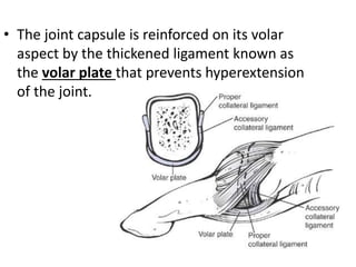
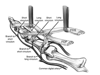
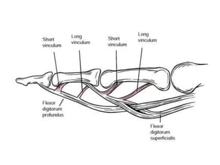
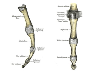
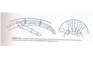
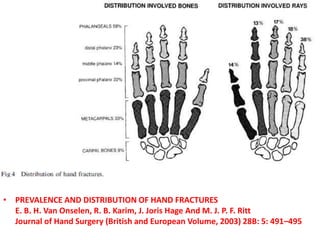
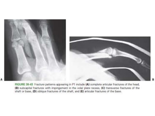
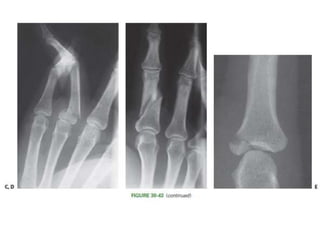
This document summarizes principles for the management of hand fractures presented by Dr. REJUL K RAJ. It discusses anatomy of the hand bones, common fracture patterns, mechanisms of injury, signs and symptoms, imaging, classification systems, treatment principles including splinting and various operative fixation methods, and postoperative care. Key points covered include fracture patterns of the distal phalanx, middle phalanx, proximal phalanx and metacarpals as well as treatment approaches for each. Studies on outcomes of K-wire fixation versus ORIF for metacarpal fractures are summarized.
conventional plates including different functions of screws, modes of plate application, Compression Mode.
Neutralization Mode.
Buttress plate.
Antiglide plate.
Bridge plating or span plating.
Tension band.
prebending precountouring
working length
lag screw
AO principles
biological fixation
MIPO
Distal femur fractures what makes it complex ,dr mohamed ashraf,hod orthopae...drashraf369
distal femur fractures are notorious for post operative complications due to malreduction and improper fixation.unless plan and execute a sound and stable fixation,this injury will lead to undesirable results.dr mohamed ashraf HOD orthopaedics govt TD medical college is presenting how to avoid complications in surgical management of these fractures..
Surgical Approaches to Acetabulum and PelvisBijay Mehta
Important surgical approaches to acetabulum and pelvis are described.
Ilioinguinal approach, Modified Stoppa Approach, Kocher lagenbeck Approach, Ilifemoral approach and extensile approaches are well illustrated and described.
ANATOMY
Meatcarpophalangeal joint- Condyloid joints
ROM at MCPJ- flexion and extension of the digits, as well as a very small degree of abduction and adduction when the digits are extended.
• Phalanges - has a base, shaft, neck and head that is formed from two condyles.
• PIPJ, DIPJ - Hinge joints,
ROM at PIP and DIP joint : flexion and extension.
VERDAN’S ZONES OF HANDS
VOLAR PLATE
Vinculum breve and Vinculum longum
MECHANISMS OF INJURY
Intramedullary nailing of fractures.dr mohamed ashraf.HOD.govt TD medical co...drashraf369
presentation of biology,biomechanics and practice of intramedullary nailing of long bone fractures by dr mohamed ashraf,govt TD medical college,alleppey,kerala,india
Title: Sense of Smell
Presenter: Dr. Faiza, Assistant Professor of Physiology
Qualifications:
MBBS (Best Graduate, AIMC Lahore)
FCPS Physiology
ICMT, CHPE, DHPE (STMU)
MPH (GC University, Faisalabad)
MBA (Virtual University of Pakistan)
Learning Objectives:
Describe the primary categories of smells and the concept of odor blindness.
Explain the structure and location of the olfactory membrane and mucosa, including the types and roles of cells involved in olfaction.
Describe the pathway and mechanisms of olfactory signal transmission from the olfactory receptors to the brain.
Illustrate the biochemical cascade triggered by odorant binding to olfactory receptors, including the role of G-proteins and second messengers in generating an action potential.
Identify different types of olfactory disorders such as anosmia, hyposmia, hyperosmia, and dysosmia, including their potential causes.
Key Topics:
Olfactory Genes:
3% of the human genome accounts for olfactory genes.
400 genes for odorant receptors.
Olfactory Membrane:
Located in the superior part of the nasal cavity.
Medially: Folds downward along the superior septum.
Laterally: Folds over the superior turbinate and upper surface of the middle turbinate.
Total surface area: 5-10 square centimeters.
Olfactory Mucosa:
Olfactory Cells: Bipolar nerve cells derived from the CNS (100 million), with 4-25 olfactory cilia per cell.
Sustentacular Cells: Produce mucus and maintain ionic and molecular environment.
Basal Cells: Replace worn-out olfactory cells with an average lifespan of 1-2 months.
Bowman’s Gland: Secretes mucus.
Stimulation of Olfactory Cells:
Odorant dissolves in mucus and attaches to receptors on olfactory cilia.
Involves a cascade effect through G-proteins and second messengers, leading to depolarization and action potential generation in the olfactory nerve.
Quality of a Good Odorant:
Small (3-20 Carbon atoms), volatile, water-soluble, and lipid-soluble.
Facilitated by odorant-binding proteins in mucus.
Membrane Potential and Action Potential:
Resting membrane potential: -55mV.
Action potential frequency in the olfactory nerve increases with odorant strength.
Adaptation Towards the Sense of Smell:
Rapid adaptation within the first second, with further slow adaptation.
Psychological adaptation greater than receptor adaptation, involving feedback inhibition from the central nervous system.
Primary Sensations of Smell:
Camphoraceous, Musky, Floral, Pepperminty, Ethereal, Pungent, Putrid.
Odor Detection Threshold:
Examples: Hydrogen sulfide (0.0005 ppm), Methyl-mercaptan (0.002 ppm).
Some toxic substances are odorless at lethal concentrations.
Characteristics of Smell:
Odor blindness for single substances due to lack of appropriate receptor protein.
Behavioral and emotional influences of smell.
Transmission of Olfactory Signals:
From olfactory cells to glomeruli in the olfactory bulb, involving lateral inhibition.
Primitive, less old, and new olfactory systems with different path
- Video recording of this lecture in English language: https://youtu.be/lK81BzxMqdo
- Video recording of this lecture in Arabic language: https://youtu.be/Ve4P0COk9OI
- Link to download the book free: https://nephrotube.blogspot.com/p/nephrotube-nephrology-books.html
- Link to NephroTube website: www.NephroTube.com
- Link to NephroTube social media accounts: https://nephrotube.blogspot.com/p/join-nephrotube-on-social-media.html
TEST BANK for Operations Management, 14th Edition by William J. Stevenson, Ve...kevinkariuki227
TEST BANK for Operations Management, 14th Edition by William J. Stevenson, Verified Chapters 1 - 19, Complete Newest Version.pdf
Explore natural remedies for syphilis treatment in Singapore. Discover alternative therapies, herbal remedies, and lifestyle changes that may complement conventional treatments. Learn about holistic approaches to managing syphilis symptoms and supporting overall health.
Report Back from SGO 2024: What’s the Latest in Cervical Cancer?bkling
Are you curious about what’s new in cervical cancer research or unsure what the findings mean? Join Dr. Emily Ko, a gynecologic oncologist at Penn Medicine, to learn about the latest updates from the Society of Gynecologic Oncology (SGO) 2024 Annual Meeting on Women’s Cancer. Dr. Ko will discuss what the research presented at the conference means for you and answer your questions about the new developments.
Prix Galien International 2024 Forum ProgramLevi Shapiro
June 20, 2024, Prix Galien International and Jerusalem Ethics Forum in ROME. Detailed agenda including panels:
- ADVANCES IN CARDIOLOGY: A NEW PARADIGM IS COMING
- WOMEN’S HEALTH: FERTILITY PRESERVATION
- WHAT’S NEW IN THE TREATMENT OF INFECTIOUS,
ONCOLOGICAL AND INFLAMMATORY SKIN DISEASES?
- ARTIFICIAL INTELLIGENCE AND ETHICS
- GENE THERAPY
- BEYOND BORDERS: GLOBAL INITIATIVES FOR DEMOCRATIZING LIFE SCIENCE TECHNOLOGIES AND PROMOTING ACCESS TO HEALTHCARE
- ETHICAL CHALLENGES IN LIFE SCIENCES
- Prix Galien International Awards Ceremony
ARTIFICIAL INTELLIGENCE IN HEALTHCARE.pdfAnujkumaranit
Artificial intelligence (AI) refers to the simulation of human intelligence processes by machines, especially computer systems. It encompasses tasks such as learning, reasoning, problem-solving, perception, and language understanding. AI technologies are revolutionizing various fields, from healthcare to finance, by enabling machines to perform tasks that typically require human intelligence.
1. JOURNAL CLUB - 12.01.17
PRINCIPLES OF MANAGEMENT
OF HAND FRACTURES
Presentor- Dr. REJUL K RAJ
CMCH LUDHIANA
2. REFERENCES
• ROCK WOOD AND GREENS,
FRACTURES IN ADULTS – VIII th Edition.
• Campbell’s Operative Orthopaedics, XII th Edition.
3. ANATOMY
• 27 bones
• Carpals (8), metacarpals (5) and phalanges(14).
• The shaft of each metacarpal is curved - characteristic cup
shape.
• MCPJ- Condyloid joints , flexion and extension of the digits,
as well as a very small degree ofabduction and adduction
when the digits are extended.
• Phalanges - has a base, shaft, neck and head that is formed
from two condyles.
• PIPJ, DIPJ - Hinge joints, flexion and extension.
4.
5. • The joint capsule is reinforced on its volar
aspect by the thickened ligament known as
the volar plate that prevents hyperextension
of the joint.
6.
7.
8.
9.
10. INTRODUCTION.
• One of the most commmon encountered
injuries.
• Phalangeal (23%) and metacarpal (18%)
fractures to be the second and third most
common fractures below the elbow.
11. • PREVALENCE AND DISTRIBUTION OF HAND FRACTURES
E. B. H. Van Onselen, R. B. Karim, J. Joris Hage And M. J. P. F. Ritt
Journal of Hand Surgery (British and European Volume, 2003) 28B: 5: 491–495
12. Injury Mechanisms
• The mechanism of injury description should include ,
– magnitude
– direction
– point of contact
– type of force
13. 1. Axial load or “jamming” injuries -
– shearing articular fractures or metaphyseal compression
fractures-
– catching a falling object-
2. Bending –
– Diaphyseal fractures and joint dislocations-
– ball-handling sports or when the hand is trapped
3. Torsional –
– spiral fractures
– Individual digits can easily be caught in clothing, furniture, or
workplace equipment
4. Crushing
– Bending + shearing + torsion-
– with significant soft tissue injury.
14. Injuries associated.
• Open injuries.
– Greatest challenge is thin and supple soft tissue
coverage.
• Tendons.
– Dislocations.
• Nerves and vessels.
• Massive hand trauma.
• Bone loss
15. Signs and symptoms
• Pain, swelling, deformity, stiffness, weakness,
and loss of coordination.
• Numbness and tingling - nerve involvement
• Signs - tenderness, swelling, ecchymosis,
deformity, crepitus, and instability.
19. Total active movement: TAM
• by sum of the angles formed by MCP, PIP and DIP joints in maximum
active flexion,
• minus total extension deficit at the MCP, PIP and DIP joints during active
finger extension.
• MCP joint can flex up to 85°
• PIP joint can flex up to 110°
• DIP joint can flex up to 65°.
• If the finger is capable of full extension, the loss of extension is 0°.
• As a result, TAM is measured as 260° (260°-0°: 260°).
• In a normal first finger, the MCP joint - 85° and the IP joint - 90°.
• TAM is measured as 175°.
20. FUNCTIONAL STABILITY
• Pun WK, Chow SP, So YC, et al.
A prospective study on 284 digital fractures of the hand.
J Hand Surg Am. 1989;14(3):474-81.
• fractures as functionally stable
• if patients could actively move the adjacent joint more
than 30% of the expected range while the alignment of
fracture remained within acceptable range.
• Unstable fracture
• If the patient was not able to move the adjacent joint
more than 30% of the expected range or
• movement resulted in malalignment.
21. IMAGING
• Anteroposterior
• Lateral
• Splay lateral views
– varying amounts of flexion to prevent phalangeal override
– best show only one digit in a true lateral projection.
• Oblique views -assessing reduction of articular fractures.
• True lateral views - ring and small finger MCs- 10 degrees of
supination
• Index and middle fingers - 10 degrees of pronation.
22. Description of fractures
• Location within the bone
– (HEAD, NECK, SHAFT, BASE)
• Direction of the fracture plane
– (TRANSVERSE, SPIRAL, OBLIQUE, COMMINUTED)
• Degree of displacement.
• Dislocations -direction the distal segment travels
– (DORSAL, VOLAR, ROTATORY)
• Capacity for closed reduction –
– SIMPLE OR COMPLEX
23. PRINCIPLES
• Negative effects of surgery on the tissues should not exceed
the negative effects of the original injury.
• Difficult decision making centers on management of the soft
tissues.
• The injured part must not be considered in isolation.
24. AIM
1. Sufficient stability of the bone or joint injury
2. To permit early motion rehabilitation
3. Without resulting in malunion or residual
instability
• The preferred treatment option is the least
invasive technique that can accomplish these
goals.
25. 5 major treatment alternatives
1. Immediate motion
2. Temporary splinting
3. CRIF
4. ORIF
5. Immediate reconstruction
• External fixation is a variation.
26. NON operative treatment.
• Advantages
– lower cost
– avoidance of the risks and complications
• Disadvantage
– is that stability is less assured
27. Closed reduction internal fixation.
• Prevent overt deformity
• Not to achieve an anatomically perfect
reduction.
• Pin tract infection is the prime complication.
28. Nonoperative Treatment- Principles
• Assessments of rotational malalignment and stability.
• Stability- maintenance of fracture reduction when the
adjacent joints are taken through at least 30% of their
normal motion.
• Contraction of soft tissues begins approximately 72 hours
• Motion should be instituted by this time for all joints stable
enough to tolerate rehabilitation.
29. • Elevation and elastic compression.
• The more aggressive the surgeon’s management of the injury has
been, the more aggressive must be the rehabilitation.
• Low-energy isolated injuries have far less risk of stiffness.
• If the injury is reducible at all, gentle manipulation will accomplish
the reduction far more successfully than forceful longitudinal
traction.
• The principle is relaxation of deforming forces through proximal
joint positioning such as MP joint flexion to relax the intrinsics or
wrist flexion to relax the digital flexor tendons. part.
30. • Splints should immobilize the minimum number of joints
• Wrist immobilization - 25 to 35 degrees of extension.
• “Wounded paw” - wrist flexion + MP joint extension–
interphalangeal (IP) joint flexion (the position).
• Full motion of the IP joints should be encouraged
throughout.
• The total duration of immobilization - 3 to 4 weeks.
• Stable enough by this time to tolerate active range of
motion (AROM) with further remodeling by 8 to 10 weeks.
31.
32.
33. Open reduction internal fixation.
• Add the morbidity of surgical tissue
trauma.
• Titrated against the presumed advantage of
achieving the most anatomic and stable
reduction
34. Introduction to Distal Phalanx (P3) Fractures
• Terminal point of contact.
• Soft tissue coverage is limited.
• Soft tissue injury is of greater significance
• Hematoma can be seen beneath the nail plate- Open fracture.
• Mechanism – crushing.
• Radiographs - isolated views of the injured digit.
35.
36. PATHOANATOMY
• Fractures in three primary regions:
– the tuft, shaft, and base
• The two mechanisms .
– sudden axial load (as in ball sports)
– crush injury
37.
38. • Crush fractures of the tuft are often stable.
• Proximally, the digital flexor and terminal extensor
tendons insert on the volar and dorsal bases of the
distal phalanx.
• Majority of bone flakes at the volar base P3 are FDP
tendon ruptures
39. Distal Phalanx Fracture- Treatment Options
CONSERVATIVE.
• Digital splints
• should leave the PIP joint free
• needs to cross the DIP joint simply to gain enough
foundation
40.
41.
42.
43.
44.
45.
46.
47.
48. Middle Phalanx (P2) Fractures
• Intra-articular fractures that occur at the base of the middle
phalanx.
• Most functionally devastating of all fractures.
• the most technically difficult to treat.
• head, neck, shaft, and base.
49. Axial loading patterns ,
• Unicondylar or bicondylar fractures of the head.
• Intra-articular fractures of the base.
• Partial articular fractures
– Dorsal base
– Volar base
– Lateral base
• Complete articular fractures
– “pilon” fractures. “
– Unstable in every direction including axially.
53. Middle Phalanx Fracture- Treatment Options
• Static Splinting.
– Crushing- comminution with no significant displacement.
• Dynamic Extension Block Splinting.
– Volar base of P2 - less than 40% of the articular surface
• Condylar fractures
– CRIF- converging or diverging.
• Unstable shaft fractures
– CRIF – K wiring
– ORIF – Lag screw fixation – if rotational instabiity.
– Plate and screw fixation – if axial instability.
54. • Temporary Transarticular Pinning for Partial Articular Base
Fractures.
• Volar Base Fractures
– CRIF /ORIF
• Pilon fractures.
– Highly unstable,stifness of PIPJ.
– Dynamic traction / dorsal spring mechanism.
– The general principle is to establish a foundation at the center
of rotation in the head of P1.
– traction (adjustable or elastic) is applied along the axis of P2
to hold the metaphyseal component of the fracture out to
length
– while allowing early motion to remodel the articular surface
55.
56.
57.
58.
59.
60.
61.
62.
63.
64.
65.
66.
67.
68.
69.
70. Proximal Phalanx (P1) Fractures
• Head - Intraarticular fractures
– partial or complete articular
• Neck - extra-articular fractures
– (extreme PIP limitation)
• Base - extra-articular and intra-articular.
• Shaft extra-articular fractures
– transverse, short oblique, long oblique, or spiral
71.
72.
73. PATHO ANATOMY – P 1 #
• sheet-like extensor mechanism with a
complex array of decussating collagen fibers
76. P 1 – Treatment options.
• Non operative.
– Stable proximal fractures, Transverse shaft.
– Dorsal splinting with the MP joint in flexion.
– discontinued at 3 weeks, followed by AROM .
– Stable + undisplaced – immediate AROM with
buddy strapping.
– Weekly folllow up.
• Operative – CRIF/ORIF
77. Closed Reduction + Internal Fixation.
• Reducible but unstable isolated fractures.
• For long oblique and spiral fractures
– three K-wires- perpendicular to the fracture
• For neck fractures-
– retrograde pinning may be necessary
• For short oblique and transverse fractures,
– longitudinal K-wires .
78.
79.
80.
81. Open Reduction and Internal Fixation
• Indications :
– Open fractures
– multiple fractures
– intra-articular fractures with displacement
– Spiral fractures
• lag screws
• to achieve precise control over rotation.
82.
83.
84.
85.
86.
87.
88.
89.
90.
91. Postoperative Care – P 1 #
• Non operative –
– Restrict splinting to 3 weeks followed by AROM.
• CRIF –
– pin removal at 3 weeks strat AROM.
• ORIF –
– AROM should begin within 72 hours of surgery
and edema control
92. • Prospective study - 43 fractures
• Inclusion criteria
– Functionally unstable fractures
– Unacceptable radiographic alignment (>10° in both AP and lat)
93.
94.
95.
96.
97. • RESULTS.
– Good function - 35 patient
– Fair – 6
– Poor – 2 (TAM less than 180)
• Nail traction with digital splint is an effective
and safe technique
101. • 32 patients.
• Inclusion criteria
– (1) single-digit, closed, proximal phalangeal
– fracture;
– (2) displaced, extra-articular involvement;
– (3) non-pathological, fresh (<1 week) injury
– (4) noassociated injuries
• The results were excellent in 72%, good in 22%, and poor in
6%.
• Skeletal stability, not rigidity, is necessary for functional
movements of the hand.
102.
103. • 32 patients.
• Used splint only when the fracture line was stable.
• In case of unstable fractures - K-wire.
• TAM scores of 20 fingers were perfect (≥220°
• for D2-5, ≥150° for D1), for 7 fingers were good (180-
• 220° for D2-5, 120-150° for D1), and for 5 fingers were
• either moderate or poor.
• Kirschner wire fixation is a reliable and simple method of treating
unstable proximal phalangeal fractures.
• In stable proximal phalanx fractures, splints provide sufficient
treatment.
104. METACARPAL FRACTURES- INTRODUCTION
• Fracture patterns - head, neck, and shaft.
• Transverse Neck and shaft fractures - apex dorsal angulation.
• normal anatomic neck to shaft angle of 15 degrees.
• Evaluation of rotation .
• Ten degrees of malrotation ( 2 cm of overlap at the digital tip)
is the upper tolerable limit.
105.
106.
107.
108.
109. Pathoanatomy and Applied Anatomy
• formation of the three arches of the hand.
• 20% thicker volar cortex.
• bound to each other by strong interosseous ligaments at their
bases and by the deep transverse intermetacarpal ligaments
distally.
• 2 mm of metacarpal shortening, 7 degrees of extensor lag
• In the sagittal plane, the primary deforming forces are the
intrinsic muscles
– counteracted through MP joint flexion,
– reduction maneuver
110. Treatment - Nonoperative Management
• Intra-articular fractures of the head and base
– stable and minimally displaced.
• dorsal splint in full MP joint flexion.
• Greater degrees of angulation are tolerable in neck
• Greater angulation is tolerable in the ring and small metacarpals
than in the index and long metacarpals
– because of the increased mobility of the ulnar-sided CMC joints.
• 30 degree at small MT.
111. Closed Reduction and Internal Fixation
• Isolated metacarpal fractures not meeting the criteria for
nonoperative.
• Extra-articular and intra-articular fractures
• anatomically reducible and stable to the stress of motion
112.
113.
114. Intramedullary Fixation
• Transverse and short oblique fracture patterns
• large diameter rod such as a Steinmann pin, an expandable
intramedullary device, multiple prebent K-wires
115.
116.
117.
118. Open Reduction and Internal Fixation
• Indications :
– Intra-articular fractures that cannot be reduced
– multiple fractures without inherent stability
– open fractures especially when associated with tendon
disruptions.
• Internal fixation can be accomplished with
– intraosseous wiring,
– composite wiring,
– screws only, or
– Screws and plates
119.
120.
121.
122.
123.
124.
125. Post op care- MT #.
• The importance of early motion must be considered in direct
proportion to the magnitude of the injury or the surgical
procedure performed.
• When internal fixation has been required, one must anticipate
the development of an extensor lag at the MP joint.
126.
127.
128.
129.
130. • The degrees of angulation for each digit that may for surgical
fixation are
– - 15 for the index finger
– - 25 for the long finger
– - 35 for the ring finger
– - 45 for the small finger
131. • A systematic review.
• Five non-comparative studies were found.
– Two studies reported on 36 ORIF-treated patients.
– Three studies reported on 65 K-wire-treated patients
132.
133. • Complications
– 8 ORIF-treated patients (22 %)
– 23 K-wire-treated patients (35 %)
• Functional outcome
– functional impairment requiring reoperation was reported
in 6 ORIF-treated patients (17 %) and in none ofthe K-
wire-treated patients.
• Conclusions
– ORIF to be a less favorable technique for single, closed
metacarpal shaft fractures
134. Carpometacarpal (CMC) Fractures
• The normal ROM at the thumb CMC joint
• 50 degrees of flexion-extension
• 40 degrees of abduction-adduction
• 15 degrees of pronation-supination.
153. Ten Years Stable Internal Fixation of Metacarpal and Phalangeal
Hand Fractures—Risk Factor and Outcome Analysis .
Journal of Trauma-Injury Infection & Critical Care
Bannasch, Holger MD; Heermann, Anne K. MD; Iblher, Niklas MD;
Momeni, Arash MD; Schulte-Mönting, Jürgen Dr. rer. nat;
Stark, G. Bjoern MD
March 2010 - Volume 68 - Issue 3 - pp 624-628
• 365 patients treated during the last 10 years
• Results: Uneventful bony consolidation was observed in 91.2%.
• The functional results were excellent to acceptable in 85.2%, whereas in
14.8% (n = 54), the result was unsatisfactory, the latter group
presenting with concominant soft tissue injury.
• Conclusion:
• These results confirm that most patients with open metacarpal and
phalangeal fractures can be treated by stable internal fixation.
154. • Clinical Orthopaedics & Related Research:
• April 2006 - Volume 445 - Issue - pp 133-145
• doi: 10.1097/01.blo.0000205888.04200.c5
• SECTION I: SYMPOSIUM: Problem Fractures of the Hand and Wrist
• Extraarticular Hand Fractures in Adults: A Review of New Developments.
• Freeland, Alan E MD*; Orbay, Jorge L MD†
• Section Editor(s): Meals, Roy A MD, Guest Editor; Harness, Neil G MD, Guest Editor
• Abstract
• This report cites new developments in the treatment of extra-articular hand fractures in adults. Recent reports
confirm that small amounts of metacarpal shortening or dorsal angulation cause minimal functional impairment.
Unilateral excision of the lateral band and oblique fibers of the extensor apparatus of the metacarpophalangeal
joint facilitates proximal phalangeal fracture exposure and may improve functional recovery. Results using open
mini screw fixation of oblique extra-articular metacarpal and phalangeal fractures may be comparable to those
of percutaneous Kirschner wire fixation. Bicortical self-tapping mini screw fixation of extra-articular oblique
metacarpal and phalangeal fractures simplifies screw insertion and provides stability comparable to that of
fractures fixed with lag screws. Percutaneous intramedullary wire fixation may afford suitable fixation for
unstable extra-articular oblique as well as transverse metacarpal fractures. Locked intramedullary nails may offer
similar advantages. Unicortical screw fixation of mini plates securing transverse extra-articular metacarpal
fractures affords stability comparable to that of bicortical screw fixation while creating less bone damage. The
dissection required for plate fixation and the small surface area of transverse fractures delay and occasionally
impair bone healing. Primary bone grafting of diaphyseal defects in clean stable wounds may shorten and
simplify treatment and decrease morbidity. As little as 1.7 mm of flexor tendon excursion during the first 4 weeks
after reduction or repair may substantially diminish peritendonous adhesions at the fracture site. Synchronous
wrist and digital exercises may also reduce peritendonous fracture adhesions. Early motion of adjacent joints in
closed simple metacarpal fractures expedites recovery of motion and strength without adversely affecting
fracture alignment and leads to earlier return to work.
• Level of Evidence: Level V (expert opinion). See the Guidelines for Authors for a complete description of levels of
evidence.
155. • Journal of Trauma-Injury Infection & Critical Care:
• March 2002 - Volume 52 - Issue 3 - pp 535-539
• Original Articles
• Complications of Plate Fixation in Metacarpal Fractures
• Fusetti, Cesare MD; Meyer, Henning MD; Borisch, Nicola MD; Stern, Richard MD; Santa, Dominique
Della MD; Papaloïzos, Michael MD
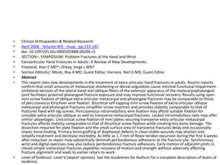
• Abstract
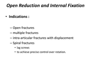
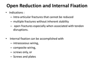
• Background : The objective of this study is to assess the complications after open reduction and
plate fixation of extra-articular metacarpal fractures.
• Methods : We retrospectively reviewed the clinical and radiologic records of 129 consecutive
patients with 157 metacarpal fractures treated by open reduction and internal fixation with plates
between 1993 and 1999. Intra-articular fractures and fractures of the thumb metacarpal were
excluded. Eighty-one patients (64 men and 17 women) with 104 fractures were available for
review, at an average follow-up of 13.6 months (range, 6–27 months).
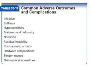
• Results : Twenty-eight patients (35%) and 33 fractures (32%) had one or more complications,
including difficulty with fracture healing (12 patients [15%]), stiffness (eight patients [10%]), plate
loosening or breakage (seven patients [8%]), complex regional pain syndrome (two patients), and
one patient who developed a deep infection.
• Conclusion : Despite technical advances in implant material, design, and instrumentation, plate
fixation of metacarpal fractures remains fraught with complications and unsatisfactory results.
Editor's Notes
Axial Loading -
No acceptable classification.
nonoperative treatment plays a significant role in the management of fractures and dislocations of the hand.
even though fractures and dislocations are fundamentally skeletal injuries.
The multiple joints of the hand are maintained in a delicate balance by the intrinsic and extrinsic tendon systems such that a disturbance in one set of tissues will often significantly affect others.
Critical elements in selecting between nonoperative and operative
than those created by high-energy trauma with large zones of injury.
One controversial point concerns the need to immobilize the wrist.
Setting appropriate length–tension relationships in the extrinsic motors (in cases where they are deforming forces) is most easily accomplished
Wrist splinting in extension is extremely helpful in patients with low pain tolerance who tend to place the hand in a characteristic dysfunctional posture
As the terminal point of contact with the environment, experiences stress loading with nearly every use of the hand.
When fractures accompany a nail bed injury, hematoma can be seen beneath the nail plate
Soft tissue injury is frequently of greater significance for long-term prognosis.
When the seal between the nail plate and the hyponychium is also broken- the fracture is open.
tuft is an anchoring point for the architecture of the digital pulp,
a honeycomb structure of fibrous septae that contains pockets of fat in each compartment.
The proximal portion of a tuft fracture may become entrapped in the septae of the pulp and prove irreducible.
The dorsal surface of the distal phalanx is the direct support for the germinal matrix and sterile matrix of the nail.
The bone volarly and the nail plate dorsally create a three-layered sandwich with the matrix in the middle.
Crush fractures of the tuft are stable - fibrous network of the pulp volarly and the splinting effect of the nail plate dorsally.
all fracture planes occurring distal to these tendon insertions have been separated from any internal deforming forces.
volar and dorsal base fractures are unstable, with the entire force of a tendon pulling the small base fragment away from the remainder of the bone.
Tendon insertions that play a role in fracture deformation include the central slip at the dorsal base and the terminal tendon acting through the DIP joint.
The FDS has a long insertion along the volar lateral margins of the shaft of P2 from the proximal onefourth to the distal one-fourth.
Fractures at the neck of P2 will usually angulate apex volar as the proximal fragment is flexed by the FDS.
Those at the base will usually angulate apex dorsal as the distal fragment is flexed by the FDS and the proximal extended by the central slip.
Actual P2 fracturesare less predictable and subject to any variety of displacement patterns.
When the volar fragment constitutes greater than around 40% of the articular surface, this fragment carries the majority of the proper collateral
ligament insertion in addition to the accessory ligament and volar plate insertions.
The dorsal fragment and remainder of P2 will thus sublux proximally and dorsally with displacement being driven by the pull of the FDS and the
central slip.
Relatively intact periosteum- The inherent stability of the fracture is more related to the degree of displacement thanthe direction or number of fracture planes.
The
key to success with this treatment is absolute maintenance of
a congruent reduction, avoiding the hinge motion that occurs
with dorsal and proximal subluxation of the major fragment.
Correct application of a dorsal extension block splint requires
maintenance of contact between the dorsum of the proximal
phalangeal segment and the splint. If the digit is allowed to
“pull away” from the splint volarly, the PIP joint can extend
beyond the safe range, sublux, and negate the desired effect of
the splint.
Dynamic traction / dorsal spring mechanism.
The general principle is to establish a foundation at the center of rotation in the head of P1.
From this foundation, traction (adjustable or elastic) is applied along the axis of P2 to hold the metaphyseal component of the fracture out to length
while allowing early motion to remodel the articular surface
Fracture patterns appearing in P1 include (A) complete articular fractures of the head,
(B) subcapital fractures with impingement in the volar plate recess
(C) transverse fractures of the shaft or base,
(D) oblique fractures of the shaft,
(E) articular fractures of the base.
Lateral mid axial approach is preferred than the dorsal approach.
As dorsal approach violates the permanently the extensor mechanism.
Flexing the MP joints fully causes the extensor apparatus to function as a tension band to a transverse fracture in the P1 shaft, helping to reduce the deformityand stabilize the fracture when the PIP joint is actively flexed.
Forms the basis of conservative management.
Spiral and long oblique fractures tend to shorten and rotate rather than angulate, not easily controlled by the joined positioning.
For long oblique and spiral fracture - three K-wires- perpendicular to the fracture
For neck fractures-retrograde pinning may be necessary
For short oblique and transverse fractures, -longitudinal K-wires
Fractures of the proximal phalangeal neck angulated apex volarly (A), can be stabilized by (B) antegrade pinning with a rotational
control crosswire if the fracture is sufficiently proximal,
but very distal fractures (C, D) usually require retrograde pinning.
Transverse shaft fractures of P1 are
best stabilized by 0.045-inch K-wires passed longitudinally
through the metacarpal head and removed at
3 weeks.
Figure 30-50
In long oblique fractures of the shaft with
shortening an exact reduction and stability sufficient to
withstand early motion can be achieved through lag screw
fixation only.
Figure 30-51
More complex fractures of the shaft
can be well stabilized by (B) lateral plating.
Specific care should be taken to (C) contour the plate
meticulously to fit the cortex and to place the hardware
in (D) the true midlateral position.
Figure 30-52 A:
Unicondylar fractures of the head of P1 benefit from compression between the
articular fragments through (B, C) lag screw fixation.
Figure 30-58
Transverse P1 fractures without comminution should achieve sufficient interfragmentary
stability to have axial rotational control with a single wire alone that targets the
(A) intercondylar notch and going (B) all the way to the subchondral bone.
Figure 30-59
Partial articular fractures that can be rendered stable
by interfragmentary compression are excellent candidates for lag
screw fixation.
Figure 30-61
Small locking plates can span zones of comminution and obviate the need to use the fixed angle blade plate.
When placing the splint, the PIP joints are held in nearly full extension to prevent the collateral ligament and volar plate contracture that occurs
in flexion.
The normal anatomic neck to shaft angle of 15 degrees should be recalled when assessing the amount of angulation in subcapital fractures.
Pseudoclawing is a term used to describe a dynamic imbalance ,hyperextension
deformity of the MP joint and a flexion deformity of the PIP
joint (Fig. 30-68).
This occurs as a compensatory response to the apex dorsal angulation of the metacarpal fracture (usually
at the neck) and represents a clinical indication for correcting the fracture angulation.
Figure 30-70 (A) the Brewerton view for the metacarpal bases,
(B) the Mehara view for the index CMC relationships,
(C) the reverse oblique view for angulation in the index metacarpal neck, and
(D) The skyline view for vertical impaction fractures of the metacarpal head.
Long oblique spiral fracture.
Introduce a Kirschner wire at the fracture site, and drill it out through the skin at the metacarpal base;
■ Reduce the fracture, and drill the wire in the opposite direction into the distal fragment, stopping just proximalto the metacarpophalangeal joint.
■ Apply a splint holding the wrist in extension.
For partial articular metacarpal head fractures, screw-only fixation
is the treatment of choice with up to 79 degrees of ROM
achieved.If sufficient interlock of bone spicules occurs, a
single 1.2- to1.5-mm countersunk screw can control rotation
of angulation increases for the more ulnar digits owing to the compensatory movement of the 4th and 5th carpometacarpal joints.
ROBERT S VIEW, hand in pronation.
Although the deep anterior obliquewas previously considered the primary stabilizer, more recent research has effectively demonstrated that
the dorsoradial ligament is the prime restraint to dislocation.
The distal metacarpal is adducted and supinated by the adductor pollicis.
At the same time, the APL pulls the metacarpal radially and proximally.
(WAGNER)
■ Maintaining fracture reduction by manual traction and pressure, drill a 0.045- or 0.062-inch Kirschner wire into the base of the metacarpal across the joint and into the trapezium.
Apply a forearm cast, holding the wrist in extension and the thumb in abduction; leave the thumb interphalangealjoint free.
Infection.
Segmental bone loss.
Soft tissue injuries.