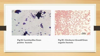
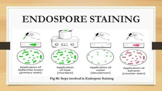
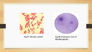
This document discusses various staining techniques used to visualize bacteria, including Gram staining, endospore staining, and flagella staining. Gram staining is used to differentiate between Gram-positive and Gram-negative bacteria based on their cell wall structure. Endospore staining targets bacterial endospores within certain genera like Bacillus. Flagella staining allows visualization of flagella which aid in bacterial identification and motility. Diagrams and microscopy images are included to illustrate the staining procedures and bacterial structures observed with each technique.