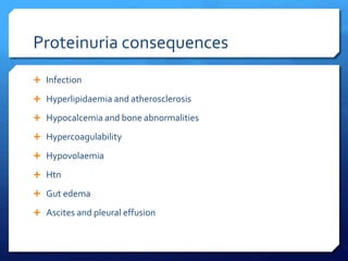
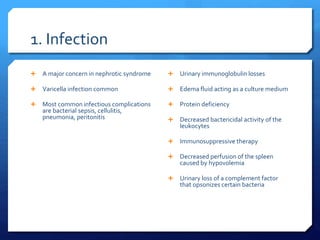
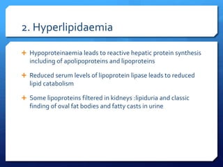
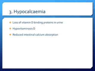
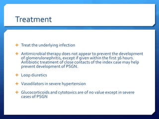
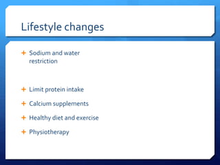
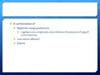
2. AGN
A specific set of kidney diseases in which an immunologic
mechanism triggers inflammation and proliferation of
glomerular tissue that can result to damage of the basement
membrane or capillary endothelium
Acute nephritic syndrome is the most serious of these
syndromes
AGN progresses to chronic GN in -30%
APSGN is the archetype of AGN: incidences have fallen
3. GN associated with Staph aureus increasing due to increased
antibiotic resistance to staph aureus
5. AGN Manifests with sudden onset of haematuria ,
proteinuria, and RBC casts in urine
Also usually accompanied by htn , edema, azotemia, renal
salt and water retention
Can be due to primary or sec renal disease
6. AGN Pathophysiology
Result from glomerular deposition or in situ formation of
immune complexes : kidneys may be enlarged upto 50% of
cases
PSGN: Streptococcal protein neuraminidase may alter host
IgG
IgG/ antiIgG immune complexes form and collect in the
glomeruli
Other antibody titers to other antigens elevations (evidence
of a recent poststreptococcal infection)=ASOT,
antihyaluronidase, DNAase-B, streptokinase
7. Structural and functional changes
Cellular proliferation of endothelial, mesangial and epithelial cells
in the glomerular tuft
Proliferation is in the glomerulus and bowman space
Proliferation of parietal epithelial cells leads to the formation of
crescents, a feature xstic of rapidly progressive GN
Leukocytes proliferation within the glomerular capillary
Thickening of basement memb
Functional changes: proteinuria, hematuria, low GFR, urine
sedments with RBCs and RBC casts
8. Etiology
1. Infectious
Strep spp GABH
Serotype 12: upper airway
infection
Serotype 49: skin
infection
Usually dvps 1-3 wks post
infection
Incidence 5-10% and 25%
in pharyngitis and skin
infection respectively
Staph
Other strep
Mycobacteria
Salmonella typhosa
Brucella suis
T.pllidum
9. CMC
EBV
COXSACKIE
Hep b
Rubella
Mumps
Hep A
C. immitis
P.Malariae
Schistosoma mansoni
Toxoplasma gondii
Wuchereria bancrofti
Trichinella
Trypanasomes
10. Aetiology
2. Non infectious
Primary kidney disease
Membranoproliferative GN: Due to expansion & proliferation of
mesangial cells as a consequence of complement deposition
IgA Nephropathy:diffuse mesangial deposition of IgA and IgG
Idiopathic rapidly progressive GN-xcterised by presence of
glemerular crescents
Sytemic diseases
Vasculitis
Collagen vascular disease e.g. SLE
Polyarteritis nodosa
Good pasture syndrome: autoantb against lungs and kidneys
12. Prognosis
Most epidemic cases follow a course ending in complete recovery
Sporadic cases of acute nephritis often progress to chronic
In PSGN long term prognosis is generally good , more than 98% of
individuals are asymptomatic after 5 yrs
Within a week of onset , most PSGN patients experience
spontaneous resolution of fluid retention, and HTN.Proteinuria
may persist for 6 months and microscopic haematuria upto 1 yr
Eventually, all urinary abnormalities should disappear,
hypertension should subside, and kidney function should return to
normal
GN is the most common cause of CKD
13. The prognosis for nonstreptococcal postinfectious
glomerulonephritis depends on the underlying agent, which
must be identified and addressed
MRSA associated nephritis usually resolves after treatment
14. Patient education
Salt restriction during acute phase
Bp monitoring
Ongoing long term monitoring
Consideration of protein restriction and ACE
Early antibiotic rx for close contacts
15. Presentation
History:Identification of underlying cause, abrupt onset,
Usually a boy, 2-14 yrs, sudden puffiness,edema of face with hx of
poststreptococcal infection. Dark urine, scanty, high BP,,,fevers,
weakness,malaise, abdominal pains
Anorexia
Itching , easy bruising, nose bleeds,
Sob , leg edema , dyspnoea
Haematuria, oliguria,
Flank pain- stretching of renal capsule
16. Symptoms specific to underling diseases
Triad of sinusitis, pulmonary infiltrates, and nephritis, suggesting
granulomatosis with polyangiitis (Wegener granulomatosis)- a rare
disorder where blood vessels become inflammed leading to
damage in major organs
Nausea and vomiting, abdominal pain, and purpura, observed with
Henoch-Schönlein purpura-inflammation of the small blood vessels
of the skin, joints, bowels and kidneys
Arthralgias, associated with systemic lupus erythematosus (SLE)
Hemoptysis, occurring with Goodpasture syndrome or idiopathic
progressive glomerulonephritis
Skin rashes, observed with SLE
17. PE
Normal including BP
Often a combination of edema,
HTN and oliguria
Periorbital / pedal edema
Crackles
Elevated JVP
Ascites and pleural effusion
Rash
pallor
CVA tenderness
Hematuria, microscopic or
gross
Abnormal neurological exam:
HTN, Urea
Arthritis
Pharyngitis, impetigo, murmur
Weight gain
19. DDX
Renal syndromes commonly
mimick early stage AGN
Chronic GN with an acute
exacerbation
Idiopathic hematuria
Familial nephritis –Alport
syndrome(hearing
loss/ocular abnormalities)
AKI
Goodpasture syndrome
Lupus nephritis
Membrenoproliferative GN
PSGN
20. Workup
Urinalysis
Protein
Blood
Red blood cells (RBCs)
White blood cells (WBCs)
Cellular (ie, RBC,WBC)
casts
Oval fat bodies
Presence of RBC casts is
almost pathognomonic of
GN
Urine sodium
CBC
BUN/C/E
Complement levels
21. The antistreptolysin O (ASO) titer is increased in 60-80% of
patients.
The increase begins in 1-3 weeks, peaks in 3-5 weeks, and
returns to normal in 6 months.ASO titer is unrelated to severity,
duration, or prognosis of kidney disease.
Increasing ASO titers confirm recent infection.
In patients with skin infection, anti-DNAase B (ADB) titers
are more sensitive than ASO titers for infection with
Streptococcus
22. Cultures throat and skin
Blood culture: IVDU, iss, indwelling shunts
Triglyceride levels
Hepatitis B and C serologies
Antineutrophil cytoplasmic antibody (ANCA)
24. Treatment
Treat the underlying infection
Antimicrobial therapy does not appear to prevent the development
of glomerulonephritis, except if given within the first 36 hours.
Antibiotic treatment of close contacts of the index case may help
prevent development of PSGN.
Loop diuretics
Vasodilators in severe hypertension
Glucocorticoids and cytotoxics are of no value except in severe
cases of PSGN
25. Lifestyle changes
Sodium and water
restriction
Limit protein intake
Calcium supplements
Healthy diet and exercise
Physiotherapy
27. A combination of
Nephrotic range proteinuria
>3g/day or on a single spot urine collection the presence of 2g/g of
urine creatinine
Low serum albumin
Edema
28. May occur in typical form or
in association with nephritic
syndrome
Classification
Primary
Minimal-change
nephropathy
Focal glomerulosclerosis
Membranous nephropathy
Hereditary nephropathies
secondary
Diabetes mellitus
Lupus erythematosus
Viral infections (eg,
hepatitis B, hepatitis C,
human immunodeficiency
virus [HIV] )
Amyloidosis and
paraproteinemias
Preeclampsia
Allo-antibodies from
enzyme replacement
therapy
30. PATHOPHYSIOLOGY
<0.1% plasma albumin pass glomerular
filtration barrier in healthy persons
Urine albumin in health is <50mg/day
>500mg/day point to glomerular disease
Glomerular caps endothelium are
fenestrated
Sits on GBM
GBM covered by glomerular
epithelium(podocytes)
In nephrotic syndrome there is damage to
endothelial surface, GBM or the podocytes
Glomerular filtration barrier –filtration slits
32. Theories for edema
Underfill hypothesis –albuminuria-
hypoalbuminaemia
Overfill hypothesis-filtered intraluminal protein
stimulate renal epithelial sodium reabsorption
2 facts favour this
sodium retention is observed even before the
serum albumin level starts falling
Intravascular volume is normal or even
increased in most patients with nephrotic
syndrome.
33. Proteinuria consequences
Infection
Hyperlipidaemia and atherosclerosis
Hypocalcemia and bone abnormalities
Hypercoagulability
Hypovolaemia
Htn
Gut edema
Ascites and pleural effusion
34. 1. Infection
A major concern in nephrotic syndrome
Varicella infection common
Most common infectious complications
are bacterial sepsis, cellulitis,
pneumonia, peritonitis
Urinary immunoglobulin losses
Edema fluid acting as a culture medium
Protein deficiency
Decreased bactericidal activity of the
leukocytes
Immunosuppressive therapy
Decreased perfusion of the spleen
caused by hypovolemia
Urinary loss of a complement factor
that opsonizes certain bacteria
35. 2. Hyperlipidaemia
Hypoproteinaemia leads to reactive hepatic protein synthesis
including of apolipoproteins and lipoproteins
Reduced serum levels of lipoprotein lipase leads to reduced
lipid catabolism
Some lipoproteins filtered in kidneys :lipiduria and classic
finding of oval fat bodies and fatty casts in urine
36. 3. Hypocalcaemia
Loss of vitamin D binding proteins in urine
Hypovitaminosis D
Reduced intestinal calcium absorption
37. Hypercoagulability
Risk forVTEs-renal ven thrombosis
Urinary loss of anticoagulants
Loss of Antithrombin III and plasminogen
Increase in clotting factors I,VII,VIII and X
Protein C and S Altered levels and activity
Hyperfibrinogenemia –increased hepatic synthesis
Impaired fibrinolysis due to decreased plasminogen
Increased plt aggregability
Alteration in endothelial function
39. Etiology
Minimal change disease, membraneous nephropathy, focal
glomerulosclerosis
StimulateT cells , release cytokines, damage podocytes
Medications – NSAIDs, gold, penicillamine, lithium,
anticancer drugs,
Has been reported following Covid 19 infection and
vaccination
40. Presentation
History :
Swelling of face
Dependent edema
Foamy urine
Fatigue
Loss of appetite
Thrombotic event
Recent start of a drug e.g NSAID
42. Workup
Urinalysis
Urine protein:urine creatine ration >2g/g corresponds to >3gram
protein/day
Urine sediment examination
Urinary protein measurement
Serum albumin
Serologic studies for infection and immune abnormalities
Renal ultrasonography
Kidney biopsy
43. Important definitions
Response: protein free urine on 3 consequtive days within 7 days
Relapse: Protein +ve urine on 3 consequtive days within one week
Frequent relapsing : steroid sensitive NS with 2 or more relapses in
6 months or > 3 in 1 year
Steroid dependant:responders who relapses while steroid is being
tapered or within 14 days of stopping steroid treatment
Initial non responders: No response during initial 8 weeks of rx
Late non responders:Initial steroid responder who fails to respond
to 4 week treatment in relapse
44. Treatment
Specific treatment depends on the cause
Diuretics
Anticoagulation
Statins
ARB/ACE for secondary nephrotic syndrome
45. Specific treatment
Minimal change nephropathy 70-80% of NS: Glucocorticoids e.g
prednisolone
It is Idiopathic loss of net negative charge in cap BM
Sec causes:Infections , NSAIDs, Hodgkins lymphoma
StimulateT cells-cytokines destroy podocytes
Membraneous nephropathy:Primary:antiPLA2R receptor antibody.
Attacks a protein in podocytes. Sec: HBA/B, syphillis,Gold,
Penicillamine.
Antibodies deposit in subepithelial region, inflammation, activation
of compliment system, podocyte effacement
Watchful waiting,Rituximab or cyclophosphamide+/-
glucocorticoids
46.
47. Focal segmental glomerulosclerosis
Can be primary-idiopathic
Sec: HIV,SCD, Heroin use
Sclerosis of parts of the
glomerulous leading to
podocyte effacement
Longterm
immunosupressants
In all treat the sec cause
48. In all treat the sec cause
In primary cause start on
steroids
Good response usually
indicates MC disease.Bx
may not be necessary
Poor response ?FSGS
Longterm
immunosupressants
50. LONGTERM MONITORING
Adjustments for diuretics
Immunizations :Pneumococcal and influenza
Monitoring corticosteroid toxicity every 3 months
Supplemental vit D and Calcium may attenuate bone loss