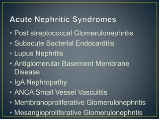
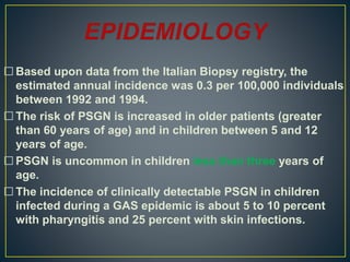
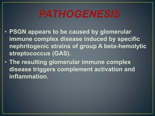
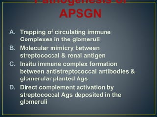
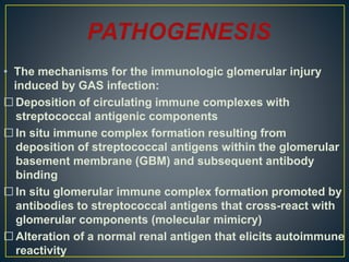
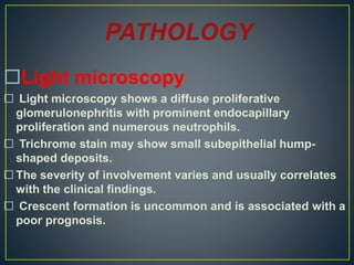
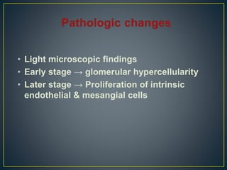
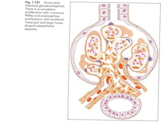
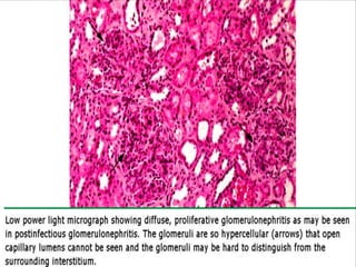
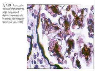
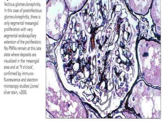
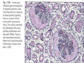
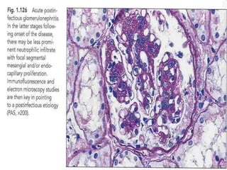
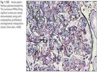
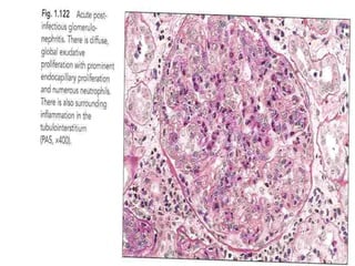
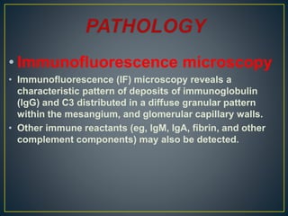
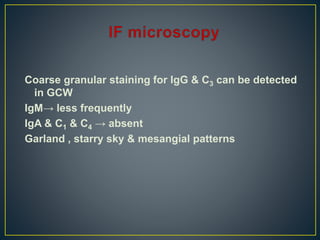
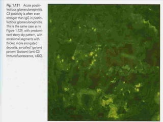
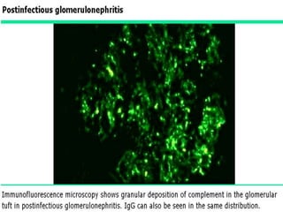
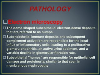
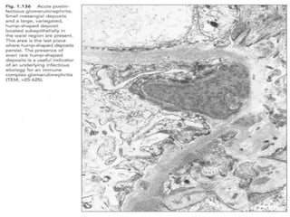
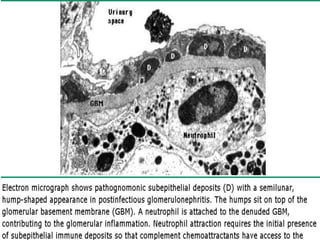
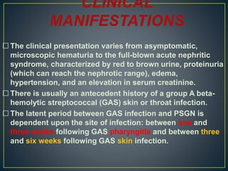
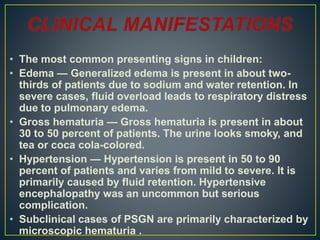
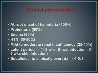
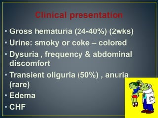
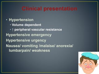
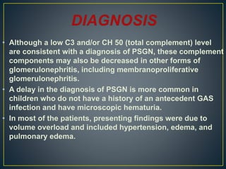
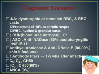
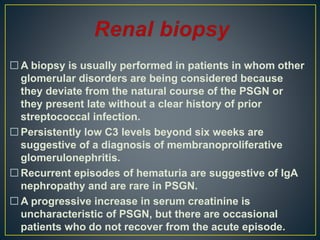
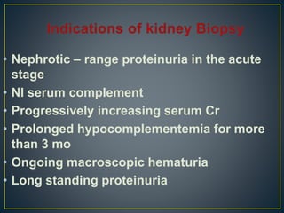
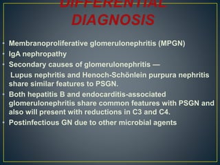
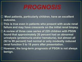
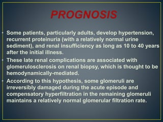
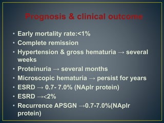
bhubaneswar Call Girls 👙 6297143586 👙 Genuine WhatsApp Number for Real Meet
Acute Nephritic Syndrome VKP.pptx
1.
2. • Acute nephritic syndromes classically
present with the following:
• Hypertension
• Hematuria
• Red blood cell casts
• Polyuria
• Mild to moderate proteinuria
3. Extensive inflammatory damage to
glomeruli can cause a fall in GFR and
eventually produce uremic symptoms
with salt and water retention, leading
to edema and hypertension.
4. • Post streptococcal Glomerulonephritis
• Subacute Bacterial Endocarditis
• Lupus Nephritis
• Antiglomerular Basement Membrane
Disease
• IgA Nephropathy
• ANCA Small Vessel Vasculitis
• Membranoproliferative Glomerulonephritis
• Mesangioproliferative Glomerulonephritis
5.
6. • Poststreptococcal glomerulonephritis (PSGN) is
caused by prior infection with specific
nephritogenic strains of group A beta-hemolytic
streptococcus.
• The clinical presentation of PSGN varies from
asymptomatic, microscopic hematuria to the full-
blown acute nephritic syndrome, characterized by
red to brown urine, proteinuria (which can reach
the nephrotic range), edema, hypertension, and
acute kidney injury.
• The prognosis is generally favorable, especially in
children, but in some cases, the long-term
7. Although PSGN continues to be the most
common cause of acute nephritis globally, it
primarily occurs in developing countries.
Of the estimated 470,000 new annual cases
of PSGN worldwide, 97 percent occur in
developing countries, with an annual
incidence that ranges from 9.5 to 28.5 per
100,000 individuals.
In more developed and industrialized
countries, the incidence has decreased over
the past three decades.
8. Based upon data from the Italian Biopsy registry, the
estimated annual incidence was 0.3 per 100,000 individuals
between 1992 and 1994.
The risk of PSGN is increased in older patients (greater
than 60 years of age) and in children between 5 and 12
years of age.
PSGN is uncommon in children less than three years of
age.
The incidence of clinically detectable PSGN in children
infected during a GAS epidemic is about 5 to 10 percent
with pharyngitis and 25 percent with skin infections.
10. • PSGN appears to be caused by glomerular
immune complex disease induced by specific
nephritogenic strains of group A beta-hemolytic
streptococcus (GAS).
• The resulting glomerular immune complex
disease triggers complement activation and
inflammation.
11. A. Trapping of circulating immune
Complexes in the glomeruli
B. Molecular mimicry between
streptococcal & renal antigen
C. Insitu immune complex formation
between antistreptococcal antibodies &
glomerular planted Ags
D. Direct complement activation by
streptococcal Ags deposited in the
glomeruli
12. • The mechanisms for the immunologic glomerular injury
induced by GAS infection:
Deposition of circulating immune complexes with
streptococcal antigenic components
In situ immune complex formation resulting from
deposition of streptococcal antigens within the glomerular
basement membrane (GBM) and subsequent antibody
binding
In situ glomerular immune complex formation promoted by
antibodies to streptococcal antigens that cross-react with
glomerular components (molecular mimicry)
Alteration of a normal renal antigen that elicits autoimmune
reactivity
13. Light microscopy
Light microscopy shows a diffuse proliferative
glomerulonephritis with prominent endocapillary
proliferation and numerous neutrophils.
Trichrome stain may show small subepithelial hump-
shaped deposits.
The severity of involvement varies and usually correlates
with the clinical findings.
Crescent formation is uncommon and is associated with a
poor prognosis.
14. • Light microscopic findings
• Early stage → glomerular hypercellularity
• Later stage → Proliferation of intrinsic
endothelial & mesangial cells
15.
16.
17.
18.
19.
20.
21.
22.
23. • Immunofluorescence microscopy
• Immunofluorescence (IF) microscopy reveals a
characteristic pattern of deposits of immunoglobulin
(IgG) and C3 distributed in a diffuse granular pattern
within the mesangium, and glomerular capillary walls.
• Other immune reactants (eg, IgM, IgA, fibrin, and other
complement components) may also be detected.
24. Coarse granular staining for IgG & C3 can be detected
in GCW
IgM→ less frequently
IgA & C1 & C4 → absent
Garland , starry sky & mesangial patterns
25.
26.
27. Electron microscopy
The dome-shaped subepithelial electron-dense deposits
that are referred to as humps.
Subendothelial immune deposits and subsequent
complement activation are responsible for the local
influx of inflammatory cells, leading to a proliferative
glomerulonephritis, an active urine sediment, and a
variable decline in glomerular filtration rate.
Subepithelial "humps" are responsible for epithelial cell
damage and proteinuria, similar to that seen in
membranous nephropathy
28. Swelling of glomerular endothelial & mesangial cells
Humps (disappear 6 wks after the clinicalonset of dx)
29.
30.
31. The clinical presentation varies from asymptomatic,
microscopic hematuria to the full-blown acute nephritic
syndrome, characterized by red to brown urine, proteinuria
(which can reach the nephrotic range), edema,
hypertension, and an elevation in serum creatinine.
There is usually an antecedent history of a group A beta-
hemolytic streptococcal (GAS) skin or throat infection.
The latent period between GAS infection and PSGN is
dependent upon the site of infection: between one and
three weeks following GAS pharyngitis and between three
and six weeks following GAS skin infection.
32. • The most common presenting signs in children:
• Edema — Generalized edema is present in about two-
thirds of patients due to sodium and water retention. In
severe cases, fluid overload leads to respiratory distress
due to pulmonary edema.
• Gross hematuria — Gross hematuria is present in about
30 to 50 percent of patients. The urine looks smoky, and
tea or coca cola-colored.
• Hypertension — Hypertension is present in 50 to 90
percent of patients and varies from mild to severe. It is
primarily caused by fluid retention. Hypertensive
encephalopathy was an uncommon but serious
complication.
• Subclinical cases of PSGN are primarily characterized by
microscopic hematuria .
36. •Urinalysis
• The urinalysis in patients with PSGN reveal
hematuria (some of the red cells are typically
dysmorphic) with or without red blood cell casts,
varying degrees of proteinuria and often pyuria.
• Nephrotic range proteinuria is uncommon and
occurs in about 5 percent of cases at
presentation.
37. Complement
In about 90 percent of patients, C3 and CH50 (total
complement activity) are significantly depressed in the
first two weeks of the disease course.
In comparison, C4 and C2 levels are usually normal or
only mildly decreased.
The C3 and CH 50 return to normal within four to eight
weeks after presentation.
The combination of a low C3 level and a normal or only
slightly decreased C4 level indicates activation of the
alternative pathway of complement.
38. •Culture
• Because PSGN presents weeks after an antecedent GAS
infection, only about 25 percent of patients will have
either a positive throat or skin culture.
• In patients with impetigo, there is an increased likelihood
of obtaining a positive skin culture.
39. Serology
The streptozyme test, which measures five different
streptococcal antibodies, is positive in more than 95
percent of patients with PSGN due to pharyngitis and
about 80 percent of those with skin infections.
It includes the following antibodies:
Anti-streptolysin (ASO)
Anti-hyaluronidase (AHase)
Anti-streptokinase (ASKase)
Anti-nicotinamide-adenine dinucleotidase (anti-NAD)
Anti-DNAse B antibodies
40. • After a pharyngeal infection, the ASO, anti-DNAse B, anti-
NAD, and AHase titers are commonly elevated.
• In comparison, only the anti-DNAse B and AHase titers are
typically increased after a skin infection.
• If only the ASO titer is used to screen for GAS infection, it
may be falsely low or negative in patients with skin
infections.
41. PSGN is usually diagnosed based upon clinical findings of
acute nephritis and demonstration of a recent group A
beta-hemolytic streptococcal (GAS) infection.
The clinical findings of acute nephritis include hematuria
with or without red blood cell casts, variable degrees of
proteinuria, edema, and hypertension.
Documentation of a recent GAS infection includes either a
positive throat or skin culture or serologic tests (eg, ASO
or streptozyme test).
42. • Although a low C3 and/or CH 50 (total complement) level
are consistent with a diagnosis of PSGN, these complement
components may also be decreased in other forms of
glomerulonephritis, including membranoproliferative
glomerulonephritis.
• A delay in the diagnosis of PSGN is more common in
children who do not have a history of an antecedent GAS
infection and have microscopic hematuria.
• In most of the patients, presenting findings were due to
volume overload and included hypertension, edema, and
pulmonary edema.
44. A biopsy is usually performed in patients in whom other
glomerular disorders are being considered because
they deviate from the natural course of the PSGN or
they present late without a clear history of prior
streptococcal infection.
Persistently low C3 levels beyond six weeks are
suggestive of a diagnosis of membranoproliferative
glomerulonephritis.
Recurrent episodes of hematuria are suggestive of IgA
nephropathy and are rare in PSGN.
A progressive increase in serum creatinine is
uncharacteristic of PSGN, but there are occasional
patients who do not recover from the acute episode.
45. • Nephrotic – range proteinuria in the acute
stage
• Nl serum complement
• Progressively increasing serum Cr
• Prolonged hypocomplementemia for more
than 3 mo
• Ongoing macroscopic hematuria
• Long standing proteinuria
46. • Membranoproliferative glomerulonephritis (MPGN)
• IgA nephropathy
• Secondary causes of glomerulonephritis —
Lupus nephritis and Henoch-Schönlein purpura nephritis
share similar features to PSGN.
• Both hepatitis B and endocarditis-associated
glomerulonephritis share common features with PSGN and
also will present with reductions in C3 and C4.
• Postinfectious GN due to other microbial agents
47. • The patients with more than 30 percent crescents
on renal biopsy are often treated with
methylprednisolone pulses.
• Management is supportive and is focused on
treating the clinical manifestations of the disease,
particularly complications due to volume overload.
• These include hypertension and, less commonly,
pulmonary edema.
• General measures include sodium and water
restriction and loop diuretics.
48. • Loop diuretics generally provide a prompt
diuresis with reduction of blood pressure and
edema.
• In our practice, intravenous furosemide is given
at an initial dose of 1 mg/kg (maximum 40 mg).
• Patients with PSGN have variable reductions in
renal function, and some patients require
dialysis during the acute episode.
• Patients with evidence of persistent group A
streptococcal infection should be given a
course of antibiotic therapy.
49. Resolution of the clinical manifestations of PSGN is
generally quite rapid, assuming concurrent resolution of
the infection.
A diuresis typically begins within one week, and the serum
creatinine returns to the previous baseline by three to four
weeks.
The urinary abnormalities disappear at differing rates.
Hematuria usually resolves within three to six months.
Proteinuria also falls during recovery, but at a much
slower rate.
A mild increase in protein excretion is still present in 15
percent at 3 years, and 2 percent at 7 to 10 years.
In severe cases with nephrotic range proteinuria, this
50. • Recurrent episodes of PSGN are rare.
• This may be due to the long-term persistence of
antibodies to nephritis-associated streptococcal antigen.
51. Most patients, particularly children, have an excellent
outcome.
This is true even in patients who present with acute renal
failure and may have crescents on the initial renal biopsy.
A review of three case series of 229 children with PSGN
found that approximately 20 percent had an abnormal
urinalysis (proteinuria and/or hematuria), but almost all
(92 to 99 percent) had normal or only modestly reduced
renal function 5 to 18 years after presentation.
However, the long-term prognosis of PSGN is not always
benign.
52. • Some patients, particularly adults, develop hypertension,
recurrent proteinuria (with a relatively normal urine
sediment), and renal insufficiency as long as 10 to 40 years
after the initial illness.
• These late renal complications are associated with
glomerulosclerosis on renal biopsy, which is thought to be
hemodynamically-mediated.
• According to this hypothesis, some glomeruli are
irreversibly damaged during the acute episode and
compensatory hyperfiltration in the remaining glomeruli
maintains a relatively normal glomerular filtration rate.
53. • Early mortality rate:<1%
• Complete remission
• Hypertension & gross hematuria → several
weeks
• Proteinuria → several months
• Microscopic hematuria → persist for years
• ESRD → 0.7- 7.0% (NAplr protein)
• ESRD →2%
• Recurrence APSGN →0.7-7.0%(NAplr
protein)