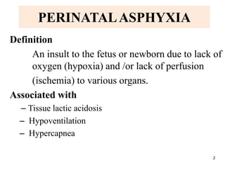
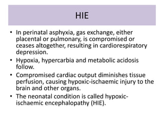
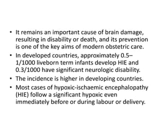
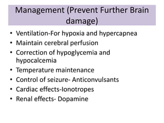
Perinatal Asphyxia
Natangwe Shimhanda
201208214
PERINATALASPHYXIA
Definition
An insult to the fetus or newborn due to lack of
oxygen (hypoxia) and /or lack of perfusion
(ischemia) to various organs.
Associated with
– Tissue lactic acidosis
– Hypoventilation
– Hypercapnea
2
Definitions
• Birth Asphyxia - failure to initiate
spontaneous and sustained respiration after
delivery (may result in severe hypoxia)
• Fetal Hypoxia - refers to inadequate
oxygenation before delivery (may predispose
the fetus to birth asphyxia)
• Hypoxia during labour in term babies is the
most common cause of neonatal brain
damage and is the leading cause of cerebral
palsy in the developing word.
• The effects of hypoxia & Ischemia inseparable
and they both contribute to tissue injury
ESSENTIAL CRITERIA FOR PERINATAL
ASPHYXIA
• Prolonged metabolic or mixed acidemia (pH <
7.00) on an umbilical cord arterial blood sample
• Persistence of an Apgar score of 0-3 for > 5
minutes
• Clinical neurological manifestations e.g. seizure,
hypotonia, coma or hypoxic-ischaemic
encephalopathy in the immediate neonatal
period
• Evidence of multiorgan system dysfunction in the
immediate neonatal periods
Phases of Asphyxia
• Primary Apnea (30-60s)
– short series of respiratory
efforts
– convulsion or a series of
clonic movements
– abrupt fall in heart rate
– then no muscle tone
– skin cyanotic and then
blotchy
• Gasping (8m)
– 3-6/minute
– heart rate and blood pressure
continue to fall
• Secondary/Terminal Apnea
Apgar score
APGAR Score:
8-10 No asphyxia
5-7 Mild asphyxia
3-4 Moderate asphyxia
0-2 Severe asphyxia
ETIOLOGY
• 90% Antepartum/ Intrapartum
• 10% Postpartum
Maternal
– Hypertension - chronic / pre-eclampsia
– Diabetes, Anemia, Malnutrition, Maternal health
– Hypoxia - Pulmonary/Cardiac Diseases, Bronchial asthma,
Hypotension
– anesthetics and analgesics
– Intrauterine infections
– Prolonged and difficult 2nd stage of labour
– Placental: Infarction/fibrosis/abruption/insufficiency
– Cord Accidents: Prolapse / True knots / compression/Abn. vessels
Fetal Factors
• Infections - Intrauterine
• Anemia - Feto-fetal or Feto-maternal transfusion
• Growth restriction (IUGR)/ Postmaturity -
Meconium aspiration, tracheal plug by bloody
mucus
• Congenital malformations - choanal atresia,
laryngeal web, diaphragmatic hernias,
• Hydrops fetalis
• Prematurity, multiple births and rhesus
isoimmunization
Pathophysiology
Diving reflex:
Shunting of blood to brain ,heart, adrenals and away
from gut, kidney, liver, spleen ,skeletal muscle and
skin
Progressive asphyxia:
Hypoxia, Hypercapnea, acidosis
Pulmonary vasoconstriction,
Decrease in HR, CO, BP
Cerebral edema- Petechial hemorrhage,
SIADH,
Na- intracellular accumulation
Acute Sequelae Of Asphyxia
Late effects
• Microcephaly
• MR
• Developmental delay
• Cerebral palsy
• Epilepsy
• Visual, hearing impairement
• Behavioral disturbances
Management
• Presence of a health professional skilled in
Neonatal resuscitation
• Necessary equipment for resuscitation: infant size
laryngoscpoe and margill’s forceps, ETT 2.5-
3.5mm, neonatal Ambu-bag mask, suction
catheters 8FG and 6FG, umbilical catheters if
peripheral IV line is not possible, necessary fluids,
etc.
• Emergency medicines e.g. naloxone, adrenaline
Management
Aimed to prevent further Brain Damage
• Determine your APGAR score
• Ensure warmth and not allowing hypothermia to occur
• Administer 100% O2, reduce as soon as the colour
improves
• Apply gentle nasopharyngeal suction if indicated
• Administer bag mask ventilation
• Administer Naloxone when indicated
• Undertake external Cardiac massage for bradycardia
HIE
• In perinatal asphyxia, gas exchange, either
placental or pulmonary, is compromised or
ceases altogether, resulting in cardiorespiratory
depression.
• Hypoxia, hypercarbia and metabolic acidosis
follow.
• Compromised cardiac output diminishes tissue
perfusion, causing hypoxic-ischaemic injury to the
brain and other organs.
• The neonatal condition is called hypoxic-
ischaemic encephalopathy (HIE).
• It remains an important cause of brain damage,
resulting in disability or death, and its prevention
is one of the key aims of modern obstetric care.
• In developed countries, approximately 0.5–
1/1000 liveborn term infants develop HIE and
0.3/1000 have significant neurologic disability.
• The incidence is higher in developing countries.
• Most cases of hypoxic-ischaemic encephalopathy
(HIE) follow a significant hypoxic even
immediately before or during labour or delivery.
TARGET ORGAN-BRAIN
HIE:PATHOPHYSIOLOGY
Hypoxia Alteration in glucose and energy
metabolism loss of autoregulation & decreased
cardiac function ischemia
Severe hypoxia and ischemia failure of oxidative
phosphorylation & ATP production
Accumulation of Na, Cl,Ca intracellular and K and
excitatory neurotransmitters extracellular
neuronal death
20
Pathology and Pattern seen in term
babies
• Selective neuronal necrosis - cerebral and
cerebellar cortex, Thalamus, brain stem nuclei
(Spastic CP)
• Status Marmoratus (Chorea, Athetoid, Dystonia)
• Parasagittal cerebral injury (Prox Spastic
Quadriparesis)
• Focal and multifocal ischemic brain injury (sp.
Hemiparesis, cognitive defects, seizure)
Pattern predominant in preterm
• Periventricular leukomalacia
Grading
The clinical manifestations start immediately or up to
48 h after asphyxia, and can be graded:
• Mild – the infant is irritable, responds excessively to
stimulation, may have staring of the eyes and
hyperventilation and has impaired feeding
• Moderate – the infant shows marked abnormalities of
tone and movement, cannot feed and may have seizures
• Severe – there are no normal spontaneous movements
or response to pain; tone in the limbs may fluctuate
between hypotonia and hypertonia; seizures are
prolonged and often refractory to treatment; multi-organ
failure is present.
Management (Prevent Further Brain
damage)
• Ventilation-For hypoxia and hypercapnea
• Maintain cerebral perfusion
• Correction of hypoglycemia and
hypocalcemia
• Temperature maintenance
• Control of seizure- Anticonvulsants
• Cardiac effects-Ionotropes
• Renal effects- Dopamine
Treatment of seizures
• Correction of hypoglycemia, hypocalcemia &
electrolyte
• Prophylactic Phenobarbitone ?
• Therapeutic Phenobarbitone
20 mg / kg (loading), 5 mg / kg / d
(maintenance)
• Lorazepam – 0.05 – 0.1 mg / kg
• Diazepam to be avoided
Cerebral Edema
• Avoid fluid overload (SIADH, ATN)
• 30 degrees Head raise
• Maintain PaCo2 25-30mm Hg in ventilated
infants
• Mannitol 20% (0.5 - 1g / kg) 6 hrly. x 24 hrs.
• Frusemide 1.0 mg / kg every 12 hrs.
Prognosis
• Overall mortality - 10-20%
• Neurological sequele - 20-45%
Sarnat Stages
• 1- 100% Normal neurological outcome
• 2- 80% Normal neurological outcome
• 3- 50% deaths, 50% major neurological sequelae
Risk for CP - 5-10%
• Subtle school problems in neurologically and
mentally normal survivors of HIE stage 1-nil, stage 2-
18-35%
References
• Lissauer T, Clayden G, 2012. Illustrated
Textbook Of Paediatrics. Fourth edition. ©
2012 Elsevier Ltd
• Wittenberg D.F. (2009), Coovadia’s Paediatrics
and Child Health. Oxford University Press
Southern Africa (pty) Ltd

Perinatal asphyxia

  • 1.
  • 2.
    PERINATALASPHYXIA Definition An insult tothe fetus or newborn due to lack of oxygen (hypoxia) and /or lack of perfusion (ischemia) to various organs. Associated with – Tissue lactic acidosis – Hypoventilation – Hypercapnea 2
  • 3.
    Definitions • Birth Asphyxia- failure to initiate spontaneous and sustained respiration after delivery (may result in severe hypoxia) • Fetal Hypoxia - refers to inadequate oxygenation before delivery (may predispose the fetus to birth asphyxia)
  • 4.
    • Hypoxia duringlabour in term babies is the most common cause of neonatal brain damage and is the leading cause of cerebral palsy in the developing word. • The effects of hypoxia & Ischemia inseparable and they both contribute to tissue injury
  • 5.
    ESSENTIAL CRITERIA FORPERINATAL ASPHYXIA • Prolonged metabolic or mixed acidemia (pH < 7.00) on an umbilical cord arterial blood sample • Persistence of an Apgar score of 0-3 for > 5 minutes • Clinical neurological manifestations e.g. seizure, hypotonia, coma or hypoxic-ischaemic encephalopathy in the immediate neonatal period • Evidence of multiorgan system dysfunction in the immediate neonatal periods
  • 6.
    Phases of Asphyxia •Primary Apnea (30-60s) – short series of respiratory efforts – convulsion or a series of clonic movements – abrupt fall in heart rate – then no muscle tone – skin cyanotic and then blotchy • Gasping (8m) – 3-6/minute – heart rate and blood pressure continue to fall • Secondary/Terminal Apnea
  • 7.
    Apgar score APGAR Score: 8-10No asphyxia 5-7 Mild asphyxia 3-4 Moderate asphyxia 0-2 Severe asphyxia
  • 8.
    ETIOLOGY • 90% Antepartum/Intrapartum • 10% Postpartum Maternal – Hypertension - chronic / pre-eclampsia – Diabetes, Anemia, Malnutrition, Maternal health – Hypoxia - Pulmonary/Cardiac Diseases, Bronchial asthma, Hypotension – anesthetics and analgesics – Intrauterine infections – Prolonged and difficult 2nd stage of labour – Placental: Infarction/fibrosis/abruption/insufficiency – Cord Accidents: Prolapse / True knots / compression/Abn. vessels
  • 9.
    Fetal Factors • Infections- Intrauterine • Anemia - Feto-fetal or Feto-maternal transfusion • Growth restriction (IUGR)/ Postmaturity - Meconium aspiration, tracheal plug by bloody mucus • Congenital malformations - choanal atresia, laryngeal web, diaphragmatic hernias, • Hydrops fetalis • Prematurity, multiple births and rhesus isoimmunization
  • 10.
    Pathophysiology Diving reflex: Shunting ofblood to brain ,heart, adrenals and away from gut, kidney, liver, spleen ,skeletal muscle and skin
  • 11.
    Progressive asphyxia: Hypoxia, Hypercapnea,acidosis Pulmonary vasoconstriction, Decrease in HR, CO, BP Cerebral edema- Petechial hemorrhage, SIADH, Na- intracellular accumulation
  • 12.
  • 13.
    Late effects • Microcephaly •MR • Developmental delay • Cerebral palsy • Epilepsy • Visual, hearing impairement • Behavioral disturbances
  • 14.
    Management • Presence ofa health professional skilled in Neonatal resuscitation • Necessary equipment for resuscitation: infant size laryngoscpoe and margill’s forceps, ETT 2.5- 3.5mm, neonatal Ambu-bag mask, suction catheters 8FG and 6FG, umbilical catheters if peripheral IV line is not possible, necessary fluids, etc. • Emergency medicines e.g. naloxone, adrenaline
  • 15.
    Management Aimed to preventfurther Brain Damage • Determine your APGAR score • Ensure warmth and not allowing hypothermia to occur • Administer 100% O2, reduce as soon as the colour improves • Apply gentle nasopharyngeal suction if indicated • Administer bag mask ventilation • Administer Naloxone when indicated • Undertake external Cardiac massage for bradycardia
  • 18.
    HIE • In perinatalasphyxia, gas exchange, either placental or pulmonary, is compromised or ceases altogether, resulting in cardiorespiratory depression. • Hypoxia, hypercarbia and metabolic acidosis follow. • Compromised cardiac output diminishes tissue perfusion, causing hypoxic-ischaemic injury to the brain and other organs. • The neonatal condition is called hypoxic- ischaemic encephalopathy (HIE).
  • 19.
    • It remainsan important cause of brain damage, resulting in disability or death, and its prevention is one of the key aims of modern obstetric care. • In developed countries, approximately 0.5– 1/1000 liveborn term infants develop HIE and 0.3/1000 have significant neurologic disability. • The incidence is higher in developing countries. • Most cases of hypoxic-ischaemic encephalopathy (HIE) follow a significant hypoxic even immediately before or during labour or delivery.
  • 20.
    TARGET ORGAN-BRAIN HIE:PATHOPHYSIOLOGY Hypoxia Alterationin glucose and energy metabolism loss of autoregulation & decreased cardiac function ischemia Severe hypoxia and ischemia failure of oxidative phosphorylation & ATP production Accumulation of Na, Cl,Ca intracellular and K and excitatory neurotransmitters extracellular neuronal death 20
  • 21.
    Pathology and Patternseen in term babies • Selective neuronal necrosis - cerebral and cerebellar cortex, Thalamus, brain stem nuclei (Spastic CP) • Status Marmoratus (Chorea, Athetoid, Dystonia) • Parasagittal cerebral injury (Prox Spastic Quadriparesis) • Focal and multifocal ischemic brain injury (sp. Hemiparesis, cognitive defects, seizure) Pattern predominant in preterm • Periventricular leukomalacia
  • 22.
    Grading The clinical manifestationsstart immediately or up to 48 h after asphyxia, and can be graded: • Mild – the infant is irritable, responds excessively to stimulation, may have staring of the eyes and hyperventilation and has impaired feeding • Moderate – the infant shows marked abnormalities of tone and movement, cannot feed and may have seizures • Severe – there are no normal spontaneous movements or response to pain; tone in the limbs may fluctuate between hypotonia and hypertonia; seizures are prolonged and often refractory to treatment; multi-organ failure is present.
  • 23.
    Management (Prevent FurtherBrain damage) • Ventilation-For hypoxia and hypercapnea • Maintain cerebral perfusion • Correction of hypoglycemia and hypocalcemia • Temperature maintenance • Control of seizure- Anticonvulsants • Cardiac effects-Ionotropes • Renal effects- Dopamine
  • 24.
    Treatment of seizures •Correction of hypoglycemia, hypocalcemia & electrolyte • Prophylactic Phenobarbitone ? • Therapeutic Phenobarbitone 20 mg / kg (loading), 5 mg / kg / d (maintenance) • Lorazepam – 0.05 – 0.1 mg / kg • Diazepam to be avoided
  • 25.
    Cerebral Edema • Avoidfluid overload (SIADH, ATN) • 30 degrees Head raise • Maintain PaCo2 25-30mm Hg in ventilated infants • Mannitol 20% (0.5 - 1g / kg) 6 hrly. x 24 hrs. • Frusemide 1.0 mg / kg every 12 hrs.
  • 26.
    Prognosis • Overall mortality- 10-20% • Neurological sequele - 20-45% Sarnat Stages • 1- 100% Normal neurological outcome • 2- 80% Normal neurological outcome • 3- 50% deaths, 50% major neurological sequelae Risk for CP - 5-10% • Subtle school problems in neurologically and mentally normal survivors of HIE stage 1-nil, stage 2- 18-35%
  • 27.
    References • Lissauer T,Clayden G, 2012. Illustrated Textbook Of Paediatrics. Fourth edition. © 2012 Elsevier Ltd • Wittenberg D.F. (2009), Coovadia’s Paediatrics and Child Health. Oxford University Press Southern Africa (pty) Ltd

Editor's Notes

  • #10 *choanal atresia – congenital disorder where by the back of the nasal passage (choana) is blocked * Hydrops fetalis- abnormal accumulation of fluid in 2/more fetal compartments e.g ascities, pleural effusion, pericardial efusion
  • #12 Syndrome of inappropriate antidiuretic Hormone secretion - SIADH