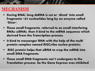
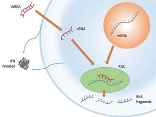
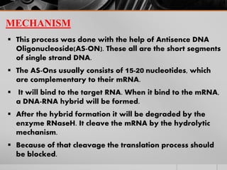
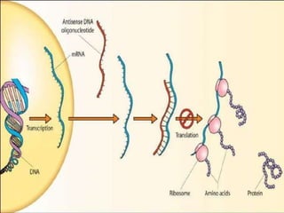
This document discusses two methods of gene silencing: RNA interference (RNAi) and antisense therapy. RNAi involves long double-stranded RNA being cut into small interfering RNAs (siRNAs) that bind to messenger RNA and prevent translation. Antisense therapy uses short antisense DNA oligonucleotides that are complementary to mRNA and form DNA-RNA hybrids that are degraded by RNaseH, blocking translation. Both methods allow targeted inhibition of gene expression for research and potential therapeutic applications.
RNA interference (RNAi): Cellular process by which an mRNA is targeted for degradation by a dsRNA with a strand complementary to a fragment of such mRNA.
RNA interference (RNAi) is a system within living cells that takes part in controlling which genes are active and how active they are. RNA interference has an important role in defending cells against parasitic genes – viruses and transposons – but also in directing development as well as gene expression in general.
RNA interference (RNAi) is a mechanism that inhibits gene expression at the stage of translation or by hindering the transcription of specific genes.
RNAi targets include RNA from viruses and transposons.
INTERFERENCE means the act of interfering with something, here, with RNA. RNAi is an evolutionarily conserved mechanism triggered by dsRNA molecules, to prevent the expression of specific genes or the translation, causes sequence-specific degradation of the targeted mRNA molecules of that particular gene. It was also known as CO-SUPPRESSION, POST TRANSCRIPTIONAL GENE SILENCING [PTGS] in plants and QUELLING in fungi.
The increased availability of biomedical data, particularly in the public domain, offers the opportunity to better understand human health and to develop effective therapeutics for a wide range of unmet medical needs. However, data scientists remain stymied by the fact that data remain hard to find and to productively reuse because data and their metadata i) are wholly inaccessible, ii) are in non-standard or incompatible representations, iii) do not conform to community standards, and iv) have unclear or highly restricted terms and conditions that preclude legitimate reuse. These limitations require a rethink on data can be made machine and AI-ready - the key motivation behind the FAIR Guiding Principles. Concurrently, while recent efforts have explored the use of deep learning to fuse disparate data into predictive models for a wide range of biomedical applications, these models often fail even when the correct answer is already known, and fail to explain individual predictions in terms that data scientists can appreciate. These limitations suggest that new methods to produce practical artificial intelligence are still needed.
In this talk, I will discuss our work in (1) building an integrative knowledge infrastructure to prepare FAIR and "AI-ready" data and services along with (2) neurosymbolic AI methods to improve the quality of predictions and to generate plausible explanations. Attention is given to standards, platforms, and methods to wrangle knowledge into simple, but effective semantic and latent representations, and to make these available into standards-compliant and discoverable interfaces that can be used in model building, validation, and explanation. Our work, and those of others in the field, creates a baseline for building trustworthy and easy to deploy AI models in biomedicine.
Bio
Dr. Michel Dumontier is the Distinguished Professor of Data Science at Maastricht University, founder and executive director of the Institute of Data Science, and co-founder of the FAIR (Findable, Accessible, Interoperable and Reusable) data principles. His research explores socio-technological approaches for responsible discovery science, which includes collaborative multi-modal knowledge graphs, privacy-preserving distributed data mining, and AI methods for drug discovery and personalized medicine. His work is supported through the Dutch National Research Agenda, the Netherlands Organisation for Scientific Research, Horizon Europe, the European Open Science Cloud, the US National Institutes of Health, and a Marie-Curie Innovative Training Network. He is the editor-in-chief for the journal Data Science and is internationally recognized for his contributions in bioinformatics, biomedical informatics, and semantic technologies including ontologies and linked data.
Seminar of U.V. Spectroscopy by SAMIR PANDASAMIR PANDA
Spectroscopy is a branch of science dealing the study of interaction of electromagnetic radiation with matter.
Ultraviolet-visible spectroscopy refers to absorption spectroscopy or reflect spectroscopy in the UV-VIS spectral region.
Ultraviolet-visible spectroscopy is an analytical method that can measure the amount of light received by the analyte.
Observation of Io’s Resurfacing via Plume Deposition Using Ground-based Adapt...Sérgio Sacani
Since volcanic activity was first discovered on Io from Voyager images in 1979, changes
on Io’s surface have been monitored from both spacecraft and ground-based telescopes.
Here, we present the highest spatial resolution images of Io ever obtained from a groundbased telescope. These images, acquired by the SHARK-VIS instrument on the Large
Binocular Telescope, show evidence of a major resurfacing event on Io’s trailing hemisphere. When compared to the most recent spacecraft images, the SHARK-VIS images
show that a plume deposit from a powerful eruption at Pillan Patera has covered part
of the long-lived Pele plume deposit. Although this type of resurfacing event may be common on Io, few have been detected due to the rarity of spacecraft visits and the previously low spatial resolution available from Earth-based telescopes. The SHARK-VIS instrument ushers in a new era of high resolution imaging of Io’s surface using adaptive
optics at visible wavelengths.
Richard's aventures in two entangled wonderlandsRichard Gill
Since the loophole-free Bell experiments of 2020 and the Nobel prizes in physics of 2022, critics of Bell's work have retreated to the fortress of super-determinism. Now, super-determinism is a derogatory word - it just means "determinism". Palmer, Hance and Hossenfelder argue that quantum mechanics and determinism are not incompatible, using a sophisticated mathematical construction based on a subtle thinning of allowed states and measurements in quantum mechanics, such that what is left appears to make Bell's argument fail, without altering the empirical predictions of quantum mechanics. I think however that it is a smoke screen, and the slogan "lost in math" comes to my mind. I will discuss some other recent disproofs of Bell's theorem using the language of causality based on causal graphs. Causal thinking is also central to law and justice. I will mention surprising connections to my work on serial killer nurse cases, in particular the Dutch case of Lucia de Berk and the current UK case of Lucy Letby.
Cancer cell metabolism: special Reference to Lactate PathwayAADYARAJPANDEY1
Normal Cell Metabolism:
Cellular respiration describes the series of steps that cells use to break down sugar and other chemicals to get the energy we need to function.
Energy is stored in the bonds of glucose and when glucose is broken down, much of that energy is released.
Cell utilize energy in the form of ATP.
The first step of respiration is called glycolysis. In a series of steps, glycolysis breaks glucose into two smaller molecules - a chemical called pyruvate. A small amount of ATP is formed during this process.
Most healthy cells continue the breakdown in a second process, called the Kreb's cycle. The Kreb's cycle allows cells to “burn” the pyruvates made in glycolysis to get more ATP.
The last step in the breakdown of glucose is called oxidative phosphorylation (Ox-Phos).
It takes place in specialized cell structures called mitochondria. This process produces a large amount of ATP. Importantly, cells need oxygen to complete oxidative phosphorylation.
If a cell completes only glycolysis, only 2 molecules of ATP are made per glucose. However, if the cell completes the entire respiration process (glycolysis - Kreb's - oxidative phosphorylation), about 36 molecules of ATP are created, giving it much more energy to use.
IN CANCER CELL:
Unlike healthy cells that "burn" the entire molecule of sugar to capture a large amount of energy as ATP, cancer cells are wasteful.
Cancer cells only partially break down sugar molecules. They overuse the first step of respiration, glycolysis. They frequently do not complete the second step, oxidative phosphorylation.
This results in only 2 molecules of ATP per each glucose molecule instead of the 36 or so ATPs healthy cells gain. As a result, cancer cells need to use a lot more sugar molecules to get enough energy to survive.
introduction to WARBERG PHENOMENA:
WARBURG EFFECT Usually, cancer cells are highly glycolytic (glucose addiction) and take up more glucose than do normal cells from outside.
Otto Heinrich Warburg (; 8 October 1883 – 1 August 1970) In 1931 was awarded the Nobel Prize in Physiology for his "discovery of the nature and mode of action of the respiratory enzyme.
WARNBURG EFFECT : cancer cells under aerobic (well-oxygenated) conditions to metabolize glucose to lactate (aerobic glycolysis) is known as the Warburg effect. Warburg made the observation that tumor slices consume glucose and secrete lactate at a higher rate than normal tissues.
(May 29th, 2024) Advancements in Intravital Microscopy- Insights for Preclini...Scintica Instrumentation
Intravital microscopy (IVM) is a powerful tool utilized to study cellular behavior over time and space in vivo. Much of our understanding of cell biology has been accomplished using various in vitro and ex vivo methods; however, these studies do not necessarily reflect the natural dynamics of biological processes. Unlike traditional cell culture or fixed tissue imaging, IVM allows for the ultra-fast high-resolution imaging of cellular processes over time and space and were studied in its natural environment. Real-time visualization of biological processes in the context of an intact organism helps maintain physiological relevance and provide insights into the progression of disease, response to treatments or developmental processes.
In this webinar we give an overview of advanced applications of the IVM system in preclinical research. IVIM technology is a provider of all-in-one intravital microscopy systems and solutions optimized for in vivo imaging of live animal models at sub-micron resolution. The system’s unique features and user-friendly software enables researchers to probe fast dynamic biological processes such as immune cell tracking, cell-cell interaction as well as vascularization and tumor metastasis with exceptional detail. This webinar will also give an overview of IVM being utilized in drug development, offering a view into the intricate interaction between drugs/nanoparticles and tissues in vivo and allows for the evaluation of therapeutic intervention in a variety of tissues and organs. This interdisciplinary collaboration continues to drive the advancements of novel therapeutic strategies.
THE IMPORTANCE OF MARTIAN ATMOSPHERE SAMPLE RETURN.Sérgio Sacani
The return of a sample of near-surface atmosphere from Mars would facilitate answers to several first-order science questions surrounding the formation and evolution of the planet. One of the important aspects of terrestrial planet formation in general is the role that primary atmospheres played in influencing the chemistry and structure of the planets and their antecedents. Studies of the martian atmosphere can be used to investigate the role of a primary atmosphere in its history. Atmosphere samples would also inform our understanding of the near-surface chemistry of the planet, and ultimately the prospects for life. High-precision isotopic analyses of constituent gases are needed to address these questions, requiring that the analyses are made on returned samples rather than in situ.
3. INTRODUCTION
Gene silencing is the regulation of gene expression
in a cell to prevent the expression of a certain gene.
Gene silencing can occur during either transcription
or translation and is often used in research.
Gene silencing methods used to silence genes are
being increasingly used to produce therapeutics to
combat cancer and diseases, such as infectious
diseases and neurodegenerative disorders.
4. There are mainly two types of Gene silencing
Transcriptional Gene Silencing
Post Transcriptional Gene Silencing
Transcriptional gene silencing is the process of silence
the gene by modify the Gene.
Post transcriptional gene silencing means double-
stranded RNA (dsRNA) suppress the expression of
the gene.
It have two type of mechanisms
RNA Interference (RNAi)
Antisense Therapy
5. RNA INTERFERENCE (RNAi)
Andrew Fire and C. Mello shared the 2006 Nobel Prize in
Physiology or Medicine for their work on RNA
interference in the nematode worm Caenorhabditis
elegans, which they published in 1998.
RNA interference (RNAi) is a biological process in
which RNA molecules inhibit gene expression or
translation, by neutralizing targeted mRNA molecules.
RNAi was known by other names, including Co-
suppression, Post-transcriptional gene silencing (PTGS),
and Quelling.
6. MECHANISM
During RNAi, long dsRNA is cut or "diced" into small
fragments ~21 nucleotides long by an enzyme called
"Dicer".
These small fragments, referred to as small interfering
RNAs (siRNA), then it bind to the mRNA sequence which
derived from the Trancription process.
It bind to messenger RNA with the help of the multi
protein complex named RISC(ribo nucleo protein).
RISC protein helps that siRNA to crop the mRNA into
small DNA fragments.
These small DNA fragments can’t undergoes to the
Translation process. So the Gene Express was inhibited.
7.
8. ANTISENSE THERAPY
Antisense therapy was first demonstrated in 1970s
Zamecnik and Stephenson in Rous sarcoma virus.
It is slightly differed from the RNAi process.
Instead of gene cutting mechanism, the
Oligonucleotide was introduced to silence the
gene.
Antisense therapy helps in the study of Gene
function.
It uses to prevent the synthesis of specific protein.
9. MECHANISM
This process was done with the help of Antisence DNA
Oligonucleoside(AS-ON). These all are the short segments
of single strand DNA.
The AS-Ons usually consists of 15-20 nucleotides, which
are complementary to their mRNA.
It will bind to the target RNA. When it bind to the mRNA,
a DNA-RNA hybrid will be formed.
After the hybrid formation it will be degraded by the
enzyme RNaseH. It cleave the mRNA by the hydrolytic
mechanism.
Because of that cleavage the translation process should
be blocked.