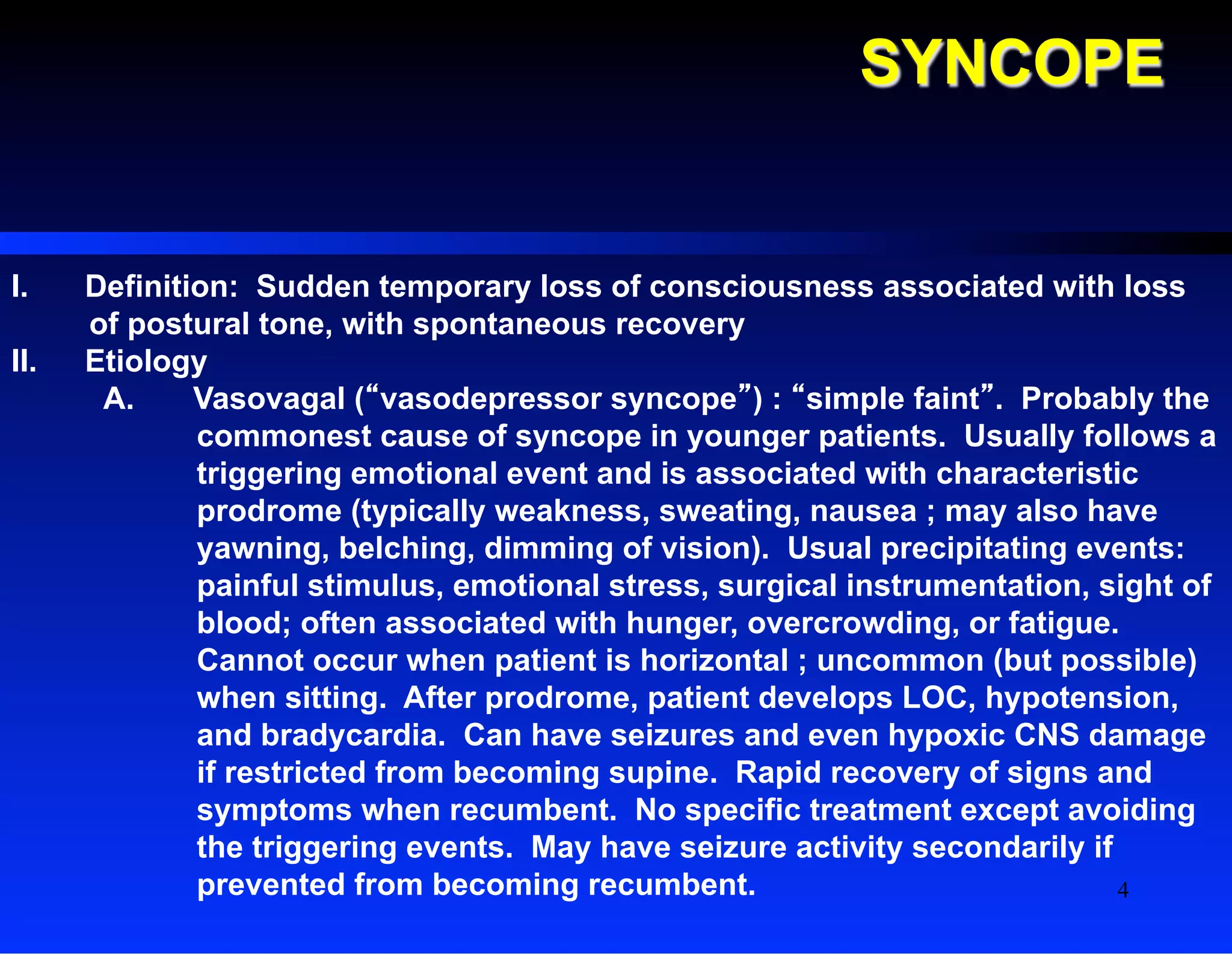
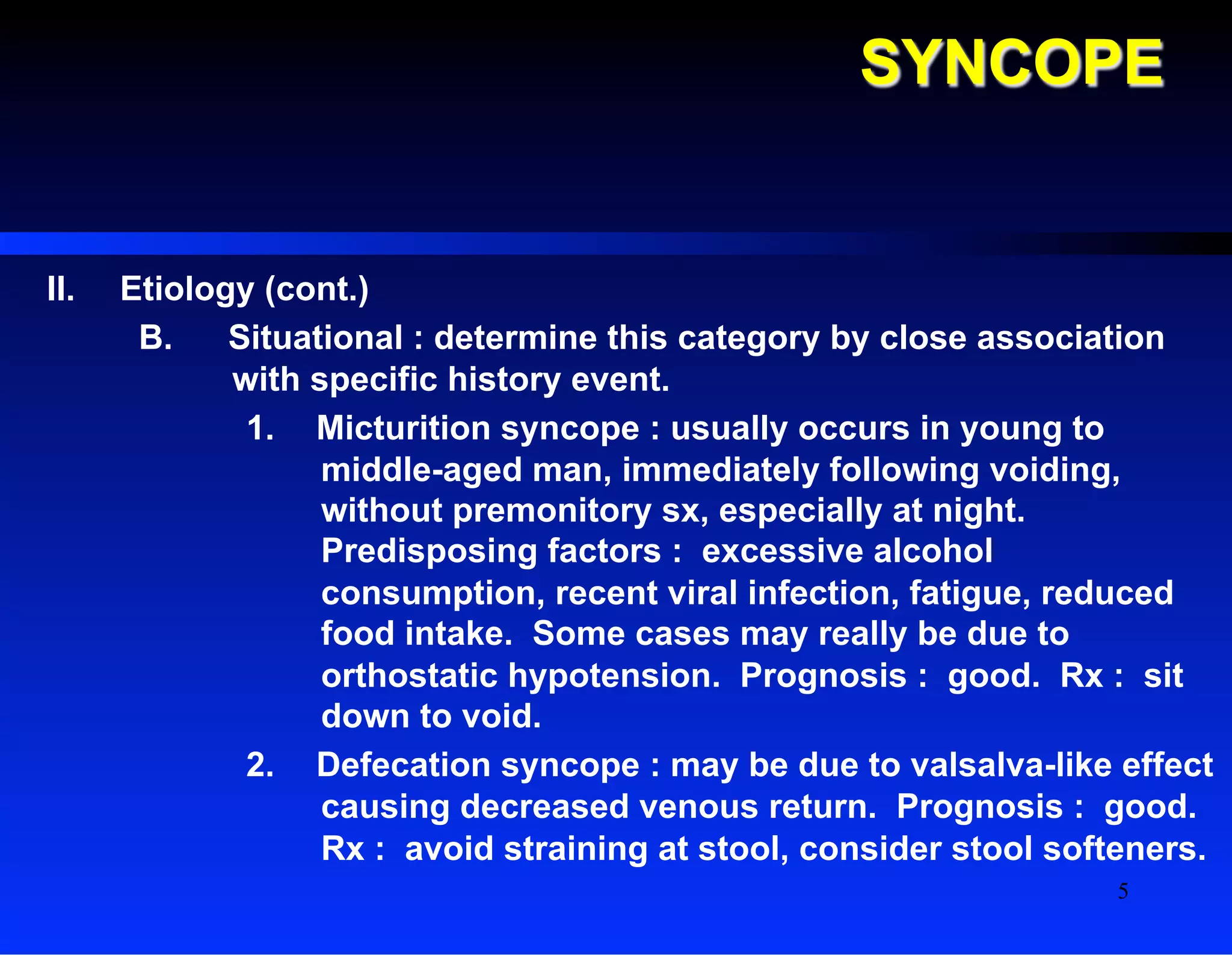
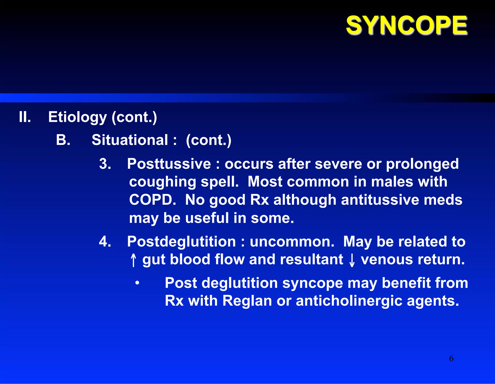
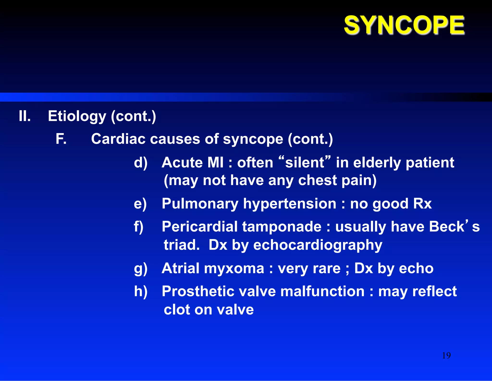
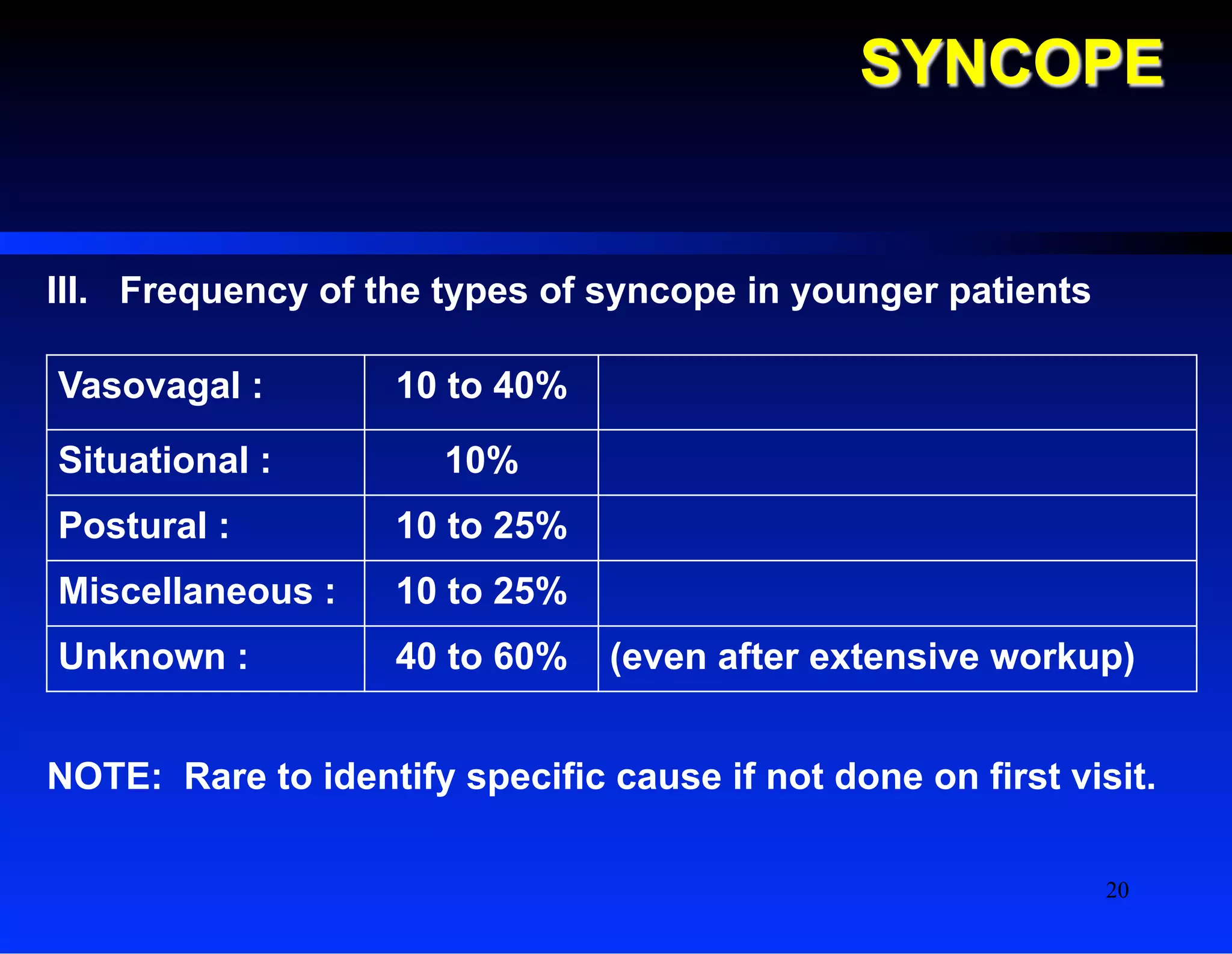
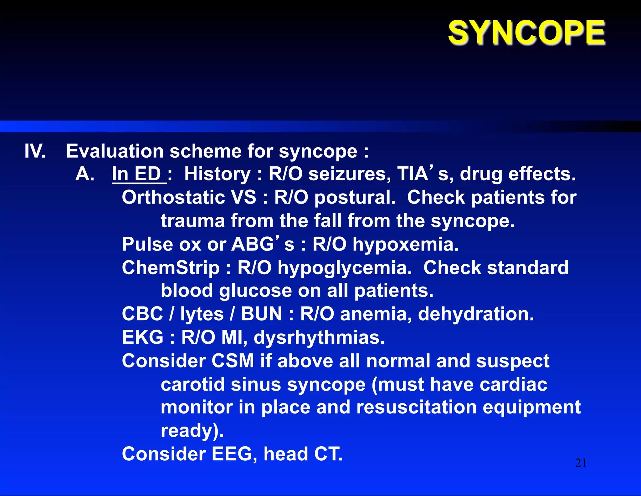
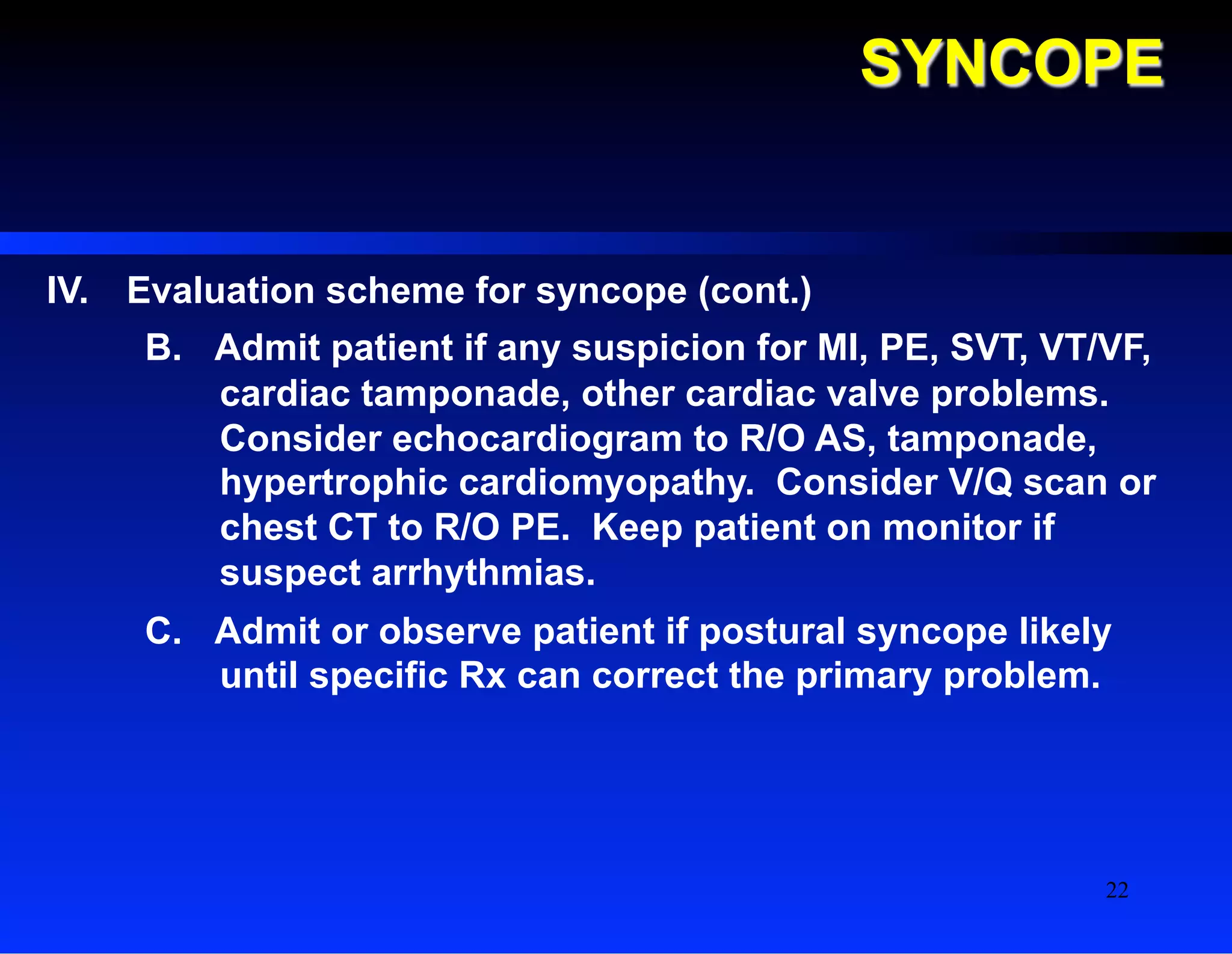
Project: Ghana Emergency Medicine Collaborative
Document Title: Syncope
Author(s): C. James Holliman, M.D. (Penn State University), 2008
License: Unless otherwise noted, this material is made available under
the terms of the Creative Commons Attribution Share Alike-3.0
License:
http://creativecommons.org/licenses/by-sa/3.0/
We have reviewed this material in accordance with U.S. Copyright Law and have tried to maximize your
ability to use, share, and adapt it. These lectures have been modified in the process of making a publicly
shareable version. The citation key on the following slide provides information about how you may share and
adapt this material.
Copyright holders of content included in this material should contact open.michigan@umich.edu with any
questions, corrections, or clarification regarding the use of content.
For more information about how to cite these materials visit http://open.umich.edu/privacy-and-terms-use.
Any medical information in this material is intended to inform and educate and is not a tool for self-diagnosis
or a replacement for medical evaluation, advice, diagnosis or treatment by a healthcare professional. Please
speak to your physician if you have questions about your medical condition.
Viewer discretion is advised: Some medical content is graphic and may not be suitable for all viewers.
1
Attribution Key
for more information see: http://open.umich.edu/wiki/AttributionPolicy
Use + Share + Adapt
Make Your Own Assessment
Creative Commons – Attribution License
Creative Commons – Attribution Share Alike License
Creative Commons – Attribution Noncommercial License
Creative Commons – Attribution Noncommercial Share Alike License
GNU – Free Documentation License
Creative Commons – Zero Waiver
Public Domain – Ineligible: Works that are ineligible for copyright protection in the U.S. (17 USC § 102(b)) *laws in
your jurisdiction may differ
Public Domain – Expired: Works that are no longer protected due to an expired copyright term.
Public Domain – Government: Works that are produced by the U.S. Government. (17 USC § 105)
Public Domain – Self Dedicated: Works that a copyright holder has dedicated to the public domain.
Fair Use: Use of works that is determined to be Fair consistent with the U.S. Copyright Act. (17 USC § 107) *laws in your
jurisdiction may differ
Our determination DOES NOT mean that all uses of this 3rd-party content are Fair Uses and we DO NOT guarantee that
your use of the content is Fair.
To use this content you should do your own independent analysis to determine whether or not your use will be Fair.
{ Content the copyright holder, author, or law permits you to use, share and adapt. }
{ Content Open.Michigan believes can be used, shared, and adapted because it is ineligible for copyright. }
{ Content Open.Michigan has used under a Fair Use determination. }
2
C. James Holliman, M.D., F.A.C.E.P.
Professor of Emergency Medicine
Director, Center for International Emergency Medicine
M. S. Hershey Medical Center
Penn State University
Hershey, PA, U.S.A.
SYNCOPE
3
SYNCOPE
I.  Definition: Sudden temporary loss of consciousness associated with loss
of postural tone, with spontaneous recovery
II.  Etiology
A.  Vasovagal (“vasodepressor syncope”) : “simple faint”. Probably the
commonest cause of syncope in younger patients. Usually follows a
triggering emotional event and is associated with characteristic
prodrome (typically weakness, sweating, nausea ; may also have
yawning, belching, dimming of vision). Usual precipitating events:
painful stimulus, emotional stress, surgical instrumentation, sight of
blood; often associated with hunger, overcrowding, or fatigue.
Cannot occur when patient is horizontal ; uncommon (but possible)
when sitting. After prodrome, patient develops LOC, hypotension,
and bradycardia. Can have seizures and even hypoxic CNS damage
if restricted from becoming supine. Rapid recovery of signs and
symptoms when recumbent. No specific treatment except avoiding
the triggering events. May have seizure activity secondarily if
prevented from becoming recumbent. 4
SYNCOPE
II.  Etiology (cont.)
B.  Situational : determine this category by close association
with specific history event.
1.  Micturition syncope : usually occurs in young to
middle-aged man, immediately following voiding,
without premonitory sx, especially at night.
Predisposing factors : excessive alcohol
consumption, recent viral infection, fatigue, reduced
food intake. Some cases may really be due to
orthostatic hypotension. Prognosis : good. Rx : sit
down to void.
2.  Defecation syncope : may be due to valsalva-like effect
causing decreased venous return. Prognosis : good.
Rx : avoid straining at stool, consider stool softeners.
5
SYNCOPE
II.  Etiology (cont.)
B.  Situational : (cont.)
3.  Posttussive : occurs after severe or prolonged
coughing spell. Most common in males with
COPD. No good Rx although antitussive meds
may be useful in some.
4.  Postdeglutition : uncommon. May be related to
↑ gut blood flow and resultant ↓ venous return.
•  Post deglutition syncope may benefit from
Rx with Reglan or anticholinergic agents.
6
SYNCOPE
II.  Etiology (cont.)
B.  Situational : (cont.)
5.  Postprandial : more common in elderly patients
with cardiac disease. Probably related to
increased gut blood flow postprandially and
resultant decreased cerebral blood flow due to
limited cardiac reserve.
•  Rx : smaller meals, adequate fluid with
meals, avoid standing soon after meals.
7
SYNCOPE
II.  Etiology (cont.)
C.  Carotid sinus syncope : caused by “hyper-sensitivity”
of carotid sinus (or carotid body) to external pressure
resulting in ↑ vagal tone with bradycardia and / or
hypotension. Commoner in elderly men. May be
caused by sudden head turning, shaving, or firm shirt
collar pressing on neck when bending over. Dx : by
CSM reproducing sx or bradycardia or hypotension.
•  Rx: avoid sudden head turning or pressure on
neck. Frequent severe recurrent sx might need
carotid sinus denervation on one side.
8
SYNCOPE
II.  Etiology (cont.)
D.  Postural syncope (basically due to decreased venous
return)
1.  Volume depletion : Dx : by hx of intravascular
volume loss (internal or external bleeding, profuse
or extended vomiting or diarrhea, or just poor PO
intake in elderly patients). May occur in elderly
patients on diuretics without obvious sx. Signs :
poor skin turgor, pallor, “sunken” eyes or
fontanelle, ± hypotension ; usually show
orthostatic (lying >sitting or standing) BP ↓ more
than 20 mmHg and pulse ↑ more than 20 BPM.
•  Rx : IV fluids or blood transfusion, correct
the underlying problem.
9
SYNCOPE
II.  Etiology (cont.)
D.  Postural syncope (cont.)
2.  Autonomic insufficiency : can occur in diabetics
or with other peripheral neuropathies, or as Shy-
Drager syndrome (primary autonomic
insufficiency, due to degeneration of the lateral
horn cells and basal ganglions).
•  Rx : support stockings, maintain fluid status,
avoid sudden postural shifts, flourinated
steroids PO, ephedrine PO.
10
SYNCOPE
II.  Etiology (cont.)
D.  Postural syncope (cont.)
3.  Drug-induced : most commonly from prazosin
(Minipres) and nitrates. Can also occur from
hydralazine, alphamethyldopa, phenothiazines,
tricyclics, ganglionic blocking agents ; possibly
also from calcium channel blockers.
11
SYNCOPE
II.  Etiology (cont.)
E.  Uncommon Miscellaneous
1.  Takayasu’s aortitis : due to ↓ flow in carotid &
vertebral arteries
2.  Subclavian steal syndrome : high grade stenosis
proximal to origin of vertebral artery ; symptoms
occur with arm exercise
3.  Glossopharyngeal neuralgia : pain with
swallowing may induce syncope
4.  Systemic mastocytosis : usually have hives and
other “allergic” manifestations ; Rx by H1 and H2
blockers and salicylates
12
SYNCOPE
II.  Etiology (cont.)
E.  Uncommon Miscellaneous (cont.)
5.  Hysteria
6.  Hyperventilation : most cases may really be
vasovagal. May be due to ↓ cerebral blood flow
from ↓ arterial PCO2. Rx by rebreathing expired
air, and / or benzodiazepines or hydroxyzine
7.  Basilar artery transient ischemic attacks ; very
rare to have syncope alone with this (nearly
always have associated diplopia, vertigo,
dysarthria, hemiparesis, or other sx of brainstem
ischemia).
13
SYNCOPE
II.  Etiology (cont.)
E.  Uncommon Miscellaneous (cont.)
8.  Hypoxemia : usually not sudden enough to cause
true syncope. May however be a more common
cause in the elderly than currently thought due to
their small “reserve” level of cerebral blood flow
over what is needed to supply the brain (3.5 ml
O2 /100 g brain tissue / min). Superimposed
pneumonia, anemia, CHF, etc., may drop the
cerebral O2 supply < 3.5 ml O2 / 100 g/min so that
syncope results.
14
SYNCOPE
II.  Etiology (cont.)
E.  Uncommon Miscellaneous (cont.)
9.  Hypoglycemia : doesn’t usually cause true
syncope because resolution is either very slow or
not spontaneous without Rx, but certainly can
cause sudden LOC.
10.  Seizure disorder : again does not cause true
syncope (because resolution is slow), but if
unwitnessed, can present in similar fashion to
true syncope. Requires EEG for Dx confirmation.
15
SYNCOPE
II.  Etiology (cont.)
F.  Cardiac causes of syncope : can cause syncope when
patient lying down. Account for 20 to 30 % of cases in
the elderly.
1.  Stokes-Adams attacks : asystolic episodes > 3
second duration
2.  Other bradyarrhythmias : sick sinus syndrome,
heart blocks, (Mobitz II, CHB) effects of drugs
such as beta-blockers or Class Ia antiarrhythmics
(quinidine, procainamide, disopyramide). Rx with
permanent pacemaker.
16
SYNCOPE
II.  Etiology (cont.)
F.  Cardiac causes of syncope (cont.) :
3.  Supraventricular tachycardias : PAT, WPW. Rx
with drugs, overdrive pacing, or surgical ablation
of abnormal focus or bypass tract.
4.  Ventricular arrhythmias : most common cardiac
cause but can be hard to prove. Rx by drugs,
surgical ablation of foci, ± CABG if ventricular
ischemia causative ; AID (automatic implantable
defibrillator) if refractory to drug regimens. May
require special electrophysiologic studies for Dx.
17
SYNCOPE
II.  Etiology (cont.)
F.  Cardiac causes of syncope (cont.)
5.  “Obstructive” problems :
a)  Aortic stenosis : perhaps most common of
these. Survival average only 1 to 3 years
after development of syncope. Rx by valve
replacement
b)  Hypertrophic cardiomyopathy : Rx : beta
blockers
c)  Mitral valve prolapse : associated
dysrhythmias may be the real cause of
associated syncope
18
SYNCOPE
II.  Etiology (cont.)
F.  Cardiac causes of syncope (cont.)
d)  Acute MI : often “silent” in elderly patient
(may not have any chest pain)
e)  Pulmonary hypertension : no good Rx
f)  Pericardial tamponade : usually have Beck’s
triad. Dx by echocardiography
g)  Atrial myxoma : very rare ; Dx by echo
h)  Prosthetic valve malfunction : may reflect
clot on valve
19
SYNCOPE
III.  Frequency of the types of syncope in younger patients
Vasovagal : 10 to 40%
Situational : 10%
Postural : 10 to 25%
Miscellaneous : 10 to 25%
Unknown : 40 to 60% (even after extensive workup)
NOTE: Rare to identify specific cause if not done on first visit.
20
SYNCOPE
IV.  Evaluation scheme for syncope :
A.  In ED : History : R/O seizures, TIA’s, drug effects.
Orthostatic VS : R/O postural. Check patients for
trauma from the fall from the syncope.
Pulse ox or ABG’s : R/O hypoxemia.
ChemStrip : R/O hypoglycemia. Check standard
blood glucose on all patients.
CBC / lytes / BUN : R/O anemia, dehydration.
EKG : R/O MI, dysrhythmias.
Consider CSM if above all normal and suspect
carotid sinus syncope (must have cardiac
monitor in place and resuscitation equipment
ready).
Consider EEG, head CT. 21
SYNCOPE
IV.  Evaluation scheme for syncope (cont.)
B.  Admit patient if any suspicion for MI, PE, SVT, VT/VF,
cardiac tamponade, other cardiac valve problems.
Consider echocardiogram to R/O AS, tamponade,
hypertrophic cardiomyopathy. Consider V/Q scan or
chest CT to R/O PE. Keep patient on monitor if
suspect arrhythmias.
C.  Admit or observe patient if postural syncope likely
until specific Rx can correct the primary problem.
22
SYNCOPE
IV.  Evaluation scheme for syncope (cont.)
D.  May usually discharge patient from ED if etiology
clearly vasovagal or situational. May still need
outpatient Holter monitor or EEG for further
confirmation.
E.  Secondary workup tests to consider :
CT scan, aortic / cerebral angiography,
electrophysiologic testing (PES, or programmed
electrical stimulation, in cath lab)
23

GEMC: Syncope: Resident Training

  • 1.
    Project: Ghana EmergencyMedicine Collaborative Document Title: Syncope Author(s): C. James Holliman, M.D. (Penn State University), 2008 License: Unless otherwise noted, this material is made available under the terms of the Creative Commons Attribution Share Alike-3.0 License: http://creativecommons.org/licenses/by-sa/3.0/ We have reviewed this material in accordance with U.S. Copyright Law and have tried to maximize your ability to use, share, and adapt it. These lectures have been modified in the process of making a publicly shareable version. The citation key on the following slide provides information about how you may share and adapt this material. Copyright holders of content included in this material should contact open.michigan@umich.edu with any questions, corrections, or clarification regarding the use of content. For more information about how to cite these materials visit http://open.umich.edu/privacy-and-terms-use. Any medical information in this material is intended to inform and educate and is not a tool for self-diagnosis or a replacement for medical evaluation, advice, diagnosis or treatment by a healthcare professional. Please speak to your physician if you have questions about your medical condition. Viewer discretion is advised: Some medical content is graphic and may not be suitable for all viewers. 1
  • 2.
    Attribution Key for moreinformation see: http://open.umich.edu/wiki/AttributionPolicy Use + Share + Adapt Make Your Own Assessment Creative Commons – Attribution License Creative Commons – Attribution Share Alike License Creative Commons – Attribution Noncommercial License Creative Commons – Attribution Noncommercial Share Alike License GNU – Free Documentation License Creative Commons – Zero Waiver Public Domain – Ineligible: Works that are ineligible for copyright protection in the U.S. (17 USC § 102(b)) *laws in your jurisdiction may differ Public Domain – Expired: Works that are no longer protected due to an expired copyright term. Public Domain – Government: Works that are produced by the U.S. Government. (17 USC § 105) Public Domain – Self Dedicated: Works that a copyright holder has dedicated to the public domain. Fair Use: Use of works that is determined to be Fair consistent with the U.S. Copyright Act. (17 USC § 107) *laws in your jurisdiction may differ Our determination DOES NOT mean that all uses of this 3rd-party content are Fair Uses and we DO NOT guarantee that your use of the content is Fair. To use this content you should do your own independent analysis to determine whether or not your use will be Fair. { Content the copyright holder, author, or law permits you to use, share and adapt. } { Content Open.Michigan believes can be used, shared, and adapted because it is ineligible for copyright. } { Content Open.Michigan has used under a Fair Use determination. } 2
  • 3.
    C. James Holliman,M.D., F.A.C.E.P. Professor of Emergency Medicine Director, Center for International Emergency Medicine M. S. Hershey Medical Center Penn State University Hershey, PA, U.S.A. SYNCOPE 3
  • 4.
    SYNCOPE I.  Definition: Suddentemporary loss of consciousness associated with loss of postural tone, with spontaneous recovery II.  Etiology A.  Vasovagal (“vasodepressor syncope”) : “simple faint”. Probably the commonest cause of syncope in younger patients. Usually follows a triggering emotional event and is associated with characteristic prodrome (typically weakness, sweating, nausea ; may also have yawning, belching, dimming of vision). Usual precipitating events: painful stimulus, emotional stress, surgical instrumentation, sight of blood; often associated with hunger, overcrowding, or fatigue. Cannot occur when patient is horizontal ; uncommon (but possible) when sitting. After prodrome, patient develops LOC, hypotension, and bradycardia. Can have seizures and even hypoxic CNS damage if restricted from becoming supine. Rapid recovery of signs and symptoms when recumbent. No specific treatment except avoiding the triggering events. May have seizure activity secondarily if prevented from becoming recumbent. 4
  • 5.
    SYNCOPE II.  Etiology (cont.) B. Situational : determine this category by close association with specific history event. 1.  Micturition syncope : usually occurs in young to middle-aged man, immediately following voiding, without premonitory sx, especially at night. Predisposing factors : excessive alcohol consumption, recent viral infection, fatigue, reduced food intake. Some cases may really be due to orthostatic hypotension. Prognosis : good. Rx : sit down to void. 2.  Defecation syncope : may be due to valsalva-like effect causing decreased venous return. Prognosis : good. Rx : avoid straining at stool, consider stool softeners. 5
  • 6.
    SYNCOPE II.  Etiology (cont.) B. Situational : (cont.) 3.  Posttussive : occurs after severe or prolonged coughing spell. Most common in males with COPD. No good Rx although antitussive meds may be useful in some. 4.  Postdeglutition : uncommon. May be related to ↑ gut blood flow and resultant ↓ venous return. •  Post deglutition syncope may benefit from Rx with Reglan or anticholinergic agents. 6
  • 7.
    SYNCOPE II.  Etiology (cont.) B. Situational : (cont.) 5.  Postprandial : more common in elderly patients with cardiac disease. Probably related to increased gut blood flow postprandially and resultant decreased cerebral blood flow due to limited cardiac reserve. •  Rx : smaller meals, adequate fluid with meals, avoid standing soon after meals. 7
  • 8.
    SYNCOPE II.  Etiology (cont.) C. Carotid sinus syncope : caused by “hyper-sensitivity” of carotid sinus (or carotid body) to external pressure resulting in ↑ vagal tone with bradycardia and / or hypotension. Commoner in elderly men. May be caused by sudden head turning, shaving, or firm shirt collar pressing on neck when bending over. Dx : by CSM reproducing sx or bradycardia or hypotension. •  Rx: avoid sudden head turning or pressure on neck. Frequent severe recurrent sx might need carotid sinus denervation on one side. 8
  • 9.
    SYNCOPE II.  Etiology (cont.) D. Postural syncope (basically due to decreased venous return) 1.  Volume depletion : Dx : by hx of intravascular volume loss (internal or external bleeding, profuse or extended vomiting or diarrhea, or just poor PO intake in elderly patients). May occur in elderly patients on diuretics without obvious sx. Signs : poor skin turgor, pallor, “sunken” eyes or fontanelle, ± hypotension ; usually show orthostatic (lying >sitting or standing) BP ↓ more than 20 mmHg and pulse ↑ more than 20 BPM. •  Rx : IV fluids or blood transfusion, correct the underlying problem. 9
  • 10.
    SYNCOPE II.  Etiology (cont.) D. Postural syncope (cont.) 2.  Autonomic insufficiency : can occur in diabetics or with other peripheral neuropathies, or as Shy- Drager syndrome (primary autonomic insufficiency, due to degeneration of the lateral horn cells and basal ganglions). •  Rx : support stockings, maintain fluid status, avoid sudden postural shifts, flourinated steroids PO, ephedrine PO. 10
  • 11.
    SYNCOPE II.  Etiology (cont.) D. Postural syncope (cont.) 3.  Drug-induced : most commonly from prazosin (Minipres) and nitrates. Can also occur from hydralazine, alphamethyldopa, phenothiazines, tricyclics, ganglionic blocking agents ; possibly also from calcium channel blockers. 11
  • 12.
    SYNCOPE II.  Etiology (cont.) E. Uncommon Miscellaneous 1.  Takayasu’s aortitis : due to ↓ flow in carotid & vertebral arteries 2.  Subclavian steal syndrome : high grade stenosis proximal to origin of vertebral artery ; symptoms occur with arm exercise 3.  Glossopharyngeal neuralgia : pain with swallowing may induce syncope 4.  Systemic mastocytosis : usually have hives and other “allergic” manifestations ; Rx by H1 and H2 blockers and salicylates 12
  • 13.
    SYNCOPE II.  Etiology (cont.) E. Uncommon Miscellaneous (cont.) 5.  Hysteria 6.  Hyperventilation : most cases may really be vasovagal. May be due to ↓ cerebral blood flow from ↓ arterial PCO2. Rx by rebreathing expired air, and / or benzodiazepines or hydroxyzine 7.  Basilar artery transient ischemic attacks ; very rare to have syncope alone with this (nearly always have associated diplopia, vertigo, dysarthria, hemiparesis, or other sx of brainstem ischemia). 13
  • 14.
    SYNCOPE II.  Etiology (cont.) E. Uncommon Miscellaneous (cont.) 8.  Hypoxemia : usually not sudden enough to cause true syncope. May however be a more common cause in the elderly than currently thought due to their small “reserve” level of cerebral blood flow over what is needed to supply the brain (3.5 ml O2 /100 g brain tissue / min). Superimposed pneumonia, anemia, CHF, etc., may drop the cerebral O2 supply < 3.5 ml O2 / 100 g/min so that syncope results. 14
  • 15.
    SYNCOPE II.  Etiology (cont.) E. Uncommon Miscellaneous (cont.) 9.  Hypoglycemia : doesn’t usually cause true syncope because resolution is either very slow or not spontaneous without Rx, but certainly can cause sudden LOC. 10.  Seizure disorder : again does not cause true syncope (because resolution is slow), but if unwitnessed, can present in similar fashion to true syncope. Requires EEG for Dx confirmation. 15
  • 16.
    SYNCOPE II.  Etiology (cont.) F. Cardiac causes of syncope : can cause syncope when patient lying down. Account for 20 to 30 % of cases in the elderly. 1.  Stokes-Adams attacks : asystolic episodes > 3 second duration 2.  Other bradyarrhythmias : sick sinus syndrome, heart blocks, (Mobitz II, CHB) effects of drugs such as beta-blockers or Class Ia antiarrhythmics (quinidine, procainamide, disopyramide). Rx with permanent pacemaker. 16
  • 17.
    SYNCOPE II.  Etiology (cont.) F. Cardiac causes of syncope (cont.) : 3.  Supraventricular tachycardias : PAT, WPW. Rx with drugs, overdrive pacing, or surgical ablation of abnormal focus or bypass tract. 4.  Ventricular arrhythmias : most common cardiac cause but can be hard to prove. Rx by drugs, surgical ablation of foci, ± CABG if ventricular ischemia causative ; AID (automatic implantable defibrillator) if refractory to drug regimens. May require special electrophysiologic studies for Dx. 17
  • 18.
    SYNCOPE II.  Etiology (cont.) F. Cardiac causes of syncope (cont.) 5.  “Obstructive” problems : a)  Aortic stenosis : perhaps most common of these. Survival average only 1 to 3 years after development of syncope. Rx by valve replacement b)  Hypertrophic cardiomyopathy : Rx : beta blockers c)  Mitral valve prolapse : associated dysrhythmias may be the real cause of associated syncope 18
  • 19.
    SYNCOPE II.  Etiology (cont.) F. Cardiac causes of syncope (cont.) d)  Acute MI : often “silent” in elderly patient (may not have any chest pain) e)  Pulmonary hypertension : no good Rx f)  Pericardial tamponade : usually have Beck’s triad. Dx by echocardiography g)  Atrial myxoma : very rare ; Dx by echo h)  Prosthetic valve malfunction : may reflect clot on valve 19
  • 20.
    SYNCOPE III.  Frequency ofthe types of syncope in younger patients Vasovagal : 10 to 40% Situational : 10% Postural : 10 to 25% Miscellaneous : 10 to 25% Unknown : 40 to 60% (even after extensive workup) NOTE: Rare to identify specific cause if not done on first visit. 20
  • 21.
    SYNCOPE IV.  Evaluation schemefor syncope : A.  In ED : History : R/O seizures, TIA’s, drug effects. Orthostatic VS : R/O postural. Check patients for trauma from the fall from the syncope. Pulse ox or ABG’s : R/O hypoxemia. ChemStrip : R/O hypoglycemia. Check standard blood glucose on all patients. CBC / lytes / BUN : R/O anemia, dehydration. EKG : R/O MI, dysrhythmias. Consider CSM if above all normal and suspect carotid sinus syncope (must have cardiac monitor in place and resuscitation equipment ready). Consider EEG, head CT. 21
  • 22.
    SYNCOPE IV.  Evaluation schemefor syncope (cont.) B.  Admit patient if any suspicion for MI, PE, SVT, VT/VF, cardiac tamponade, other cardiac valve problems. Consider echocardiogram to R/O AS, tamponade, hypertrophic cardiomyopathy. Consider V/Q scan or chest CT to R/O PE. Keep patient on monitor if suspect arrhythmias. C.  Admit or observe patient if postural syncope likely until specific Rx can correct the primary problem. 22
  • 23.
    SYNCOPE IV.  Evaluation schemefor syncope (cont.) D.  May usually discharge patient from ED if etiology clearly vasovagal or situational. May still need outpatient Holter monitor or EEG for further confirmation. E.  Secondary workup tests to consider : CT scan, aortic / cerebral angiography, electrophysiologic testing (PES, or programmed electrical stimulation, in cath lab) 23