Download to read offline
![Inflammatory Conditions Mimicking Tumours In Calabar: A 30 Year Study (1978-2007).
www.iosrjournals.org 43 | Page
The study shows that onchocerciasis is still endemic in the study area with continued transmission of
infection from long term carriers. Other lesions seen are histoplamosis, mycetoma, molloscum contagiosum etc.
More study is required to ascertain the pathogenesis of the two infectious agents, histoplasma and
onchocerca. Importantly, the finding of high number of onchocerciasis is an indication that community infection
is still rife despite efforts of eradication with ivermectin. Also, control efforts may be waning for this parasitic
infection as more efforts are concentrated in the emerging public health infections such as HIV.
Reference
[1] Woodward B H, Rosenberg S I, Farnham R and Adams D O. Incidence and nature of primary granulomatous inflammation in
surgically removed material. Am Journal of Surgical Pathology. 1982; 6 (2): 119-129.
[2] Adams, D. O. ―The granulomatous inflammatory response. A review‖. Am J of Pathology. 1976; 84(1): 164-191.
[3] Granuloma. From Wikipedia, the free encyclopedia. Last modified 6 August, 2011 at 21.52.
[4] Andraea R., Edson RS, Kern EB. Rhinoscleroma: A growing concern in the United States? Mayo Clinic experience. Mayo
Clinic Proc. 1993; 68: 1151 – 1157.
[5] DiBartolomeo, Joseph R. Scleroma of the nose and pharynx. Western Journal of Medicine. 1976; vol. 124., pp 13-17.
[6] Palmer, PES and Reader MM. The imaging of tropical diseases. 2000. Heidelberg: Springer Verlachi; vols. 1 and 2 (ISBN 3 –
540 – 66219 – 7).
[7] Saint Andre A., Blackwell NM, Hall LR, et al. The role of endosymbiotic Wolbachia bacteria in the pathogenesis of river
blindness. Science. 2002; 295:1892 – 1895. In Mandell, Douglas and Bennett’s Principles and Practice of infectious Diseases:
6th
Ed, ELSEVIER Churchill Livingstone. Pp. 3273.
[8] American Academy of Paediatrics eBooks; accessed 22 July, 2011 on http: //aaparebook.apphblications.org.
[9] Taylor H R, Duke B O L, Munoz B. The selection of communities for treatment of onchocerciasis with ivermectin. Trop Med
Parasitol. 1992; 43:267.
[10] Elder D., Rosalie E., Christine J., Bernett J, Lever’s Histopathology of the skin. 1990; 60-61.
[11] John H, Arnold O, Neil P. Textbook of Paediatric dermatology. 2002; vol.1: 516-517.
[12] Rideley D.S. The pathogenesis of cutaneous leishmaniasis. Trans. R Soc Trop. Med Hyg. 1979; 73:150-160
[13] Mandell, Douglas and Bennett’s Principles and Practice of Infectious Diseases: 6th
Ed, ELSEVIER Churchill Livingstone. Pg
[14] Keath, E.J., Kobayashi G S, Medoff G. Classification of Histoplasma capsulatum by restriction fragrant length polymorphism in a
nuclear gene. J Clin Microbiol. 1992; 30:2104-2107.
[15] Manfredi R, Mazzoni A, Nanetti A, et al. Histoplasma capsulatum and duboisii in Europe: The impact of the HIV pandemic ,
travel, and immigration. Eur J Epidemiol. 1994; 10: 675-681.
[16] Mandell, Douglas and Bennett’s Principles and Practice of Infectious Diseases: 6th
Ed, ELSEVIER Churchill Livingstone. Pg
[17] Mercurio MG, Eleroski B E. Cutaneous blastomycosis. Cutic 1992; 50:422-424.](https://image.slidesharecdn.com/g0153743-140516042730-phpapp01/85/G0153743-7-320.jpg)

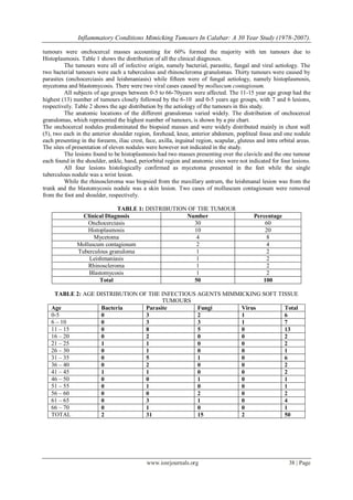
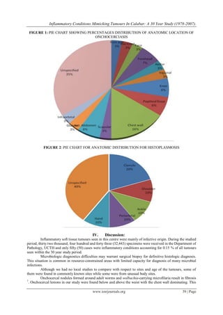
This study reviewed 50 inflammatory soft tissue lesions surgically removed and diagnosed histologically over a 30-year period from 1978-2007 at the University of Calabar Teaching Hospital in Nigeria. Onchocerciasis accounted for 60% of the lesions, followed by histoplasmosis at 20%. Other identified conditions included mycetoma, molluscum contagiosum, tuberculosis, leishmaniasis, rhinoscleroma, and blastomycosis. The lesions presented most commonly in children and young adults aged 5-15 years. Onchocercal nodules were widely distributed but predominantly in the chest wall. Histoplasmosis lesions were found in the clavicle, shoulder, ankle,