
Recommended
Recommended
More Related Content
What's hot
What's hot (20)
Similar to Comparison of tzanck smear with viral serology in varicella
Similar to Comparison of tzanck smear with viral serology in varicella (20)
Recently uploaded
Recently uploaded (20)
Comparison of tzanck smear with viral serology in varicella
- 1. Journal of Pakistan Association of Dermatologists. 2016;26 (4):306-309. 306 Address for correspondence Dr. Muhammad Irfan Anwar Assistant Professor Department of Dermatology, Bahria University Medical & Dental College, Karachi Email: doctorirfananwar@gmail.com Original Article Comparison of Tzanck smear with viral serology in varicella Introduction Varicella infection (chickenpox) is caused by varicella-zoster virus (VZV), which is distributed worldwide with overall seroprevalence of 83.6%.1 In Pakistan, its prevalence in adult population is 1.70%.2 Clinical diagnosis of varicella can be made by its characteristic appearance. Laboratory tests may be used to confirm the clinical diagnosis. Tzanck smear is an easily performed, sensitive, specific, cost-effective and rapid test that is useful in clinical settings in dermatologist’s daily practice.3,4 Arnault Tzanck first used this method for the diagnosis of cutaneous disorders in 1947.5 It can also be performed at the bedside with minimal patient discomfort.6,7 In this method, scrapings from the floor of the vesicles are transferred to a glass slide, stained with various dyes, and then examined under the light microscope, that reveals multinucleated giant cells in herpetic infection, acantholytic cells in pemphigus, dyskeratotic acantholytic cells and Musarat Shahid*, Dilawar Abbas Rizvi*, M Shahid Aslam**, Asher Ahmed Mashood¶, Agha Babar¶¶, Muhammad Irfan Anwar† * Department of Dermatology, Military Hospital, Rawalpindi ** Department of Dermatology, Combined Military Hospital, Gujranwala ¶ Department of Dermatology, Combined Military Hospital, Peshawar ¶¶ Armed Forces Institute of Pathology, Rawalpindi † Department of Dermatology, Bahria University Medical & Dental College, Karachi Abstract Objective To compare Tzanck smear with viral serology in terms of concurrence of results in patients with varicella. Methods It was a descriptive case-series done at Dermatology Department, Military Hospital, Rawalpindi. We studied 50 patients of the varicella infection in 6 months. Tzanck smear was taken from each patient at presentation and stained by Giemsa stain for giant cells and evaluated by histopathologist. Viral serology was done by complement fixation method at Virology Department. The results were statistically analyzed with SPSS 16. Results Out of 50 patients of varicella 39 (78%) were males while 11 (22%) were females. Mean age was 30.52 ± 9.763 years. Tzanck smear was positive in 33 (66%) and viral serology in 45 (90%) patients. The concurrence of results in both Tzanck smear and viral serology was seen in 31 patients (62%). Conclusion Tzanck smear is a quick and valuable tool in the diagnosis of patients suffering from varicella. Key words Varicella, Tzanck smear, viral serology.
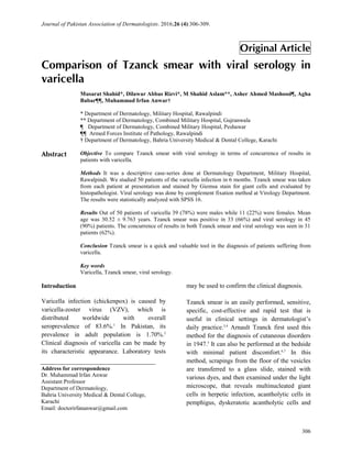
- 2. Journal of Pakistan Association of Dermatologists. 2016;26 (4):306-309. 307 cocci in bullous impetigo, pseudohyphae in candidiasis and necrotic basal cells in toxic epidermal necrolysis.7 Although it was suggested as a simple, rapid, and reliable technique to be used in the diagnosis of many diseases, but during the following six decades, its practical use has been limited to a few diseases.8 To date, therefore, only a few studies have examined the dermatological use and diagnostic value of this method.8 Methodology It was a descriptive study, conducted at Dermatology Department of Military Hospital, Rawalpindi, in collaboration with Virology Department of Armed Forces Institute of Pathology (AFIP) from August 2010 to February 2011. We included 50 patients of any age and either sex who presented with the skin lesions clinically suggestive of varicella. Sample size was calculated by using WHO sample size calculator taking confidence level 95% and true positive proportion 84.7%.8 Absolute precision = 10%. Nonprobability consecutive sampling technique was used. After informed consent blood sample for antibodies against VZV was drawn and sent to laboratory. Tzanck smear was prepared under aseptic conditions. Lesions were first cleaned with 70% alcohol swab. When there was a vesicle or a pustule, the intact roof of the lesion was incised along one side and folded back, and the base of the lesion was gently scraped with a No. 15 scalpel. The cellular material obtained was then spread as a thin layer onto at least four microscopic slides and fixed with absolute alcohol. Samples of Tzanck smear were stained with Giemsa and the stained preparations were examined under low and then high magnification of light microscope for presence of giant cells by the same histopathologist at AFIP (Figure 1). Two weeks later a second blood sample was collected for determining the levels of VZV antibodies. Both the samples were labeled, mentioning the Figure 1 Tzanck smear showing multinucleated giant cell on Giemsa staining (magnification 100x). patient’s particulars, and sent to Armed Forces Institute of Pathology, Rawalpindi (AFIP). Evaluation of the samples for viral serology was done by using complement fixation method by the virologist at AFIP. Follow-up at 2 weeks after the initial visit was ensured by taking telephonic contact. Data were analyzed using SPSS version 16. Quantitative variables like age was presented as mean and standard deviation. The qualitative variable like the gender, results of Tzanck smear, results of viral serology and concurrence of results in both Tzanck smear and viral serology were presented in the form of percentages and frequencies. Results The study group comprised of 50 patients with female (n=11) to male (n=39) ratio of 1:3.5. Their ages were between 13-70 years (mean 30.52 years, SD 9.763). The maximum number of patients was in their third decade. Despite adequate follow-up measures, 5 patients (3 males and 2 females) were lost to follow-up at two weeks after their initial visit. The Tzanck smear was positive in 33 (67%) patients, and was negative in 17 (33%) patients, (Table 1). The viral serology was positive in all 45 patients
- 3. Journal of Pakistan Association of Dermatologists. 2016;26 (4):306-309. 308 (90%) as 5 patients were lost to follow-up despite adequate measures (Table 2). The concurrence of results of both Tzanck smear and Table 1 Results of Tzanck smear (n=100). Tzanck smear Frequency Cumulative percent Positive 33 (66.0%) 66.0 Negative 17 (34.0%) 100.0 Table 2 Results of viral serology Viral serology Frequency Cumulative Percent Positive 45 (90.0%) 90.0 Missing 5 (10.0%) 100.0 Table 3 Concurrence of results of Tzanck smear and viral serology Concurrence of results Frequency Cumulative Percent Yes 31 (62.0%) 62.0 No 14 (28.0%) 90.0 Missing 5 (10.0%) 100.0 viral serology was seen in 31 patients (62%) and in 14 patients (28%) viral serology was positive but Tzanck smear was not conclusive (Table 3). Discussion Most varicella-zoster virus (VZV) infections can be readily diagnosed by characteristic clinical appearance. However, because vesico-bullous eruptions of other viral and non-viral skin infections can resemble those of VZV, the infection can be misdiagnosed.9 Also the rapidity of obtaining a conclusive diagnosis is important for adequate antiviral therapy, because delay in treatment can increase the risk of complications.10 Laboratory tests are used to confirm the clinical diagnosis. Because the classical viral culture usually requires several days, cytologic methods such as the Tzanck test are used to support the clinical diagnosis in an early stage, although the Tzanck test cannot distinguish between VZV and HSV infections. Serologic tests can be used to detect rises in VZV antibody titers, but they can only confirm a diagnosis retrospectively. The Tzanck smear as a diagnostic tool can reliably support a clinical diagnosis of herpetic skin infections.5 In a study which was conducted in a pediatric clinic and focused on herpetic and non-herpetic vesicular and bullous skin disorders, indicated the high sensitivity (80%) and specificity (90%) of the Tzanck smear.11 Durdu et al.8 conducted a study on 400 patients in Turkey to prove the value of Tzanck smear test in diagnosis of erosive, vesicular, bullous, and pustular skin lesions, indicated the sensitivity of 84.7% and specificity of 100% in herpetic infections. The percentage of positivity of Tzanck test for HSV has been reported between 53.1%12 and 86%,13 and positivity of the Tzanck test for VZV infections has been reported between 64%14 and 79%15 in different studies. In our study, positivity of Tzanck test for varicella was 66%. We also found negative results in 34% patients, who had pustular and crusted skin lesions. The two most important factors in obtaining positive Tzanck smear results were the stage of the infection at the time of sampling and the type of the lesion. Viral shedding decreases with the duration of herpetic lesions. A decrease in viral shedding from herpetic lesions of longer duration may correlate with the lower sensitivity of the Tzanck smear. Solomon et al.11 found that the percentage of positivity in Tzanck smears prepared from herpetic infections at an earlier stage tended to be higher than the ones prepared at later stages. Namely, the percentage of positivity in vesicles was much higher than that of pustules.11 Clinicians can easily obtain experience in using the Tzanck smear effectively in office practice with the help of supplemental screening by a cytopathologist or cytotechnologist. We also compared the findings of Tzanck smear with viral serology, which was positive in all 90% of patients as 10% patients were lost to follow-up despite adequate
- 4. Journal of Pakistan Association of Dermatologists. 2016;26 (4):306-309. 309 measures. We found the concurrence of results of both Tzanck smear and viral serology in 62% patients and in 28% of patients viral serology was positive but Tzanck smear was not conclusive. The major disadvantage of viral serology is that it can only confirm a diagnosis retrospectively and also the time duration, it requires minimum of two weeks duration to be interpreted. No patient found to be true negative in our study. Conclusion Tzanck smear is a reliable diagnostic tool in confirming the clinical diagnosis of varicella. It is easy to perform, quick, simple and inexpensive, and cause minimum discomfort to the patient. Also a positive Tzanck smear result in patients with equivocal clinical features may even obviate the need for the other classical confirmatory laboratory tests. References 1. Shrerifi Z, Ghanjin SE. The Seroepidemiology of varicella-zoster virus. Allergy Asthma Immunol. 2005;4;95-8. 2. Raza N, Tariq WZ, Zaidi SK. Onset of adult varicella in relation to rural or urban origin and its complications. J Coll Physicians Surg Pak. 2008;18;95-7. 3. Nikkels AF, Pierard GE. The Tzanck smear: Heading the right way. J Am Acad Dermatol. 2009;61;152-3. 4. Senol M, Saglam H, Seyhan M, Durmaz R, Aktas E, Ozerol IH. Comparison of the Tzanck test and polymerase chain reaction in the diagnosis of cutaneous herpes simplex and varicella virus infections. Int J Dermatol. 2007:46:1177-9. 5. Gupta LK, Singhi MK. Tzanck smear: a useful diagnostic tool. Indian J Dermatol Venereol Leprol. 2005;71:295-9. 6. Prabhu S, Sripathi H, Gupta S, Prabhu M. Childhood herpes zoster: A clustering of ten cases. Indian J Dermatol. 2009:54:62-4. 7. Kelly B, Shimoni T. Reintroducing the Tzanck smear. Am J Clin Dermatol. 2009;10:141-5. 8. Durdu M, Baba M, Seckin D. The value of Tzanck smear test in diagnosis of erosive, vesicular, bullous, and pustular skin lesions. J Am Acad Dermatol. 2008;59:958-64. 9. Folkers E, Vreeswjk J, Oranje AP, Wagenaar F, Duivenvoorden JN. Improved detection of HSV by electron microscopy in clinical specimens using ultracentrifugation and colloidal gold immune electron microscopy: comparison with viral culture and cytodiagnosis. J Virol Methods. 1991;34:273-89. 10. Folkers E, Vreeswijk J, Wagenaar F, Kapsenberg JG, Hulsebosch HJ, Oranje AP. Immunoelectron microscopy for rapid diagnosis of varicella-zoster virus in a complicated case of human T-cell lymphotropic virus type 1 infection. J Clin Microbiol. 1992;30:2487-91. 11. Oranje AP, Folkers E, Choufoer-Habova J, Duivenvoorden JN. Diagnostic value of Tzanck smear in herpetic and non-herpetic vesicular and bullous skin disorders in pediatric practice. Acta Derm Venereol (Stockh). 1986;66:127-33. 12. Solomon AR, Rasmussen JE, Varani J, Pierson CL. The Tzanck smear in the diagnosis of cutaneous herpes simplex. JAMA. 1984;251:633-5. 13. Motyl MR, Bottone EJ, Janda JM. Diagnosis of herpes virus infections: correlation of Tzanck preparations with viral isolation. Diagn Microbiol Infect Dis. 1984;2:157-60. 14. Sadick NS, Swenson PD, Kaufman RL, Kaplan MH. Comparison of detection of varicella-zoster virus by the Tzanck smear, direct immunofluorescence with a monoclonal antibody, and virus isolation. J Am Acad Dermatol. 1987;17:64-9. 15. Schirm J, Meulenberg JJM, Pastoor GW, van Voorst Vader PC, Schroder FP. Rapid detection of varicella-zoster virus in clinical specimens using monoclonal antibodies on shell vials and smears. J Med Virol. 1989;28:1-6.