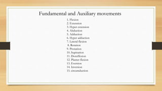
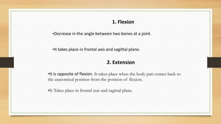
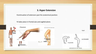
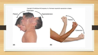
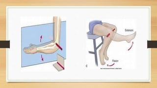
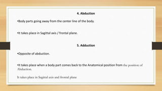
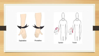
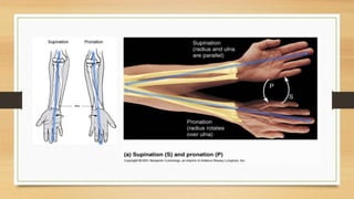
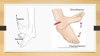
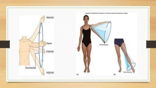
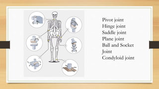
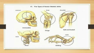
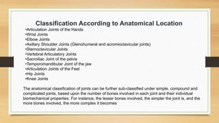
Fundamental and
Auxiliary movements
BY
CHANDRAKANT BARIK
NIS COACH
KALINGA STADIUM COMPLEX IN-CHARGE
Fundamental and Auxiliary movements
1. Flexion
2. Extension
3. Hyper extension
4. Abduction
5. Adduction
6. Hyper adduction
7. Lateral flexion
8. Rotation
9. Pronation
10. Supination
11. Dorsiflexion
12. Planter flexion
13. Eversion
14. Inversion
15. circumduction
2. Extension
•It is opposite of flexion. It takes place when the body part comes back to
the anatomical position from the position of flexion.
•It Takes place in frontal axis and sagittal plane.
1. Flexion
•Decrease in the angle between two bones at a joint.
•It takes place in frontal axis and sagittal plane.
3. Hyper Extension
•Continuation of extension past the anatomical position.
•It takes place in frontal axis and sagittal plane.
Hyper
4. Abduction
•Body parts going away from the center line of the body.
•It takes place in Sagittal axis / frontal plane.
5. Adduction
•Opposite of abduction.
•It takes place when a body part comes back to the Anatomical position from the position of
Abduction.
It takes place in Sagittal axis and frontal plane
6. Hyper Adduction
•Continuation of adduction past the anatomical position.
•It takes place in sagittal axis and frontal plane.
Hyper
. Lateral Flexion
•Sideward bending.
•Flexion movement towards lateral side of the body.
•It takes place in Sagittal axis and frontal plane.
8. Rotation
Long axis of the bone turns around the vertical axis.
Takes place in Vertical axis and Horizontal plane.
Inward Rotation/
•Medial Rotation/
Anterior surface turns outward (laterally)
Internal Rotation
•Anterior surface turns inward (medially).
•Outward Rotation/
•Lateral Rotation/
•External Rotation
Two specific movements at elbow joint
9. Pronation :-
Type of inward rotation when the palm of the hand faces downward.
9. Supination :-
Type of outward rotation when the palm of the hand faces upward.
• It takes place in vertical axis and horizontal plane.
Specific movements at Ankle
11.Dorsi flexion :-
Toe of the foot coming close to the tibia (lower leg).
11.Planter flexion :oposite of dorsi flexion
•It takes place in Frontal axis and sagittal plane
11.Inversion :-
Lifting inside (medial) border of the sole of the foot.
11.Eversion :-
Lifting outside (Lateral) border of the sole of the foot.
It takes place in Vertical axis and horizontal plane
15. Circumduction
•It is a combination of movements that takes place in a specific
sequence.
•It involves
• Flexion
• Abduction
• Extension
• Adduction
EVALUATION
OF
KNOWLEDGE
ARE YOU READY
1
Let’s break down these key components
of understanding and
communicating by taking a look at
two seemingly simple poses through an
optimal teaching lens:
Lets understand some basic
poses
Poses like Cat can reveal postural imbalances that could be
masking potential injuries and structural limitations.
Cat Camel
Understanding
Cat is considered a low level intensity pose. The simplicity of the posture provides a great
opportunity for you and your student to see/feel smoothly and efficiently move through the kinetic
chain, or the notion that joints have a direct effect on one another during movement.
Communicating
First cue the movement from an activation point. For example, load the energy down through the
hands, and observe how the student naturally moves through their body from that starting point.
Take note of where the movement looks blocked or restricted - this visible blockage will begin to tell
you a story that will help to clearly communicate needed modifications and adjustments to the
student.
Downward Dog might seem like an easier posture to body read, but the real
skill lies in what you what do with you see
Downward Dog
Understanding
Downward Dog is a complex, advanced pose that is often used by many yoga teachers too early in a
session. Without enough physiological adaptation and preparation of the body to adapt to the
potentially challenging loading on joints and muscles, incorrect loading can occur which leads to
increased postural issues.
Communicating
First shift the load in Downward Dog to experiment with what feels more mobile or stable for you.
Now ask yourself - if I want to teach this posture to help my students feel more mobile, (or stable,
depending on what the objectives are), where do I need to shift the load to facilitate that safely? After
playing with this shifting in your individual practice, you will be able to pass this information to your
student in a confident manner.
JOINTS
N
MUSCLES
Pivot joint
Hinge joint
Saddle joint
Plane joint
Ball and Socket
Joint
Condyloid joint
Joints In the Body
The joints in the body act as the ball bearings that connect and
aid in the movement of many body parts such as the limbs,
fingers, shoulders, elbows, neck, etc. Let's take a closer look
at these joints and what they do for us.
Distribution of Human Body Joints
Part of the Body Number of Joints
Skull 86
Throat and Neck 6
Thorax 66
Spine and Pelvis 76
Hands, Arms and Fingers 64
Legs, Feet and Toes 62
Types of Joints in the Human Body
Joints can be classified in accordance with various bases such as type of movement imparted,
functions, structure, etc. Let’s take a look at these various types of joints and their different
classifications.
Classification According to Movement Imparted
•Ball and Socket Joints (shoulders and hips)
•Hinge Joints (knees and ulna of the elbows)
•Condyloid Joints (jaw and fingers)
•Gliding Joints (spine, wrists and ankles)
•Pivot Joints (neck and radius of elbows)
•Saddle Joints (thumbs)
Classification According to Function
•Synarthrosis (allow very limited or no mobility such as gomphosis, synostoses and
synchondrosis; mostly fibrous joints.)
•Amphiarthrosis (Allow somewhat limited mobility such as symphysis, syndesmosis and
interosseous membrane; mostly cartilaginous joints)
•Diarthrosis (enables a wide variety of movements and all synovial joints come under this
classification)
Classification According to Structure
•Fibrous Joints (sutures, syndesmoses and gomphosis)
•Cartilaginous Joints (synchondroses and symphyses)
•Synovial Joints (wrist carpals, acromioclavicular joint, elbow area between humerus and ulna,
Atlanto-axial joint, proximal radio-ulnar joint, distal radio-ulnar joint, wrist joint, carpometacarpal
thumb joint, Glenohumeral shoulder joint, hip joint, sternoclavicular joint, knee-joint)
Classification According to Anatomical Location
•Articulation Joints of the Hands
•Wrist Joints
•Elbow Joints
•Axillary Shoulder Joints (Glenohumeral and acromioclavicular joints)
•Sternoclavicular Joints
•Vertebral Articulatory Joints
•Sacroiliac Joint of the pelvis
•Temporomandibular Joint of the jaw
•Articulation Joints of the Feet
•Hip Joints
•Knee Joints
The anatomical classification of joints can be further sub-classified under simple, compound and
complicated joints, based upon the number of bones involved in each joint and their individual
biomechanical properties. For instance, the lesser bones involved, the simpler the joint is, and the
more bones involved, the more complex it becomes
Yoga Injury Prevention
An increasing number of Americans are turning to yoga for exercise and relaxation, as well
as relief of bone, joint, and muscle-related pain. Although yoga does offer many health benefits,
if it is practiced incorrectly, it may cause muscle strain, torn ligaments, or more serious injuries.
Fortunately, the rewards of basic yoga outweigh the potential physical risks, as long as you
take caution and perform the exercises in moderation, according to your individual flexibility
level. These rewards include improved strength, balance, and flexibility, as well as an improved
sense of well-being. Yoga may also be beneficial for certain bone and joint problems like carpal
tunnel syndrome, tennis elbow, and arthritis.
Factors that affect equilibrium and balance in the body include
breathing, vision, vestibular function, musculoskeletal alignment and proprioception.
The eyes, vestibular system and proprioceptors of the neck read and adjust head
placement in relation to the environment. The respiratory system, the musculoskeletal
system and the proprioceptors of the feet and body influence the stability and
equilibrium of the rest of the body
Factors that affect equilibrium and balance

Fundamental and Auxiliary movements.pptx

  • 1.
    Fundamental and Auxiliary movements BY CHANDRAKANTBARIK NIS COACH KALINGA STADIUM COMPLEX IN-CHARGE
  • 3.
    Fundamental and Auxiliarymovements 1. Flexion 2. Extension 3. Hyper extension 4. Abduction 5. Adduction 6. Hyper adduction 7. Lateral flexion 8. Rotation 9. Pronation 10. Supination 11. Dorsiflexion 12. Planter flexion 13. Eversion 14. Inversion 15. circumduction
  • 4.
    2. Extension •It isopposite of flexion. It takes place when the body part comes back to the anatomical position from the position of flexion. •It Takes place in frontal axis and sagittal plane. 1. Flexion •Decrease in the angle between two bones at a joint. •It takes place in frontal axis and sagittal plane.
  • 5.
    3. Hyper Extension •Continuationof extension past the anatomical position. •It takes place in frontal axis and sagittal plane. Hyper
  • 8.
    4. Abduction •Body partsgoing away from the center line of the body. •It takes place in Sagittal axis / frontal plane. 5. Adduction •Opposite of abduction. •It takes place when a body part comes back to the Anatomical position from the position of Abduction. It takes place in Sagittal axis and frontal plane
  • 9.
    6. Hyper Adduction •Continuationof adduction past the anatomical position. •It takes place in sagittal axis and frontal plane.
  • 10.
  • 11.
    . Lateral Flexion •Sidewardbending. •Flexion movement towards lateral side of the body. •It takes place in Sagittal axis and frontal plane.
  • 13.
    8. Rotation Long axisof the bone turns around the vertical axis. Takes place in Vertical axis and Horizontal plane.
  • 14.
    Inward Rotation/ •Medial Rotation/ Anteriorsurface turns outward (laterally) Internal Rotation •Anterior surface turns inward (medially). •Outward Rotation/ •Lateral Rotation/ •External Rotation
  • 16.
    Two specific movementsat elbow joint 9. Pronation :- Type of inward rotation when the palm of the hand faces downward. 9. Supination :- Type of outward rotation when the palm of the hand faces upward. • It takes place in vertical axis and horizontal plane.
  • 19.
    Specific movements atAnkle 11.Dorsi flexion :- Toe of the foot coming close to the tibia (lower leg). 11.Planter flexion :oposite of dorsi flexion •It takes place in Frontal axis and sagittal plane
  • 22.
    11.Inversion :- Lifting inside(medial) border of the sole of the foot. 11.Eversion :- Lifting outside (Lateral) border of the sole of the foot. It takes place in Vertical axis and horizontal plane
  • 24.
    15. Circumduction •It isa combination of movements that takes place in a specific sequence. •It involves • Flexion • Abduction • Extension • Adduction
  • 26.
  • 27.
  • 28.
  • 47.
    Let’s break downthese key components of understanding and communicating by taking a look at two seemingly simple poses through an optimal teaching lens:
  • 48.
  • 49.
    Poses like Catcan reveal postural imbalances that could be masking potential injuries and structural limitations. Cat Camel
  • 50.
    Understanding Cat is considereda low level intensity pose. The simplicity of the posture provides a great opportunity for you and your student to see/feel smoothly and efficiently move through the kinetic chain, or the notion that joints have a direct effect on one another during movement. Communicating First cue the movement from an activation point. For example, load the energy down through the hands, and observe how the student naturally moves through their body from that starting point. Take note of where the movement looks blocked or restricted - this visible blockage will begin to tell you a story that will help to clearly communicate needed modifications and adjustments to the student.
  • 51.
    Downward Dog mightseem like an easier posture to body read, but the real skill lies in what you what do with you see Downward Dog
  • 52.
    Understanding Downward Dog isa complex, advanced pose that is often used by many yoga teachers too early in a session. Without enough physiological adaptation and preparation of the body to adapt to the potentially challenging loading on joints and muscles, incorrect loading can occur which leads to increased postural issues. Communicating First shift the load in Downward Dog to experiment with what feels more mobile or stable for you. Now ask yourself - if I want to teach this posture to help my students feel more mobile, (or stable, depending on what the objectives are), where do I need to shift the load to facilitate that safely? After playing with this shifting in your individual practice, you will be able to pass this information to your student in a confident manner.
  • 53.
  • 54.
    Pivot joint Hinge joint Saddlejoint Plane joint Ball and Socket Joint Condyloid joint
  • 56.
    Joints In theBody The joints in the body act as the ball bearings that connect and aid in the movement of many body parts such as the limbs, fingers, shoulders, elbows, neck, etc. Let's take a closer look at these joints and what they do for us.
  • 57.
    Distribution of HumanBody Joints Part of the Body Number of Joints Skull 86 Throat and Neck 6 Thorax 66 Spine and Pelvis 76 Hands, Arms and Fingers 64 Legs, Feet and Toes 62
  • 58.
    Types of Jointsin the Human Body Joints can be classified in accordance with various bases such as type of movement imparted, functions, structure, etc. Let’s take a look at these various types of joints and their different classifications. Classification According to Movement Imparted •Ball and Socket Joints (shoulders and hips) •Hinge Joints (knees and ulna of the elbows) •Condyloid Joints (jaw and fingers) •Gliding Joints (spine, wrists and ankles) •Pivot Joints (neck and radius of elbows) •Saddle Joints (thumbs)
  • 59.
    Classification According toFunction •Synarthrosis (allow very limited or no mobility such as gomphosis, synostoses and synchondrosis; mostly fibrous joints.) •Amphiarthrosis (Allow somewhat limited mobility such as symphysis, syndesmosis and interosseous membrane; mostly cartilaginous joints) •Diarthrosis (enables a wide variety of movements and all synovial joints come under this classification) Classification According to Structure •Fibrous Joints (sutures, syndesmoses and gomphosis) •Cartilaginous Joints (synchondroses and symphyses) •Synovial Joints (wrist carpals, acromioclavicular joint, elbow area between humerus and ulna, Atlanto-axial joint, proximal radio-ulnar joint, distal radio-ulnar joint, wrist joint, carpometacarpal thumb joint, Glenohumeral shoulder joint, hip joint, sternoclavicular joint, knee-joint)
  • 60.
    Classification According toAnatomical Location •Articulation Joints of the Hands •Wrist Joints •Elbow Joints •Axillary Shoulder Joints (Glenohumeral and acromioclavicular joints) •Sternoclavicular Joints •Vertebral Articulatory Joints •Sacroiliac Joint of the pelvis •Temporomandibular Joint of the jaw •Articulation Joints of the Feet •Hip Joints •Knee Joints The anatomical classification of joints can be further sub-classified under simple, compound and complicated joints, based upon the number of bones involved in each joint and their individual biomechanical properties. For instance, the lesser bones involved, the simpler the joint is, and the more bones involved, the more complex it becomes
  • 63.
    Yoga Injury Prevention Anincreasing number of Americans are turning to yoga for exercise and relaxation, as well as relief of bone, joint, and muscle-related pain. Although yoga does offer many health benefits, if it is practiced incorrectly, it may cause muscle strain, torn ligaments, or more serious injuries. Fortunately, the rewards of basic yoga outweigh the potential physical risks, as long as you take caution and perform the exercises in moderation, according to your individual flexibility level. These rewards include improved strength, balance, and flexibility, as well as an improved sense of well-being. Yoga may also be beneficial for certain bone and joint problems like carpal tunnel syndrome, tennis elbow, and arthritis.
  • 66.
    Factors that affectequilibrium and balance in the body include breathing, vision, vestibular function, musculoskeletal alignment and proprioception. The eyes, vestibular system and proprioceptors of the neck read and adjust head placement in relation to the environment. The respiratory system, the musculoskeletal system and the proprioceptors of the feet and body influence the stability and equilibrium of the rest of the body Factors that affect equilibrium and balance