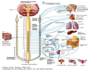
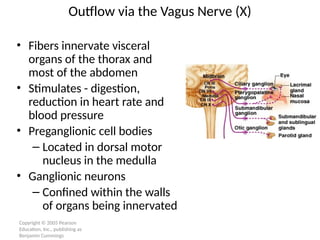
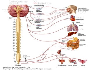
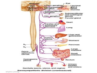
ANS FUNCTIONS AND CLINICAL
EVALUATION
Sympathetic division
• Thoracolumbar division- Preganglionic
neurons originate from the thoracic and
lumbar levels of the spinal cord (T1-L2).
• Sympathetic ganglia:
Sympathetic trunk (vertebral chain) ganglia.
Prevertebral (collateral) ganglia: celiac,
superior mesenteric, inferior mesenteric,
aorticorenal and renal.
Sympathetic nervous system
• The preganglionic neurons of the sympathetic division originate
in the intermediolateral cell column of the spinal gray matter,
from the eighth cervical to the second lumbar segments.
• Axons of the nerve fibers originating in the intermediolateral
column are of small caliber and are myelinated; form the white
communicating rami.
• These preganglionic fibers synapse with the cell bodies of the
postganglionic neurons, which are collected into two large
ganglionated chains or cords, one on each side of the vertebral
column (paravertebral ganglia), and several single prevertebral
ganglia.
• Most of the postganglionic fibers pass via gray
communicating rami to spinal nerves of T5 to L2;
they supply blood vessels, sweat glands, and hair
follicles.
• The postganglionic fibers of the prevertebral ganglia
(located in the posterior abdomen rather than
paravertebrally) form the hypogastric, splanchnic,
and mesenteric plexuses, which innervate the
glands, smooth muscle, and blood vessels of the
abdominal and pelvic viscera
Post ganglionic neurons
• An axon may synapse with postganglionic neurons in
the ganglion it first reaches or
• Sympathetic chains or
• An axon may continue, without synapsing, through the
sympathetic trunk ganglion to end at a prevertebral
ganglion and synapse with postganglionic neurons
there or
• An axon may pass through the sympathetic trunk
ganglion and a prevertebral ganglion and then to the
adrenal medulla.
Copyright © 2005 Pearson
Education, Inc., publishing as
Benjamin Cummings
Sympathetic Trunk Ganglia
Copyright © 2005 Pearson
Education, Inc., publishing as
Benjamin Cummings
Sympathetic Pathways to Periphery
Figure 15.9
S
8
• PARASYMPATHETIC DIVISION
Cranial outflow
• Preganglionic fibers run via:
– Oculomotor nerve (III)
– Facial nerve (VII)
– Glossopharyngeal nerve (IX)
– Vagus nerve (X)
• Cell bodies located in cranial nerve nuclei in
the brain stem
Copyright © 2005 Pearson
Education, Inc., publishing as
Benjamin Cummings
Outflow via the Vagus Nerve (X)
• Fibers innervate visceral
organs of the thorax and
most of the abdomen
• Stimulates - digestion,
reduction in heart rate and
blood pressure
• Preganglionic cell bodies
– Located in dorsal motor
nucleus in the medulla
• Ganglionic neurons
– Confined within the walls
of organs being innervated
Sacral outflow
• Emerges from S2-S4
• Innervates organs of the pelvis and lower
abdomen
• Preganglionic cell bodies
– Located in visceral motor region of spinal gray
matter
• Form splanchnic nerves
• Parasympathetic ganglia: terminal ganglia.
• Presynaptic neuron usually synapses with 4-5
postsynaptic neurons all of which supply a
single visceral effector.
Structure of the Parasympathetic Division
14
Copyright © 2005 Pearson
Education, Inc., publishing as
Benjamin Cummings
The Parasympathetic Division

ANS FUNCTIONS AND CLINICAL EVALUATION.pptx

  • 1.
    ANS FUNCTIONS ANDCLINICAL EVALUATION
  • 2.
    Sympathetic division • Thoracolumbardivision- Preganglionic neurons originate from the thoracic and lumbar levels of the spinal cord (T1-L2). • Sympathetic ganglia: Sympathetic trunk (vertebral chain) ganglia. Prevertebral (collateral) ganglia: celiac, superior mesenteric, inferior mesenteric, aorticorenal and renal.
  • 3.
    Sympathetic nervous system •The preganglionic neurons of the sympathetic division originate in the intermediolateral cell column of the spinal gray matter, from the eighth cervical to the second lumbar segments. • Axons of the nerve fibers originating in the intermediolateral column are of small caliber and are myelinated; form the white communicating rami. • These preganglionic fibers synapse with the cell bodies of the postganglionic neurons, which are collected into two large ganglionated chains or cords, one on each side of the vertebral column (paravertebral ganglia), and several single prevertebral ganglia.
  • 4.
    • Most ofthe postganglionic fibers pass via gray communicating rami to spinal nerves of T5 to L2; they supply blood vessels, sweat glands, and hair follicles. • The postganglionic fibers of the prevertebral ganglia (located in the posterior abdomen rather than paravertebrally) form the hypogastric, splanchnic, and mesenteric plexuses, which innervate the glands, smooth muscle, and blood vessels of the abdominal and pelvic viscera
  • 5.
    Post ganglionic neurons •An axon may synapse with postganglionic neurons in the ganglion it first reaches or • Sympathetic chains or • An axon may continue, without synapsing, through the sympathetic trunk ganglion to end at a prevertebral ganglion and synapse with postganglionic neurons there or • An axon may pass through the sympathetic trunk ganglion and a prevertebral ganglion and then to the adrenal medulla.
  • 6.
    Copyright © 2005Pearson Education, Inc., publishing as Benjamin Cummings Sympathetic Trunk Ganglia
  • 7.
    Copyright © 2005Pearson Education, Inc., publishing as Benjamin Cummings Sympathetic Pathways to Periphery Figure 15.9
  • 8.
  • 9.
  • 10.
    Cranial outflow • Preganglionicfibers run via: – Oculomotor nerve (III) – Facial nerve (VII) – Glossopharyngeal nerve (IX) – Vagus nerve (X) • Cell bodies located in cranial nerve nuclei in the brain stem
  • 11.
    Copyright © 2005Pearson Education, Inc., publishing as Benjamin Cummings Outflow via the Vagus Nerve (X) • Fibers innervate visceral organs of the thorax and most of the abdomen • Stimulates - digestion, reduction in heart rate and blood pressure • Preganglionic cell bodies – Located in dorsal motor nucleus in the medulla • Ganglionic neurons – Confined within the walls of organs being innervated
  • 12.
    Sacral outflow • Emergesfrom S2-S4 • Innervates organs of the pelvis and lower abdomen • Preganglionic cell bodies – Located in visceral motor region of spinal gray matter • Form splanchnic nerves
  • 13.
    • Parasympathetic ganglia:terminal ganglia. • Presynaptic neuron usually synapses with 4-5 postsynaptic neurons all of which supply a single visceral effector.
  • 14.
    Structure of theParasympathetic Division 14
  • 15.
    Copyright © 2005Pearson Education, Inc., publishing as Benjamin Cummings The Parasympathetic Division