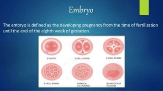
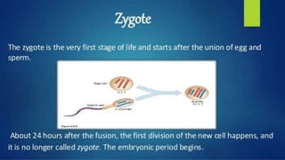
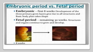
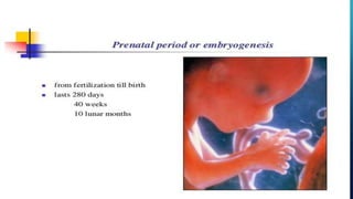
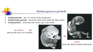
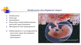
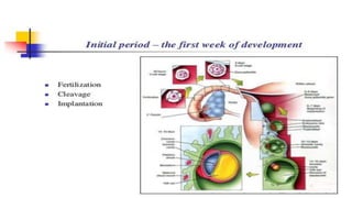
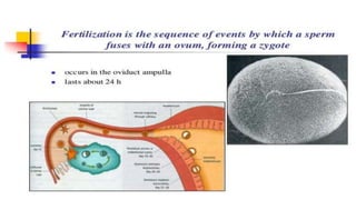
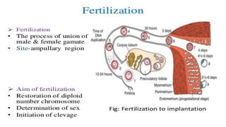
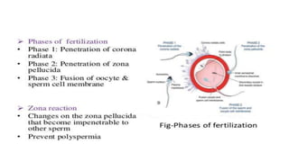
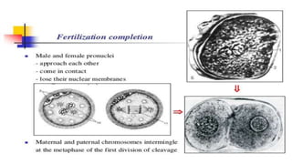
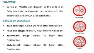
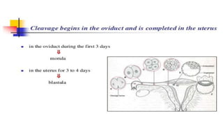
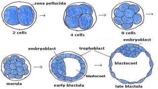
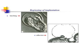
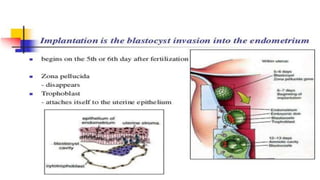
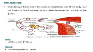
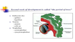
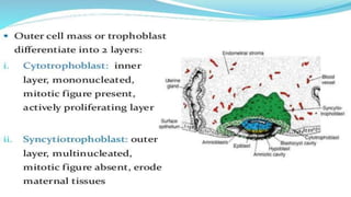
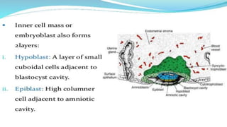
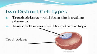
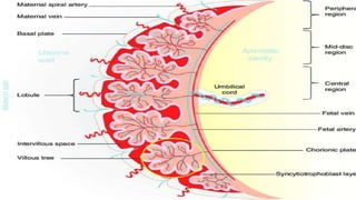
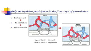
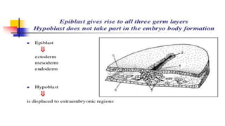
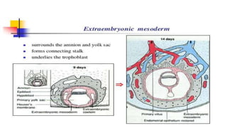
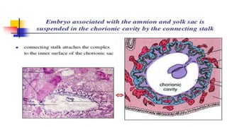
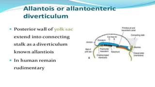
MITTAL COLLEGE OF NURSING, AJM
SUBMITTED TO :-. SUBMITTED BY:-
SNEHALATA PARASHAR. GAGANDEEP
KUMAWAT
A PRESENTATION ON
Embryology
Development of fetus and
placenta
DEFINATION
Embryology is the branch of biology that
studies the prenatal development of
gametes, fertilization, and development
of embryos and fetuses. Additionally,
embryology encompasses the study of
congenital disorders that occur before
birth, known as teratology
FERTILIZATION
IMPLANTATION
TROPHOBLAST
•Trophoblast is the outer cell mass which
forms the placenta and chorion
WHEN
When the interstitial implantation is
completed on 11th day ,the blastocyst is
surrounded on all side by lacunar spaces
around cord of syncytial cells, called
trabeculae
VILLI
•The finger like buds are called primary
stem villi surrounded by lacunar spaces
which will later form into intervillous
space.
•After the appearance of the primitive
mesenchyme and the development of the
chorion the primary stem villi are named
chorionic villi
PLACENTA
•As the growth of embryo proceeds decidua
capsularis become thinner beginning at 6th
week
•Villi and lacunar spaces in the embronic area
get obliterated converting the chorion into
chorion leave
•This is compensated (a) exuberant growth
and proliferation of the decidua basalis (b)
PLACENTA.
•These two chorion frondosum and
decidua. Basalis form the discrete
placenta .It begins at 6th week and
complete by 12th week
EMBROBLAST
•Embyroblast is the inner cell mass which
forms the fetus , amnion and umbilical
cord
EXTRAEMBRYONIC COELOM
•Extraembryonic mesenchyme. Derived
from the trophoblast appear to separate
the yolk sac from the blastocyst wall and
also the amniotic cavity from the
trophoblast of chorion.Small cystic space
(lacuna) now appear with in the
extraembryonic mesenchyme these space
gradually enlarge and fuse to form
BODY STALK
•Progressive enlargment of extraembryonic
coelom separate the amnion from the
inner aspect of the chorion except the
caudal end of the Embro. There the
mesenchymal attachment persists to form
body stalk . Umbilical cord develops from
this body stalk
4TH WEEK OF DEVELOPMENT
•The embryo grows dramatically during the
fourth week. It more than triples its length
to 3.5mm and its weight approximates 5
mg.
4TH WEEK OF DEVELOPMENT
•Elongation of the embryo has occurred,
and it has become curved pon itself with
the formation of a head and tail fold.
Lateral body folds develop making the
embryo tubular rather than flat and disc
shaped. Closure of the neural tube begins
in the area of the occiput and proceeds
upward and downward from that point.
Somites formed in a craniocaudal
sequence as the neural tube closes, can
5TH WEEK OF DEVELOPMENT
•The intestinal system is formed from the
yolk sac, and differentiation of the buds,
which will become the oesophagus,
stomach, liver and pancreas, is
progressing. The thyroid and thymus
glands are also developing. The primitive
circulatory system is established, and the
heart is beating. The budlike projections
on the surface of the embryo are the
6TH WEEK OF DEVELOPMENT
•The head has become larger than the trunk
and is bent over the heart prominence.
Elevations in the facial ectoderm are evident
and the position of the eyes, nose, mouth,
and the groove that is to become the external
acoustic meatus is established. In the upper
limbs, the elbows and wrists are identifiable,
and the hand plates develop ridges called
finger rays. The lower limbs are not developed
to the same degree. Changes are beginning to
7TH WEEK OF DEVELOPMENT
•Cerebral hemispheres appear as the head
enlarges rapidly. The eyes move from a lateral to
a more frontal position as the face elongates.
Prominences appear over the ventral body wall
from early because their function is vital to the
maintenance and survival of the embryo. As the
embryo continues to grow, the umbilical cord
shrinks. The arm and hand of the upper limbs
and the high, leg, and foot segments of the lower
limbs become apparent The fingers develop, the
their growth is critical at this point (40 to 50
days). The CRL is 14 to 20 mm; the weight is 400
8TH WEEK OF DEVELOPMENT
•During this final week of the embryonic
period, the embryo exhibits definite human
characteristics. The cerebral hemispheres
have grown so rapidly that the head now
makes up 50% of the mass of the embryo. The
face occupies the lower half of the head, and
the eyes continue to move to a more frontal
plane. Eyelids folds develop. These will
become fused during the ninth week and
FOETAL AND PLACENTA DEVLOPMENT

FOETAL AND PLACENTA DEVLOPMENT

  • 1.
    MITTAL COLLEGE OFNURSING, AJM SUBMITTED TO :-. SUBMITTED BY:- SNEHALATA PARASHAR. GAGANDEEP KUMAWAT
  • 2.
  • 3.
    DEFINATION Embryology is thebranch of biology that studies the prenatal development of gametes, fertilization, and development of embryos and fetuses. Additionally, embryology encompasses the study of congenital disorders that occur before birth, known as teratology
  • 11.
  • 30.
  • 43.
    TROPHOBLAST •Trophoblast is theouter cell mass which forms the placenta and chorion
  • 51.
    WHEN When the interstitialimplantation is completed on 11th day ,the blastocyst is surrounded on all side by lacunar spaces around cord of syncytial cells, called trabeculae
  • 52.
    VILLI •The finger likebuds are called primary stem villi surrounded by lacunar spaces which will later form into intervillous space. •After the appearance of the primitive mesenchyme and the development of the chorion the primary stem villi are named chorionic villi
  • 53.
    PLACENTA •As the growthof embryo proceeds decidua capsularis become thinner beginning at 6th week •Villi and lacunar spaces in the embronic area get obliterated converting the chorion into chorion leave •This is compensated (a) exuberant growth and proliferation of the decidua basalis (b)
  • 54.
    PLACENTA. •These two chorionfrondosum and decidua. Basalis form the discrete placenta .It begins at 6th week and complete by 12th week
  • 56.
    EMBROBLAST •Embyroblast is theinner cell mass which forms the fetus , amnion and umbilical cord
  • 61.
    EXTRAEMBRYONIC COELOM •Extraembryonic mesenchyme.Derived from the trophoblast appear to separate the yolk sac from the blastocyst wall and also the amniotic cavity from the trophoblast of chorion.Small cystic space (lacuna) now appear with in the extraembryonic mesenchyme these space gradually enlarge and fuse to form
  • 62.
    BODY STALK •Progressive enlargmentof extraembryonic coelom separate the amnion from the inner aspect of the chorion except the caudal end of the Embro. There the mesenchymal attachment persists to form body stalk . Umbilical cord develops from this body stalk
  • 76.
    4TH WEEK OFDEVELOPMENT •The embryo grows dramatically during the fourth week. It more than triples its length to 3.5mm and its weight approximates 5 mg.
  • 77.
    4TH WEEK OFDEVELOPMENT •Elongation of the embryo has occurred, and it has become curved pon itself with the formation of a head and tail fold. Lateral body folds develop making the embryo tubular rather than flat and disc shaped. Closure of the neural tube begins in the area of the occiput and proceeds upward and downward from that point. Somites formed in a craniocaudal sequence as the neural tube closes, can
  • 78.
    5TH WEEK OFDEVELOPMENT •The intestinal system is formed from the yolk sac, and differentiation of the buds, which will become the oesophagus, stomach, liver and pancreas, is progressing. The thyroid and thymus glands are also developing. The primitive circulatory system is established, and the heart is beating. The budlike projections on the surface of the embryo are the
  • 79.
    6TH WEEK OFDEVELOPMENT •The head has become larger than the trunk and is bent over the heart prominence. Elevations in the facial ectoderm are evident and the position of the eyes, nose, mouth, and the groove that is to become the external acoustic meatus is established. In the upper limbs, the elbows and wrists are identifiable, and the hand plates develop ridges called finger rays. The lower limbs are not developed to the same degree. Changes are beginning to
  • 80.
    7TH WEEK OFDEVELOPMENT •Cerebral hemispheres appear as the head enlarges rapidly. The eyes move from a lateral to a more frontal position as the face elongates. Prominences appear over the ventral body wall from early because their function is vital to the maintenance and survival of the embryo. As the embryo continues to grow, the umbilical cord shrinks. The arm and hand of the upper limbs and the high, leg, and foot segments of the lower limbs become apparent The fingers develop, the their growth is critical at this point (40 to 50 days). The CRL is 14 to 20 mm; the weight is 400
  • 81.
    8TH WEEK OFDEVELOPMENT •During this final week of the embryonic period, the embryo exhibits definite human characteristics. The cerebral hemispheres have grown so rapidly that the head now makes up 50% of the mass of the embryo. The face occupies the lower half of the head, and the eyes continue to move to a more frontal plane. Eyelids folds develop. These will become fused during the ninth week and