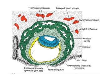
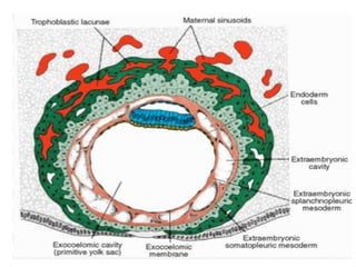
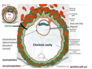
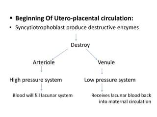
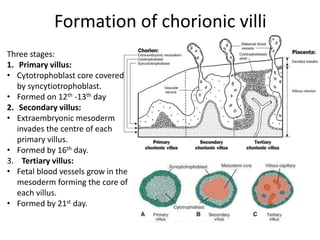
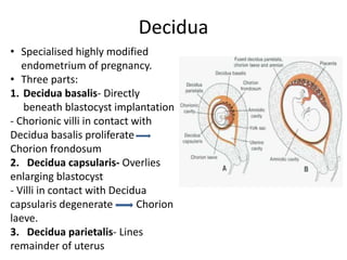
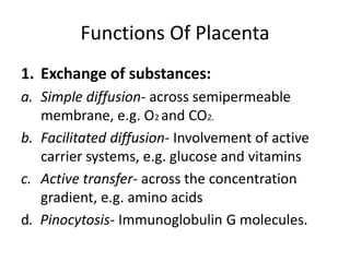
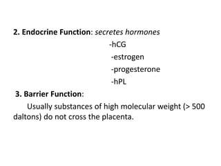
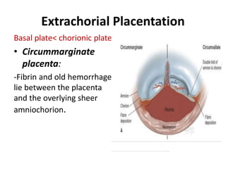
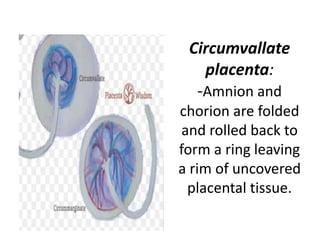
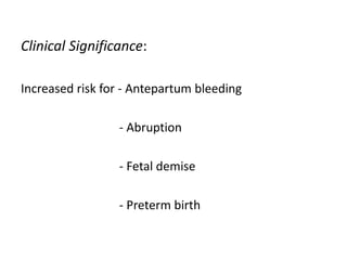
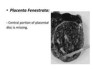
Development Of Placenta And
Abnormalities
Dr. Bhoomika
Objectives
• To describe the anatomy of placenta
• To discuss the development of placenta
• To enumerate the functions of placenta
• To describe abnormalities of placenta and
their clinical significance
The Placenta
• Discoid
• Hemochorial
• Deciduate
• Establishes connection between the mother
and fetus through the umbilical cord
Types of Placenta
• Epithelio-chorial
placenta (swine,
horse)
• Endothelio-chorial
placenta (dog, cat)
• Hemo-chorial
placenta (human,
rodents, primates)
Normal Placenta
 Shape- Round to oval
 Weight- 470g
 Diameter- 22 cm
 Central thickness- 2.5 cm
 Maternal surface
• Develop from decidua basalis
• rough, shaggy, fleshy
• 15-30 cotyledons.
 Fetal surface
• Develops from chorion frondosum
• Smooth and shiny
 Umbilical cord
• Length is 30-70 cm
• Consists 2 umbilical arteries and 1 umbilical vein
• Single umbilical artery associated with congenital
anomalies.
Development Of Placenta
Outer cell mass
Cytotrophoblast
• Well demarcated cell
border
• Single nucleus
• Undergo DNA
synthesis and mitosis
Syncytiotrophoblast
• No cell boundaries
• Amorphous cytoplasm
• Multiple nuclei varying in
size and shape
• Interconnected cavities
developed within this
syncytium called as lacunae.
 Beginning Of Utero-placental circulation:
• Syncytiotrophoblast produce destructive enzymes
Destroy
Arteriole Venule
High pressure system Low pressure system
Blood will fill lacunar system Receives lacunar blood back
into maternal circulation
Formation of chorionic villi
Three stages:
1. Primary villus:
• Cytotrophoblast core covered
by syncytiotrophoblast.
• Formed on 12th -13th day
2. Secondary villus:
• Extraembryonic mesoderm
invades the centre of each
primary villus.
• Formed by 16th day.
3. Tertiary villus:
• Fetal blood vessels grow in the
mesoderm forming the core of
each villus.
• Formed by 21st day.
Decidua
• Specialised highly modified
endometrium of pregnancy.
• Three parts:
1. Decidua basalis- Directly
beneath blastocyst implantation
- Chorionic villi in contact with
Decidua basalis proliferate
Chorion frondosum
2. Decidua capsularis- Overlies
enlarging blastocyst
- Villi in contact with Decidua
capsularis degenerate Chorion
laeve.
3. Decidua parietalis- Lines
remainder of uterus
Functions Of Placenta
1. Exchange of substances:
a. Simple diffusion- across semipermeable
membrane, e.g. O2 and CO2.
b. Facilitated diffusion- Involvement of active
carrier systems, e.g. glucose and vitamins
c. Active transfer- across the concentration
gradient, e.g. amino acids
d. Pinocytosis- Immunoglobulin G molecules.
2. Endocrine Function: secretes hormones
-hCG
-estrogen
-progesterone
-hPL
3. Barrier Function:
Usually substances of high molecular weight (> 500
daltons) do not cross the placenta.
Only IgG can cross the placental barrier
Viruses - rubella, CMV, HIV, Hepatitis B, Varicella zoster
Bacteria- treponema pallidum
Protozoa- toxoplasma gondii , malarial parasite
Drugs
4.Metabolic functions :
glycogen
lipids
Abnormalities Of Placenta
Shape and Size Variants:
• Bilobed Placenta: Cord
inserts between the two
placental lobes- either
into a connecting
chorionic bridge or into
intervening membranes.
• Multilobed Placenta:
contain three or more
equivalently sized lobes.
Succenturiate lobes:
- Small accessory lobes ≥1,
develop in the membranes at a
distant from the main placenta.
- These lobes have vessels that
course through membranes.
Clinical significance:
Retained in the uterus after delivery and may cause
PPH.
• Placenta membranacea:
- Villi cover nearly all of
the uterine cavity.
• Ring Shaped Placenta:
- Partial or complete ring
of placental tissues
present.
Clinical significance:
-APH
-PPH
-Fetal growth restriction
Extrachorial Placentation
Basal plate< chorionic plate
• Circummarginate
placenta:
-Fibrin and old hemorrhage
lie between the placenta
and the overlying sheer
amniochorion.
Circumvallate
placenta:
-Amnion and
chorion are folded
and rolled back to
form a ring leaving
a rim of uncovered
placental tissue.
Clinical Significance:
Increased risk for - Antepartum bleeding
- Abruption
- Fetal demise
- Preterm birth
• Placenta Fenestrata:
- Central portion of placental
disc is missing.
Chorioangioma
• Most common benign tumors of placenta
• Incidence is approximately 1 %
• Components- Blood vessels and stroma of chorionic villus
• Clinical significance:
 Small tumors-
Asymptomatic
 Large tumors (> 4cm)-
can create arteriovenus shunting within placenta to cause high
output failure, hydrops and fetal death.
Diagnosis:
• Ultrasonography
- well- circumscribed
- Rounded
- Hypoechoic lesion
• Colour doppler
- Increased blood flow
Cord Abnormalities
Based on length
Long cord
- More than 70 cm
Clinical significance:
• Cord prolapse
• Cord entanglement
• Fetal anomalies
Short cord
Less than 30 cm
Clinical significance:
• Congenital malformations
• Intrapartum distress
Based on insertion
Marginal
-Cord anchors at the
placental margin
-May be seen in up to 6%
cases
-Rarely causes problems
- Also called as Battledore
placenta
Velamentous
-Umbilical cord is inserted into
chorioamniotic membranes
-Incidence is 1%
-More common with twins
-Clinical significance:
• Vessels are vulnerable to
compression and may lead to
fetal hypoperfusion
• Still birth
• Preterm labour
• IUGR
Velamentous InsertionMarginal Insertion
Vasa Previa
• Umbilical vessels travel within the membrane and
overlie the cervical loss
• They can be torn with cervical dilatation or
membrane rupture and can lead to rapid fetal
blood loss
• Incidence- 2 to 6 per 10,000 pregnancies
• Antepartum diagnosis greatly improves the
perinatal survival rate
• Management- Early scheduled cesarean
delivery
Single Umbilical Artery
• When only one umbilical artery in an umbilical cord.
• Two types:
1. Isolated SUA- isolated ultrasound finding without
any other anomaly.
- Increased incidence of fetal growth restriction
2. Non-isolated SUA- Associated with structural or
chromosomal abnormalities.
-Most frequent anomalies are cardiovascular and
genitourinary.
-Karyotype abnormalities have been reported in 45 %
fetuses.
• Management: Thorough prenatal ultrasound
examination to look for all anomalies specially of
heart and kidneys.
- Fetal ECHO should be done.
- In non- isolated SUA, amniocentesis is indicated
as aneuploidies are common in them.
- Four weekly USG for fetal growth parameters .
- Weekly non- stress test and biophysical scoring
after 32 weeks till delivery.
THANK YOU

Placenta development and its abnormalities

  • 1.
    Development Of PlacentaAnd Abnormalities Dr. Bhoomika
  • 2.
    Objectives • To describethe anatomy of placenta • To discuss the development of placenta • To enumerate the functions of placenta • To describe abnormalities of placenta and their clinical significance
  • 3.
    The Placenta • Discoid •Hemochorial • Deciduate • Establishes connection between the mother and fetus through the umbilical cord
  • 4.
    Types of Placenta •Epithelio-chorial placenta (swine, horse) • Endothelio-chorial placenta (dog, cat) • Hemo-chorial placenta (human, rodents, primates)
  • 5.
    Normal Placenta  Shape-Round to oval  Weight- 470g  Diameter- 22 cm  Central thickness- 2.5 cm
  • 6.
     Maternal surface •Develop from decidua basalis • rough, shaggy, fleshy • 15-30 cotyledons.  Fetal surface • Develops from chorion frondosum • Smooth and shiny  Umbilical cord • Length is 30-70 cm • Consists 2 umbilical arteries and 1 umbilical vein • Single umbilical artery associated with congenital anomalies.
  • 7.
  • 8.
    Outer cell mass Cytotrophoblast •Well demarcated cell border • Single nucleus • Undergo DNA synthesis and mitosis Syncytiotrophoblast • No cell boundaries • Amorphous cytoplasm • Multiple nuclei varying in size and shape • Interconnected cavities developed within this syncytium called as lacunae.
  • 12.
     Beginning OfUtero-placental circulation: • Syncytiotrophoblast produce destructive enzymes Destroy Arteriole Venule High pressure system Low pressure system Blood will fill lacunar system Receives lacunar blood back into maternal circulation
  • 13.
    Formation of chorionicvilli Three stages: 1. Primary villus: • Cytotrophoblast core covered by syncytiotrophoblast. • Formed on 12th -13th day 2. Secondary villus: • Extraembryonic mesoderm invades the centre of each primary villus. • Formed by 16th day. 3. Tertiary villus: • Fetal blood vessels grow in the mesoderm forming the core of each villus. • Formed by 21st day.
  • 14.
    Decidua • Specialised highlymodified endometrium of pregnancy. • Three parts: 1. Decidua basalis- Directly beneath blastocyst implantation - Chorionic villi in contact with Decidua basalis proliferate Chorion frondosum 2. Decidua capsularis- Overlies enlarging blastocyst - Villi in contact with Decidua capsularis degenerate Chorion laeve. 3. Decidua parietalis- Lines remainder of uterus
  • 15.
    Functions Of Placenta 1.Exchange of substances: a. Simple diffusion- across semipermeable membrane, e.g. O2 and CO2. b. Facilitated diffusion- Involvement of active carrier systems, e.g. glucose and vitamins c. Active transfer- across the concentration gradient, e.g. amino acids d. Pinocytosis- Immunoglobulin G molecules.
  • 16.
    2. Endocrine Function:secretes hormones -hCG -estrogen -progesterone -hPL 3. Barrier Function: Usually substances of high molecular weight (> 500 daltons) do not cross the placenta.
  • 17.
    Only IgG cancross the placental barrier Viruses - rubella, CMV, HIV, Hepatitis B, Varicella zoster Bacteria- treponema pallidum Protozoa- toxoplasma gondii , malarial parasite Drugs 4.Metabolic functions : glycogen lipids
  • 18.
    Abnormalities Of Placenta Shapeand Size Variants: • Bilobed Placenta: Cord inserts between the two placental lobes- either into a connecting chorionic bridge or into intervening membranes. • Multilobed Placenta: contain three or more equivalently sized lobes.
  • 19.
    Succenturiate lobes: - Smallaccessory lobes ≥1, develop in the membranes at a distant from the main placenta. - These lobes have vessels that course through membranes. Clinical significance: Retained in the uterus after delivery and may cause PPH.
  • 20.
    • Placenta membranacea: -Villi cover nearly all of the uterine cavity. • Ring Shaped Placenta: - Partial or complete ring of placental tissues present. Clinical significance: -APH -PPH -Fetal growth restriction
  • 21.
    Extrachorial Placentation Basal plate<chorionic plate • Circummarginate placenta: -Fibrin and old hemorrhage lie between the placenta and the overlying sheer amniochorion.
  • 22.
    Circumvallate placenta: -Amnion and chorion arefolded and rolled back to form a ring leaving a rim of uncovered placental tissue.
  • 23.
    Clinical Significance: Increased riskfor - Antepartum bleeding - Abruption - Fetal demise - Preterm birth
  • 24.
    • Placenta Fenestrata: -Central portion of placental disc is missing.
  • 25.
    Chorioangioma • Most commonbenign tumors of placenta • Incidence is approximately 1 % • Components- Blood vessels and stroma of chorionic villus • Clinical significance:  Small tumors- Asymptomatic  Large tumors (> 4cm)- can create arteriovenus shunting within placenta to cause high output failure, hydrops and fetal death.
  • 26.
    Diagnosis: • Ultrasonography - well-circumscribed - Rounded - Hypoechoic lesion • Colour doppler - Increased blood flow
  • 27.
    Cord Abnormalities Based onlength Long cord - More than 70 cm Clinical significance: • Cord prolapse • Cord entanglement • Fetal anomalies Short cord Less than 30 cm Clinical significance: • Congenital malformations • Intrapartum distress
  • 28.
    Based on insertion Marginal -Cordanchors at the placental margin -May be seen in up to 6% cases -Rarely causes problems - Also called as Battledore placenta Velamentous -Umbilical cord is inserted into chorioamniotic membranes -Incidence is 1% -More common with twins -Clinical significance: • Vessels are vulnerable to compression and may lead to fetal hypoperfusion • Still birth • Preterm labour • IUGR
  • 29.
  • 30.
    Vasa Previa • Umbilicalvessels travel within the membrane and overlie the cervical loss • They can be torn with cervical dilatation or membrane rupture and can lead to rapid fetal blood loss • Incidence- 2 to 6 per 10,000 pregnancies • Antepartum diagnosis greatly improves the perinatal survival rate • Management- Early scheduled cesarean delivery
  • 32.
    Single Umbilical Artery •When only one umbilical artery in an umbilical cord. • Two types: 1. Isolated SUA- isolated ultrasound finding without any other anomaly. - Increased incidence of fetal growth restriction 2. Non-isolated SUA- Associated with structural or chromosomal abnormalities. -Most frequent anomalies are cardiovascular and genitourinary. -Karyotype abnormalities have been reported in 45 % fetuses.
  • 33.
    • Management: Thoroughprenatal ultrasound examination to look for all anomalies specially of heart and kidneys. - Fetal ECHO should be done. - In non- isolated SUA, amniocentesis is indicated as aneuploidies are common in them. - Four weekly USG for fetal growth parameters . - Weekly non- stress test and biophysical scoring after 32 weeks till delivery.
  • 34.