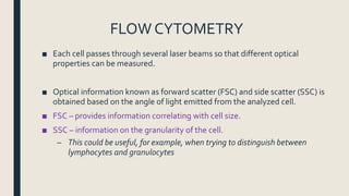
2. OBJECTIVE
■ Appreciate the mechanism and role of flow cytometry in
diagnosis and classification of various haematological
neoplasm
3. INTRODUCTION
■ Flow cytometry (FC) measures single cells “flowing” through a detector
system
■ Flow cytometer
– Flow means – flow tract
– Cytometer – device for cell analysis
■ Applications of FC range from
– common clinical laboratory tests, such as complete blood count with
differential and monitoring of CD4 cell count in HIV patients
– to advanced, multicolor flow cytometry used to identify subtypes
4. INTRODUCTION
■ Flow cytometry is a sensitive, powerful method for simultaneously obtaining
information on various cellular processes, including expression of surface
markers, intracellular cytokine and signaling proteins
■ Measures characteristics on each cell and hence play role in characterizing
heterogeneous cell populations.
5. Flow cytometry (FC)
■ FC process begins with the selection of fluorescent-labeled antibodies
specific to cell-surface markers used to characterize the cell population of
interest.
■ These cell surface markers are usually glycoproteins called cluster of
differentiation (CD) markers, and they help differentiate cell subpopulations
■ Flow cytometry can be performed on a variety of tissues, including peripheral
blood, bone marrow aspirates, skin biopsies, and tissue culture cell lines.
7. FLOW CYTOMETRY
■ Each cell passes through several laser beams so that different optical
properties can be measured.
■ Optical information known as forward scatter (FSC) and side scatter (SSC) is
obtained based on the angle of light emitted from the analyzed cell.
■ FSC – provides information correlating with cell size.
■ SSC – information on the granularity of the cell.
– This could be useful, for example, when trying to distinguish between
lymphocytes and granulocytes
9. Immunophenotyping
■ Immunophenotyping is a technique that uses monoclonal antibodies to
identify protein markers present in the cytoplasm, nucleus or surface
membrane of cells, thereby identifying the cell type and degree of
differentiation.
■ Immunophenotyping establishes clonality and determine pattern of
expression of cell markers and hence the determination of malignant
subtypes.
■ Examples;
– Chronic lymphocytic leukaemia exhibits CD5, CD23, CD19, CD20 markers
– Multiple myeloma, plasma cells exhibit – CD38 and CD138