Pediatrics notes about "Floppy infant". These notes were published in 2018.
You can download them also from
- Telegram: https://t.me/pediatric_notes_2018
- Mediafire: http://www.mediafire.com/folder/u5u60m184t9z7/Pediatric_Notes_2018
This document provides information on evaluating hypotonia in infants. It defines muscle tone and the differences between hypotonia and weakness. Hypotonia can have central or peripheral causes. The differential diagnosis for a floppy infant is extensive and includes central conditions like genetic syndromes or brain insults and peripheral conditions involving the motor unit, nerve, neuromuscular junction or muscle. A thorough evaluation includes the infant's history, development, family history, examination and potentially genetic or metabolic testing to determine the underlying cause.
Hypotonia, or low muscle tone, can have central or peripheral causes. Central hypotonia accounts for 60-80% of cases and is due to problems in the brain or spinal cord, while peripheral hypotonia accounts for 15-30% of cases and results from issues with nerves or muscles. A hypotonic infant may not display weakness. The document outlines how to classify hypotonia based on its location, potential causes, how to take a history and examine a hypotonic child, important lab investigations, and general management approaches.
A floppy infant refers to reduced muscle tone and loose joints. The main features of a floppy infant are hypotonia, abnormal postures, and delayed motor milestones. A thorough physical exam assesses tone in the limbs, neck, and trunk through various tests like leg traction and arm recoil. The cause may be central nervous system related like cerebral palsy or metabolic conditions. Alternatively, it could indicate a peripheral neuropathy, myopathy, or neuromuscular junction pathology. Further workup may include blood tests, electrophysiology, muscle biopsy, and genetic testing to determine the specific condition. Timely diagnosis and management including rehabilitation can help improve outcomes.
The document discusses hypotonia in infants. It begins by defining tone and describing central and peripheral causes of hypotonia. Central causes account for 60-80% of cases and involve the brain or spinal cord, while peripheral causes involve the motor unit, nerves, neuromuscular junction, or muscles. The document then examines the evaluation and differential diagnosis of hypotonia in infants, highlighting important history, physical exam findings, and potential etiologies to consider. Key tests include the traction response and assessment of tone, strength, and reflexes. Thorough evaluation is needed to identify the underlying cause and guide management.
Hypotonia, or low muscle tone, is common in children and can be caused by neurological conditions that affect the brain, spinal cord, nerves, or muscles. Signs of hypotonia include poor head control, slipping through caregiver's hands when held, and lying in an inverted "U" shape when placed prone or held upright. The diagnosis involves assessing prenatal risk factors, family history, signs and symptoms, and ruling out disorders of the brain, spinal cord, peripheral nerves, muscles or neuromuscular junction through examination of reflexes and extra features present. Hypotonia can be caused by various conditions like cerebral palsy, brain malformations, genetic disorders or hypoxic ischemic encephalopathy.
The document discusses hypotonia in infants and provides details on:
- The differential diagnosis of hypotonia includes both benign and serious conditions.
- Hypotonia can be caused by central nervous system issues or peripheral nervous system issues. Central causes account for 60-80% of cases.
- The evaluation of an infant with hypotonia includes a detailed history, physical exam focusing on tone and strength, and initial screening tests. Further testing may include imaging, genetic testing, and metabolic testing depending on exam findings.
The document provides information on the evaluation of hypotonia in infants. It discusses that hypotonia can be caused by central or peripheral nervous system disorders. The most common central cause is hypoxic ischemic encephalopathy, while the most common peripheral causes are congenital myopathies and spinal muscular atrophy. A thorough history, physical exam, and testing are needed to determine the underlying cause, which guides management and prognosis. The evaluation involves assessing tone, strength, reflexes and other features to localize the problem and rule out various disorders through laboratory and imaging studies.
The 5-month-old baby presented with cough and fever for 3 days. He has a known diagnosis of spinal muscular atrophy (SMA) and is unable to move his lower limbs or support his head. Examination found crepitations in both lungs and absent reflexes. A diagnosis of SMA with lower respiratory tract infection was made. SMA is caused by a defect in the SMN1 gene and results in degeneration of motor neurons. It is classified into types based on age of onset and progression, with Type 1 being the most severe and usually fatal in infancy. Currently there is no medical treatment available to slow progression.
This document discusses the approach to evaluating a hypotonic infant. It begins by defining muscle tone and how it is assessed. The causes of hypotonia are then classified based on anatomical location of the underlying pathology as paralytic or non-paralytic. A thorough history and physical exam can help differentiate between potential causes. Key diagnostic tests include bloodwork, imaging, nerve conduction studies, muscle biopsy and genetic testing to identify the specific condition causing hypotonia. The goal is to determine the anatomical location and underlying etiology to guide management.
This document provides an overview of cerebral palsy (CP), including its history, causes, risk factors, types, symptoms, diagnosis, treatment, and the potential role of kinesio taping in rehabilitation. CP is a non-progressive brain injury that causes movement disorders. It can be caused by problems before, during, or after birth. Treatment is non-curative and focuses on rehabilitation, physical therapy, medications, and surgery to improve function and prevent complications. Kinesio taping is a rehabilitation technique that may help improve motor skills in children with CP, but more research is needed on its effectiveness.
This document provides guidance on evaluating and diagnosing the cause of hypotonia or floppiness in infants. It outlines the importance of a thorough history and neurological examination to localize the lesion. The document separates causes into those involving the upper motor neuron, lower motor neuron, neuromuscular junction, or muscle. Specific clinical clues are provided to help determine if the hypotonia is accompanied by weakness and to identify patterns of weakness that may point to particular conditions. A structured approach and choice of targeted investigations is recommended based on the clinical findings. Definitive diagnosis may involve specialized tests like genetic testing, imaging, or biopsies.
This document provides guidance on evaluating floppy infants. It discusses the importance of assessing muscle tone and differentiating between central, peripheral, and mixed hypotonia. A thorough history and physical exam can help localize the cause and narrow the differential diagnosis, which includes various central nervous system disorders, peripheral nerve and muscle diseases, and metabolic conditions. The document outlines steps for evaluating tone, important exam findings for different etiologies, and differential diagnoses to consider in floppy infants.
The document discusses hypotonia in infants, which is a decreased resistance of muscles to stretch. It can be caused by central nervous system issues or peripheral motor neuron/muscle disorders. Key signs of central hypotonia include normal or brisk reflexes and cognitive/social impairment. Peripheral hypotonia is suggested by reduced reflexes, facial weakness, and relatively normal cognition. Differential diagnosis involves assessing patterns of weakness, family history, and specialized tests. Thorough neurological exam and investigations are needed to identify the specific cause.
Floppy infant syndrome is characterized by reduced muscle tone and weakness in infants. It is caused by a variety of central nervous system and neuromuscular disorders. Causes can be divided into central/CNS issues ("floppy strong") or peripheral issues involving motor neurons, neuromuscular junction, or muscle disease ("floppy weak"). Evaluation involves examining family history, assessing signs and symptoms, and testing for conditions like metabolic myopathies, spinal muscular atrophy, or myasthenia gravis. Management depends on the underlying cause but may include respiratory support, treating infections, and specialist referrals. Prognosis varies greatly depending on the specific condition causing floppiness.
Cerebral palsy is a group of permanent movement disorders caused by non-progressive disturbances that occurred in the developing fetal or infant brain. It is defined as a disorder of movement and posture with activity limitations. The seminar discussed the history and definitions of cerebral palsy, noting that William Little first described it in 1861. It reviewed the prevalence, classifications, manifestations, characteristics, and etiologies of the different types of cerebral palsy. The pathophysiology of cerebral palsy in preterm infants involves intraventricular hemorrhage and periventricular leukomalacia damaging the corticospinal tracts. The clinical features result from upper motor neuron lesions in the brain and spinal cord
Evaluation of an infant with hypotonia is described including history, examination and investigations. Clinical algorithm for such evaluation is presented.
This document discusses the approach to evaluating and diagnosing a hypotonic infant. It begins by defining hypotonia and noting that determining the cause can be challenging. A detailed history and physical exam are important to localize the cause as central or peripheral. Differential diagnosis involves considering central nervous system, genetic, infectious, metabolic, and muscular causes. Basic lab tests include screening for infection and metabolic/genetic disorders. Imaging, EMG/NCV, muscle biopsy and genetic testing can further evaluate potential peripheral and muscular etiologies. The case presented is of a newborn with hypotonia, absent reflexes, and no family history or dysmorphic features to suggest a cause.
Central nervous system involvement is a common cause of hypotonia in infants. A thorough history and physical exam seeks to determine if the origin is central or peripheral. Key aspects of the exam include assessing for proximal versus distal weakness, deep tendon reflexes, and distribution of weakness. Investigations such as EMG, nerve conduction studies, muscle biopsy and genetic testing can help characterize disorders of the motor unit to establish a diagnosis. Narrowing the likely etiology is important to guide management and prognostic expectations.
This document summarizes a seminar on cerebral palsy that included presentations from multiple speakers. It covered the epidemiology, anatomy, pathophysiology, clinical manifestations, clinical evaluation and diagnosis, and differential diagnosis and treatment of cerebral palsy. The epidemiology section provided statistics on prevalence, risk factors like preterm birth, and trends over time. The anatomy section described the pyramidal and extrapyramidal motor systems. Pathophysiology focused on causes like periventricular leukomalacia in preterm infants. Clinical manifestations included abnormal muscle tone, feeding difficulties, and lack of coordination. Assessment instruments for functional classification like the Gross Motor Function Classification System were also summarized.
Cerebral palsy can be classified in several ways:
(1) By the region of the body affected, such as hemiplegia which affects one side of the body, diplegia which primarily affects both legs, and quadriplegia which affects all four limbs.
(2) By the type of motor impairment, with spastic cerebral palsy being the most common type and affecting muscle tone, and other types including athetoid, choreiform, ataxic, and rigid.
(3) Temporally based on when the brain injury occurred such as prenatal, perinatal, or postnatal causes. Cerebral palsy results from a non-progressive
Cerebral palsy is a non-progressive brain disorder that causes impaired movement and posture. It is caused by damage to the developing brain, most often before or during birth. The document discusses the definition, prevalence, causes, classifications, clinical presentation, treatments, and management of cerebral palsy. It notes that cerebral palsy affects movement, mobility, communication, learning, self-care and more. Treatment involves a multidisciplinary team approach including physicians, physical therapists, occupational therapists and others.
This document outlines an approach to examining a floppy infant. It involves:
1) Introducing oneself and observing the infant and parents for at least 30 seconds
2) Examining the infant's posture, movements, facial features, head size, and areas affected
3) Touching the infant and examining their head, eyes, hearing, chest, abdomen, genitals, and limbs
4) Performing maneuvers like pulling the infant to sit and tests like primitive reflexes
Hypotonia can be due to central nervous system disorders above the level of the anterior horn cell ("floppy strong") or lower motor neuron disorders below that level which cause weakness ("floppy weak")
Definition, Medical and Nursing Management of the following Neurological Disorder-Cerebrovascular Disorders, Transient Ischemic Attack, Brain Attack, Cerebral Aneurysm, Subarachnoid Hemorrhage
This document provides an overview of cerebral palsy (CP), including its definition, causes, types, symptoms, diagnosis, and treatment. It begins by explaining that CP is a non-progressive brain injury occurring early in development that causes lifelong movement problems. The major types of CP are then summarized as sp
Cerebral palsy is defined as a group of permanent disorders of movement and posture caused by non-progressive disturbances in the developing brain before birth or in early childhood. It results in motor impairment and can be accompanied by sensory, cognitive, communication, perception, and/or behavioral and epilepsy problems. The document discusses the classification, epidemiology, risk factors, pathophysiology, etiology, clinical features, and subtypes of cerebral palsy.
Cerebral palsy is a static encephalopathy characterized by abnormal muscle tone and posture that is non-progressive. It is caused by brain injury during development and is often associated with epilepsy, speech problems, vision issues and cognitive dysfunction. The document discusses the various classifications, types, signs and symptoms of cerebral palsy including spastic, athetoid, ataxic and mixed variants. Prenatal, perinatal and postnatal insults can all potentially cause cerebral palsy. Early warning signs include delays in motor skills, abnormal tone, posture and presence of primitive reflexes.
This document provides information about cerebral palsy, including:
(1) It is a motor function disorder caused by permanent brain damage present at birth or shortly after.
(2) The most common types are spastic cerebral palsy (stiff muscles) and athetoid cerebral palsy (uncontrolled movements).
(3) Treatment aims to improve symptoms through physical therapy, bracing, medication, botulinum toxin injections, and sometimes surgery. The goal is improving quality of life and function rather than curing the underlying brain damage.
The document discusses the evaluation of hypotonia in infants. It begins by defining tone and describing central and peripheral causes of hypotonia. The most common central cause is hypoxic encephalopathy, while common peripheral causes include congenital myopathies and spinal muscular atrophy. A full evaluation of hypotonia includes assessing tone, strength, reflexes, the family history, and monitoring the course over time to help determine the underlying condition.
This document discusses the evaluation and management of hypotonia in infants. It begins with an introduction that defines hypotonia, weakness, and the difference between the two. The causes of hypotonia are then explored, separating them into central nervous system causes and peripheral causes. Specific central causes discussed include cerebral insults, metabolic disorders, and benign congenital hypotonia. Peripheral causes mentioned include congenital myopathies, muscular dystrophies, spinal muscular atrophy, and myasthenia gravis. The document outlines the clinical approach for evaluating a hypotonic infant, including taking a thorough history, performing a neurological examination to localize lesions, and determining if the cause is central or peripheral. Key investigations are then reviewed depending on
Cereberal palsy dr hussein abass 2019 pptHosin Abass
Cerebral palsy is caused by non-progressive disturbances in the developing fetal or infant brain that affect movement and posture. The document discusses the normal process of brain development and the critical periods of growth. The majority (80%) of causes are prenatal, such as maternal infections, drugs/alcohol exposure, genetic malformations, or complications during birth like prematurity, oxygen deprivation, or infections. While cerebral palsy was once considered static, some features like movement disorders and orthopedic complications can change over time.
Cerebral palsy (CP) is a physical disability that affects movement and posture, caused by damage to the developing brain either during pregnancy or shortly after birth. CP affects people in different ways and can affect body movement, muscle control, coordination, tone, reflexes, posture and balance. Associated problems include epilepsy, inability to walk or talk, sleep disorders, vision/hearing impairment, intellectual disability, joint/bladder problems, and learning impairments. Risk factors include prenatal issues like infections, PIH, drugs/malnutrition, breech birth, asphyxia, prematurity, and postnatal issues like encephalopathy, head trauma, infections, jaundice, and hemorrhage. Symptoms
This document discusses the approach to evaluating and diagnosing a floppy infant. A floppy infant refers to one with generalized low muscle tone or hypotonia. Common causes discussed include central nervous system issues like cerebral palsy, lower motor neuron disorders like spinal muscular atrophy, and myopathies. The document outlines examination findings that can help localize the source of the hypotonia. Initial tests may include bloodwork and imaging while EMG, genetic testing, and muscle biopsy can further evaluate certain causes. The most common causes found in one study of floppy infants were various forms of spinal muscular atrophy and congenital myopathies.
Floppy infant syndrome is caused by conditions affecting the central nervous system, spinal cord, neuromuscular junction, muscles or peripheral nerves. Key features include generalized hypotonia and weakness, with varying involvement of face, arms and legs depending on the site of involvement. Spinal muscular atrophy is a genetic disorder causing degeneration of motor neurons leading to weakness. Myasthenia gravis is an immune disorder causing weakness through antibodies blocking acetylcholine receptors.
This document discusses the evaluation of a floppy infant. It begins by defining a floppy infant as one presenting with generalized hypotonia, often arising from an insult during the fetal or neonatal period. It describes the clinical examination of a floppy infant and differential diagnosis, which includes central nervous system causes, spinal cord disorders, peripheral nerve disorders, neuromuscular transmission defects, muscle diseases, and systemic disorders. Key examination findings that help localize the cause of hypotonia are discussed. Common etiologies like cerebral palsy, spinal muscular atrophy, and myasthenia gravis are also summarized.
Ronald van Toorn obtained his medical degree from the University of Stellenbosch and completed his paediatric specialist training in both Cape Town and London. His interests include neuromuscular medicine. The document provides guidance on evaluating and diagnosing infants presenting with generalized hypotonia or floppiness, including taking a detailed history, performing a neurological examination to localize lesions, and utilizing targeted diagnostic tests and investigations.
Cerebral palsy (CP) is a group of disorders that cause limitations in movement and posture due to non-progressive disturbances that occurred in the developing fetal or infant brain. It is diagnosed based on a motor impairment, static brain lesion, and injury occurring before age 2. Common causes include prematurity, infection, stroke, and birth asphyxia. CP is classified based on affected limbs and type of movement abnormality such as spastic, dyskinetic, or ataxic. Evaluation involves history, exam assessing tone and reflexes, and neuroimaging to identify brain lesions and timing of injury.
1. Cerebral palsy is a chronic disability of the central nervous system originating early in life, characterized by abnormal control of movement and posture. 2. It can present as spastic, dyskinetic, ataxic or other types depending on the location and type of brain injury. 3. Risk factors include prematurity, low birth weight, birth asphyxia, genetic disorders, infections and other prenatal or perinatal complications.
Cerebral palsy (CP) is the most common motor disability in childhood. It is caused by non-progressive brain damage early in development and results in impaired movement and posture. Common symptoms include stiff/floppy muscles, poor head/trunk control, and developmental delays in rolling, sitting, crawling, etc. Diagnosis involves assessing risk factors, medical history, neurological exam, and developmental tests. While there is no cure, treatment aims to improve function through physical, occupational, speech and other therapies, orthotics, surgery, and special education. Managing complications and providing support are also important aspects of care.
Cerebral palsy (CP) is a group of disorders that affect movement and posture as a result of damage to the developing brain. It was first described in the 1860s and can be caused by prenatal, perinatal, or postnatal factors that damage the brain such as infection, trauma, or lack of oxygen. The main types of CP are spastic, dyskinetic, ataxic, and hypotonic. Treatment involves medical management of symptoms, surgery to improve mobility, and rehabilitative therapies like physical, occupational, and speech therapy.
An overview of cerebral palsy = الشلل الدماغيRahma ShahBahai
Cerebral palsy (CP) is a group of permanent movement disorders caused by damage to the developing brain. The damage can occur before, during, or after birth from injury or illness. CP affects muscle tone, movement, and motor skills. There are several types of CP defined by the parts of the body affected and the brain areas damaged. Common signs include poor muscle control, feeding difficulties, and developmental delays. Diagnosis involves ruling out other causes through exams and tests. Treatment is multidisciplinary and focuses on rehabilitation, physical therapy, medications, and surgery to improve function and independence over time. The earlier treatment begins, the more improvement can be made.
This document provides information about cerebral palsy (CP), including:
1. CP is a motor function disorder caused by permanent, non-progressive brain lesions present at birth or shortly after. It causes a lack of muscle control and balance issues.
2. CP has various causes like developmental malformations, neurological damage before/during/after birth from issues like lack of oxygen.
3. There are four main types of CP defined by affected movements: spastic, athetoid, ataxic, and mixed. Spastic CP is the most common.
4. Treatment aims to improve motor skills and independence through therapies, surgeries, medications, assistive devices, and family support
This document discusses seizures in children, including febrile seizures. It defines seizures and different types, like generalized seizures and focal seizures. It covers the epidemiology, causes, clinical presentation and diagnosis of seizures. Complications, both acute and chronic, are outlined. Investigations and management approaches are also summarized. The document focuses in particular on febrile seizures, their definition, causes, types, evaluation and treatment in children presenting with fever and seizures.
Cerebral palsy is a non-progressive disorder affecting movement and posture, often with associated epilepsy, vision, speech, and intellectual impairments, resulting from brain lesions or defects during development. It is the most common motor disability in childhood, affecting 2-2.5 per 1,000 children in the US. Causes include prematurity, genetic factors, infections, and brain injuries during prenatal, perinatal, or postnatal periods. Common types are spastic diplegia, hemiplegia, and quadriplegia. Diagnosis involves assessing abnormal movements, tone, reflexes and ruling out other causes through history and examination.
This document provides an overview of congenital myopathies and congenital muscular dystrophies. It defines congenital myopathies as muscle disorders presenting in infancy with generalized muscle weakness and hypotonia. Several types of congenital myopathies are described based on their histopathological features, including nemaline myopathy, central core disease, centronuclear myopathy, and congenital fiber type disproportion. The clinical features, investigations, pathology, genetics, and management are discussed for each type. Congenital muscular dystrophies are also briefly introduced.
Basics of MRI interpretation; December 2022.pptxKareem Alnakeeb
This document provides an overview of MRI basics including:
1) How MRI scanners work by using magnetic fields and radio waves to produce images mapping proton distribution and energy.
2) The differences between T1- and T2-weighted images and how they highlight different tissues.
3) How specialized sequences like STIR, FLAIR, and DWI provide additional clinical information.
4) The use of contrast agents and their role in identifying abnormal tissues.
5) The importance of a systematic approach to MRI interpretation and relating findings to clinical information.
6) Key safety considerations for MRI scanning.
The rule of 4 of the brainstem:
A simplified method for understanding brainstem anatomy and brainstem vascular syndromes
https://onlinelibrary.wiley.com/doi/10.1111/j.1445-5994.2004.00732.x
How to Read a Research Article? By Dr. Nizar Saleh Abdelfattah, 2017Kareem Alnakeeb
This presentation is created by Dr. Nizar Saleh Abdelfattah in 2017. He used it in his episodes of "Research Fundamentals For Dummies" on YouTube.
https://www.youtube.com/playlist?list=PLuDFktFSWZ_XVufo7h9bDIerKoo7s3ouA
* The original presentation on Mediafire:
http://www.mediafire.com/file/mu5dml695g5r8qf/How-to-Research-by-Nizar-Abdelfattah.pptx/file
Some notes in Cardiothoracic surgery. These notes were published in 2019.
You can download the file from:
- Mediafire: http://www.mediafire.com/file/zrxenwq4tjdnhsj/file
Refractive procedures; Ophthalmology - April 2017Kareem Alnakeeb
This document summarizes various refractive procedures for correcting vision problems. It discusses procedures for myopia, hyperopia, astigmatism, and presbyopia, including surface ablation, LASIK, phakic implants, clear lens extraction, and conductive keratoplasty. For presbyopia, multifocal lenses, monovision, and intracorneal inlays are addressed. The document was prepared by Kareem Fisal Alnakeeb for the Ophthalmology Department at Mansoura University in Egypt.
Management of twin pregnancy with single fetal demise; Obstetrics - October 2019Kareem Alnakeeb
This document summarizes the current management of single fetal demise (sIUFD) in twin pregnancies. It discusses that sIUFD occurs in 3.7-6.8% of twin pregnancies and increases risks for the surviving twin. The management approach depends on chorionicity, gestational age, and whether the demise occurred in the first, second, or third trimester. For monochorionic twins after the first trimester, the surviving twin has increased risks of death, neurological issues, and preterm birth due to shared blood flow between twins. Conservative monitoring is recommended when possible to allow further fetal development, though delivery may be considered if the in utero environment is deemed hostile.
Addisonian crisis; pharmacology - 25 March 2016Kareem Alnakeeb
An Addisonian crisis is a medical emergency caused by severe adrenal insufficiency and insufficient levels of the hormone cortisol. It can occur in patients with undiagnosed or untreated Addison's disease when they are under stress. Signs and symptoms include confusion, vomiting, diarrhea, fever, and electrolyte imbalances that can cause hypoglycemia, hyponatremia, and hyperkalemia. Treatment involves aggressive fluid resuscitation, glucose supplementation, electrolyte correction, glucocorticoid replacement, and treating any underlying infections. Prevention relies on patient education, carrying medical identification, and maintaining treatment during stressful periods. With prompt treatment, prognosis is good, but lack of treatment can lead to shock,
Referred pain, also known as reflective pain, is pain perceived in a location other than where the painful stimulus originates. There are several proposed mechanisms to explain referred pain, with the convergence-projection theory being the most widely accepted. This theory suggests that afferent nerve fibers from different structures converge on the same spinal cord neurons, resulting in pain being perceived elsewhere. Other mechanisms like central sensitization may also play a role in referred pain. Certain organs have characteristic referred pain patterns, such as cardiac pain often radiating to the left arm, helping clinicians diagnose conditions.
This document discusses the structure and development of ovarian follicles. It begins by describing the basic components of follicles: the oocyte, granulosa cells, and theca layers. It then explains the development of follicles from primordial to Graafian stage, including the roles of FSH and LH. Finally, it discusses ovulation and the formation and function of the corpus luteum, as well as clinical significance regarding cysts and ultrasound imaging of follicles.
The document summarizes the structure and development of ovarian follicles. It describes the four stages of follicular development: primordial, primary, secondary (antral), and tertiary (Graafian). Key points include that ovarian follicles contain a single oocyte and support cells, develop in response to FSH and LH, and either ovulate during each menstrual cycle or become atretic. The dominant follicle develops into a corpus luteum which secretes progesterone to support early pregnancy.
Metabolic basis of Atherosclerosis; Biochemistry - February 2015Kareem Alnakeeb
This document defines atherosclerosis and its causes and risk factors. It discusses how atherosclerosis is initiated by inflammation in artery walls in response to LDL particles. As LDL particles accumulate in arteries, they can become oxidized, attracting macrophages. If macrophages cannot process the oxidized LDL, foam cells form, which can rupture and further narrow arteries. Risk factors include older age, male sex, diabetes, high LDL and low HDL cholesterol levels, smoking, and genetic factors. Diagnosis involves medical tests, and treatment includes medications, surgery, lifestyle changes, and managing underlying conditions like high blood pressure and cholesterol.
Anatomy of the cerebrum; Anatomy - January 2015Kareem Alnakeeb
The document provides detailed information about the structure and functions of the cerebrum. It describes the lobes, sulci, gyri, poles and borders of each cerebral hemisphere. It then outlines the primary motor, sensory and association cortices and their functions. Specifically, it discusses the primary motor cortex, premotor cortex, supplementary motor cortex, frontal eye field, Broca's area, primary somatosensory cortex, primary auditory cortex, primary visual cortex, Wernicke's area and their roles in movement, speech, senses and language.
This document provides information on lung cancer, including non-small cell lung cancer (NSCLC) and small cell lung cancer (SCLC). It discusses risk factors like smoking, symptoms, diagnosis, staging, pathology, and treatment approaches. The main types of lung cancer - NSCLC subtypes like adenocarcinoma and squamous cell carcinoma, as well as SCLC - are described in terms of characteristics, histology, and prognosis. Diagnostic tests include imaging, biopsy procedures, and staging evaluations. Treatment depends on cancer type and extent of disease, and may involve surgery, radiation, chemotherapy, or a combination.
Summary notes of Anesthesia. These notes were published in 2020.
You can download them from:
-Mediafire: http://www.mediafire.com/file/wkey81yff7kv3j1/Anesthesia_Q%2526A_2020.pdf/file
Pediatrics notes about "Wheezy chest". These notes were published in 2018.
You can download them also from
- Telegram: https://t.me/pediatric_notes_2018
- Mediafire: http://www.mediafire.com/folder/u5u60m184t9z7/Pediatric_Notes_2018
TEST BANK For An Introduction to Brain and Behavior, 7th Edition by Bryan Kol...rightmanforbloodline
TEST BANK For An Introduction to Brain and Behavior, 7th Edition by Bryan Kolb, Ian Q. Whishaw, Verified Chapters 1 - 16, Complete Newest Versio
TEST BANK For An Introduction to Brain and Behavior, 7th Edition by Bryan Kolb, Ian Q. Whishaw, Verified Chapters 1 - 16, Complete Newest Version
Promoting Wellbeing - Applied Social Psychology - Psychology SuperNotesPsychoTech Services
A proprietary approach developed by bringing together the best of learning theories from Psychology, design principles from the world of visualization, and pedagogical methods from over a decade of training experience, that enables you to: Learn better, faster!
Basavarajeeyam is a Sreshta Sangraha grantha (Compiled book ), written by Neelkanta kotturu Basavaraja Virachita. It contains 25 Prakaranas, First 24 Chapters related to Rogas& 25th to Rasadravyas.
Histololgy of Female Reproductive System.pptxAyeshaZaid1
Dive into an in-depth exploration of the histological structure of female reproductive system with this comprehensive lecture. Presented by Dr. Ayesha Irfan, Assistant Professor of Anatomy, this presentation covers the Gross anatomy and functional histology of the female reproductive organs. Ideal for students, educators, and anyone interested in medical science, this lecture provides clear explanations, detailed diagrams, and valuable insights into female reproductive system. Enhance your knowledge and understanding of this essential aspect of human biology.
Here is the updated list of Top Best Ayurvedic medicine for Gas and Indigestion and those are Gas-O-Go Syp for Dyspepsia | Lavizyme Syrup for Acidity | Yumzyme Hepatoprotective Capsules etc
These lecture slides, by Dr Sidra Arshad, offer a quick overview of the physiological basis of a normal electrocardiogram.
Learning objectives:
1. Define an electrocardiogram (ECG) and electrocardiography
2. Describe how dipoles generated by the heart produce the waveforms of the ECG
3. Describe the components of a normal electrocardiogram of a typical bipolar lead (limb II)
4. Differentiate between intervals and segments
5. Enlist some common indications for obtaining an ECG
6. Describe the flow of current around the heart during the cardiac cycle
7. Discuss the placement and polarity of the leads of electrocardiograph
8. Describe the normal electrocardiograms recorded from the limb leads and explain the physiological basis of the different records that are obtained
9. Define mean electrical vector (axis) of the heart and give the normal range
10. Define the mean QRS vector
11. Describe the axes of leads (hexagonal reference system)
12. Comprehend the vectorial analysis of the normal ECG
13. Determine the mean electrical axis of the ventricular QRS and appreciate the mean axis deviation
14. Explain the concepts of current of injury, J point, and their significance
Study Resources:
1. Chapter 11, Guyton and Hall Textbook of Medical Physiology, 14th edition
2. Chapter 9, Human Physiology - From Cells to Systems, Lauralee Sherwood, 9th edition
3. Chapter 29, Ganong’s Review of Medical Physiology, 26th edition
4. Electrocardiogram, StatPearls - https://www.ncbi.nlm.nih.gov/books/NBK549803/
5. ECG in Medical Practice by ABM Abdullah, 4th edition
6. Chapter 3, Cardiology Explained, https://www.ncbi.nlm.nih.gov/books/NBK2214/
7. ECG Basics, http://www.nataliescasebook.com/tag/e-c-g-basics
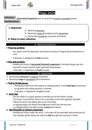
1. Floppy Infant
Kareem Alnakeeb P a g e | 1
Neurology 2018
Generalized hypotonia due to insult during fetal or neonatal period.
A. Abnormal postures:
1- Frog leg position:
- lies supine with full abduction and external rotation of legs & flaccid extension of
arms
- It denotes → Hypotonia of limb muscles.
2-Rag doll position:
- Holding the infant in horizontal (Ventral) suspension→ the back hangs over the
examiner's hand, and the limbs and head hang loosely.
- It denotes → Hypotonia of trunk muscles.
B. movements:
Diminished resistance to passive movement
Abnormal range of peripheral joint movement
1- Pull to Sit Maneuver:
- Pulling the infant from the supine to sitting position → the head lags & continues
to lag when the sitting position is reached.
- It denotes → Hypotonia of neck muscles.
2- Scarf sign:
- Put the child in a supine position and hold one of the infant’s hands.
- Try to put it around the neck as far as possible around the opposite shoulder.
- Observe how far the elbow goes across the body.
- In normal infant, the elbow does not quite reach the mid sternum
- In a floppy infant, the elbow easily crosses the midline.
3- Slip-through on vertical suspension:
- Holding the infant under the arms → the legs will be extended; decreased tone of
the shoulder girdle allows the infant to slip through the examiner's hands.
Floppy Infant
Definition:
Manifestations:
I. Hypotonia:
A. Abnormal postures
B. Abnormal range of peripheral joint movement
C. Diminished resistance to passive movement
II. Delay in motor milestones
4. Floppy Infant
Kareem Alnakeeb P a g e | 4
Neurology 2018
Figure: Anatomical-clinical correlation illustrating differential diagnosis of hypotonia in infancy
5. Floppy Infant
Kareem Alnakeeb P a g e | 5
Neurology 2018
I. History :
Family history • Affected parents or Siblings
• Consanguinity
• Stillbirths
• Childhood deaths
Maternal disease • Diabetes
• Epilepsy
• Myotonic dystrophy
Pregnancy & delivery History
( Obstetric History )
• Drug or teratogen exposure
• Decreased fetal movements
• Abnormal presentation e.g. Breech delivery
• Amount of amniotic fluid (Polyhydramnios/
oligohydramnios)
• Apgar scores
• Resuscitation requirements
• Cord gases
Since delivery History
( Developmental & Dietetic
History )
• Respiratory effort
• Ability to feed
• Level of alertness
• Level of spontaneous activity
• Character of cry
II. Identification of hypotonia
• Holding the infant in horizontal suspension – the back hangs over the examiner's hand, and
the limbs and head hang loosely. (Rag doll position)
• Passive extension of the legs at the knees – no resistance is met.
• Pulling the infant from the supine to sitting position – the head lags and continues to lag
when the sitting position is reached. (Pull to Sit Maneuver)
• Holding the infant under the arms – the legs will be extended; decreased tone of the
shoulder girdle allows the infant to slip through the examiner's hands.
(Slip-through on vertical suspension)
Diagnosis
6. Floppy Infant
Kareem Alnakeeb P a g e | 6
Neurology 2018
III. Physical Examination
Central AHCs Peripheral Nerves NMJ Muscle
Muscle power Normal Weakness
(Generalized)
Weakness
(distal > proximal)
Weakness
(face, eyes, bulbar)
Weakness
(proximal > distal)
Deep tendon
reflex ( DTR )
Normal /↑ / absent Normal / / Absent
Fasciculations Absent Prominent Absent Absent Absent
Clues & Pitfalls :
1- Localize cause of hypotonia :
Central “ UMNL “
(brain/spinal cord)
Peripheral “ LMNL “
(AHCs, peripheral nerve, NMJ, muscle)
Muscle mass • Normal bulk • Decreased bulk “muscle atrophy”
Muscle power • Normal/mild weakness “floppy
strong“
• Predominant axial weakness
• Weakness is uncommon
Except in the acute stages
• Marked weakness “floppy weak “
• Weak antigravitational limb muscles
Deep tendon reflex
( DTR )
• normal/increased reflexes • decreased reflexes
Plantar reflex • Plantar Extension “ + ve Babinski “ • Plantar Flexion
Dysmorphic features • Dysmorphisms • No dysmorphisms
CNS features • Encephalopathy, microcephaly
• Early onset seizures
• Depressed level of consciousness
• Global developmental delay
• Sustained Ankle clonus
• Scissoring on vertical suspension
• Awake, Alert
• Low-pitched weak cry
• Preserved social interaction
• Selective motor delay
• Paradoxical chest movements
• Tongue fasciculations
2- Clues to specific diagnosis :
Clues Diagnosis
Hepatosplenomegaly Storage disorders
Congenital infections
Renal cysts, high forehead, wide fontanelles Zellweger’s syndrome “cerebro-hepato-renal”
Hepatomegaly, retinitis pigmentosa Neonatal adrenoleukodystrophy
Abnormal odour metabolic disorders
Hypopigmentation, undescended testes Prader Willi
Examination of the mother Congenital myotonic dystrophy
Myasthenia gravis
7. Floppy Infant
Kareem Alnakeeb P a g e | 7
Neurology 2018
IV. Investigations:
By History and examination
► Hypotonia + a degree of strength: Central cause is most likely
► Hypotonia + weakness: Peripheral cause is possible
Central Causes Peripheral causes
o Neuroimaging
Ultrasound scan in the first instance
MRI for structural abnormality
EEG: if seizures suspected
o Genetics review
(if any dysmorphic features present)
o Karyotype (if any dysmorphic features present)
o DNA methylation studies or FISH
for Prader-Willi syndrome
(if clinically indicated after a genetics review)
o TORCH screen
o Metabolic work up
o Early review by the neurology service
o Molecular genetics – CTG repeats, deletions
in SMN gene
o Nerve conduction studies
o Creatine kinase: If elevated in an early
sample, repeat after a few days.
o Muscle biopsy
-Depending on clinical situation
-May be delayed until around 6 months of age
as neonatal results are difficult to interpret
1. Atonic Cerebral Palsy
2. Infantile Botulism
3. Werdnig-Hoffman disease (SMA I)
4. Myasthenia gravis
5. Metabolic Myopathies
Common causes of floppy infant
8. Floppy Infant
Kareem Alnakeeb P a g e | 8
Neurology 2018
1. Atonic cerebral palsy
- Mostly atonic diplegia
- Many are mentally retarded
- Near normal strength of upper limbs
- Hypotonia
- Brisk reflexes
Associated with:
• lethargy, lack of alertness, poor feeding
• Mental retardation
• poor Moro’s reflex, and seizures during the neonatal period.
2. Infantile Botulism
- Acute onset descending weakness,
- cranial neuropathies, ptosis,
- unreactive pupils, dysphagia
- Isolation of organism from stool
- Presence of toxin in the stool
C/P:
Investigations:
Forster sign:
Vertical suspension while held under the arm → flex legs at the hip.
9. Floppy Infant
Kareem Alnakeeb P a g e | 9
Neurology 2018
3. Spinal Muscle Atrophy type 1 (SMA 1)
“Werdnig-Hoffman disease”
•AR trait • AD or XR in few cases
- Degenerative disease of motor units beginning in the fetus and progressing into
infancy; denervation of muscle and atrophy
-Deletion in SMN gene on chromosome 5q13 degeneration of AHCs in spinal cord &
motor nuclei in the lower brainstem progressive muscle weakness and atrophy.
* SMA 1 presents in early infancy with:
1) Thin muscle mass, Progressive hypotonia, generalized weakness;
Infant is flaccid, has little movement & poor head control, No independent sitting
2) Absent DTRs
3) Fasciculations of the tongue (13)
4) Contractures in 10-20%.
5) Feeding difficulty: Child is alert, poor feeding and cry
6) Respiratory insufficiency
7) Normal Intelligence, cognition & facial expressions; Typically appear brighter than
others of same age
Molecular genetics: deletion in SMN gene → Simplest, most effective diagnosis
EMG—fibrillation potential and other signs of denervation
Muscle biopsy: shows neurogenic type of atrophy “perinatal denervation”
Death occurs by 2-4 years of age
AGE OF ONSET MODE OF
INHERITANCE
C/P PROGRESSION
SMA TYPE I Before birth to 6 months AR - Generalized muscle
weakness
- No independent sitting
- Progress rapidly
- Death usually by
age 2
SMA TYPE II 6 to 18 months AR - muscle weakness
- BUT many sit
independently
- Variable
- Most survive to
2nd
or 3rd
decade
SMA TYPE III Childhood to
adolescence
AR - Some muscle weakness
- BUT most ambulate
independently
- Slow Progression
- Normal lifespan
Definition:
Pathology:
C/P:
Investigations:
Prognosis:
10. Floppy Infant
Kareem Alnakeeb P a g e | 1 0
Neurology 2018
4. Myasthenia gravis
- It is an autoimmune disorder.
- Commonest cause is antibody to post synaptic acetylcholine receptors
Clinical feature frequency
Feeding/swallowing difficulties 100 %
Generalized weakness 70 %
Respiratory difficulty 66 %
Weak cry / facial weakness 60 %
Mechanical ventilation 30 %
Eye movements abnormalities 10 %
Reflexes Present
Arthrogryposis, pulmonary hypoplasia Rare
1) Congenital 2) Transient neonatal
- Not autoimmune
- It may be familial (AR) with reduced
number of Ach receptors
- Rare
- Familial disorder is permanent.
- In infant born to myasthenic mother
(10-20% affected mothers)
- Appearing within few hours: 3 days after
birth.
- After antibodies wane, they are normal and
have no risk for disease
* Characterized by:
- Generalized muscle weakness
- marked hypotonia
- poor feeding
- pooling of oral secretions
- feeble cry
Treatable (Cholinesterase inhibitors) Good response to Cholinesterase inhibitors
C/P:
Types
11. Floppy Infant
Kareem Alnakeeb P a g e | 1 1
Neurology 2018
5. Metabolic Myopathies
Mitochondrial - Biopsy Shows: Ragged red fibers
- lactic acidosis
- HSM
- systemic symptoms
Glycogen storage - Liver disease
- GSD type II “Pompe disease” or “acid maltase deficiency”;
1. profound hypotonia with progressive muscle weakness
2. Cardiomegaly
3. Macroglossia “large Tongue “
Lipid Metabolism - Hypoglycemia with low ketones
- Coma
- high NH3
• Regular physiotherapy → prevent contractures.
• Occupational therapy →facilitate daily activities .
• Annual orthopedic review is required to monitor for scoliosis and to exclude hip
dislocation/subluxation.
• Vigorous treatment of respiratory infections is indicated.
• Annual flu vaccination is necessary.
• Feeding intervention by nasogastric tube or gastrostomy For the undernourished child.
• Maintenance of ideal weight as excessive weight gain will exacerbate existing weakness.
• Children with neuromuscular disorders deserve special attention when it comes to anesthesia;
The anesthetist should be forewarned about the possibility of an underlying muscle disease
( even if the child has very mild or non-existing symptoms)
Muscle relaxants should only be used if essential
because of their more profound and prolonged effect in myopathic children.
• A family history of muscle disease or mild hyperkalemia may be of importance.
MANAGEMENT
PRINCIPLES OF MANAGEMENT:
12. Floppy Infant
Kareem Alnakeeb P a g e | 1 2
Neurology 2018
( Mainly supportive , physiotherapy )
• They require a multidisciplinary team approach with the involvement of several specialties
including: ”pediatrics, neurology, genetics, orthopedics, physiotherapy, and occupational therapy “
• Physiotherapy is mainly preventative to avoid contractures and wasting
(But will NOT increase muscle tone).
• Counseling the families about potentially preventable disorders is very important
• Consanguinity needs to be strongly discouraged to prevent inherited causes in our region.
Treatment:
Question
During the health supervision visit for a 6-week-old boy, his father expresses concern that his son
“doesn’t look like” his other children.
Growth parameters are normal except for a head circumference of 35.5 cm (<5th percentile).
On PE, you note that the infant does not appear to fixate or track your face visually. There is a “slip
through” on vertical suspension and “draping over” on horizontal suspension. DTRs are brisk. Moro
reflex is present and brisk.
* Of the following, the MOST likely cause of this infants hypotonia is:
a) AHC disease
b) Congenital brain malformation
c) Congenital myasthenic syndrome
d) Congenital myopathy
e) Spinal cord disease
Answer
B. Congenital brain malformation
- Hypotonia +
- Localize! UMN vs. LMN signs
- Take into account growth parameters, especially head circumference & features such as
tracking
* Regarding other choices:
A. anterior horn cell disease – wouldn't cause microcephaly or increased reflexes
C. Congenital myasthenic syndrome – wouldn't cause microcephaly or brisk reflexes
D. Congenital myopathy – no microcephaly or poor visual tracking
E. Spinal cord disease – wouldn't cause microcephaly or poor visual tracking