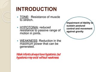
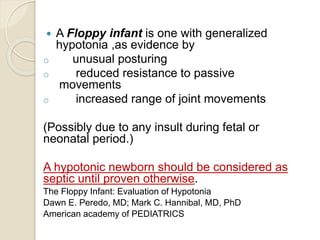
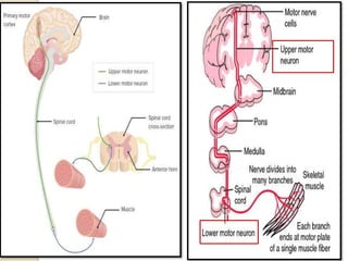
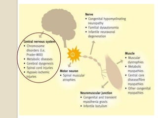
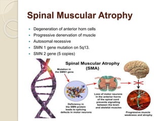
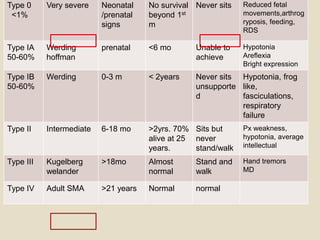
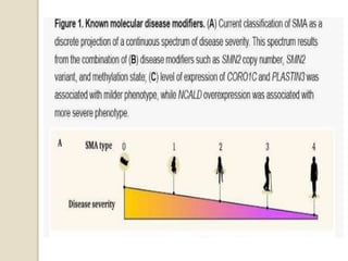
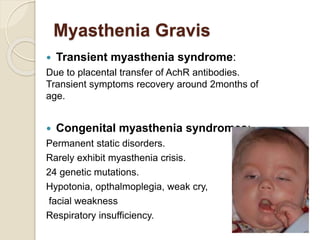
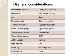
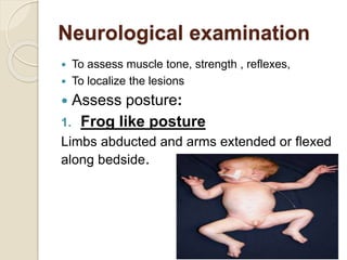
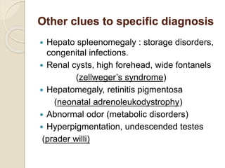
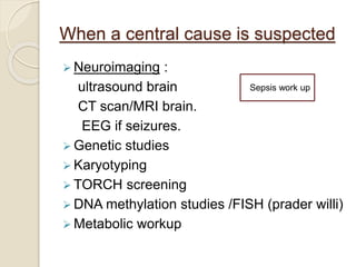
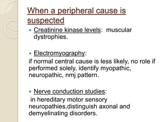
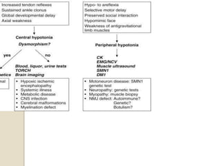
Dr HUDAZAHEER
2NDYEARRESIDENT PEDIATRICS
GMMC SUKKUR
Point of Discussion
 Introduction.
 Etiology.
 Clinical approach.
 Appreciating lesion site.
 Investigations.
 Management options.
INTRODUCTION
 TONE: Resistance of muscle
to stretch.
 HYPOTONIA: reduced
resistance to passive range of
motion in joints.
 WEAKNESS: Reduction in the
maximum power that can be
generated.
Weakinfantsalwayshavehypotonia,but
hypotoniamayexistwithoutweakness.
Impairment of Ability to
sustain postural
control and movement
against gravity
 A Floppy infant is one with generalized
hypotonia ,as evidence by
o unusual posturing
o reduced resistance to passive
movements
o increased range of joint movements
(Possibly due to any insult during fetal or
neonatal period.)
A hypotonic newborn should be considered as
septic until proven otherwise.
The Floppy Infant: Evaluation of Hypotonia
Dawn E. Peredo, MD; Mark C. Hannibal, MD, PhD
American academy of PEDIATRICS
Etiology
Central or
Peripheral
Floppy week
Floppy strong
Hypotonia in infants and young children:An etiological analysisAmirsalari S1,
Kavehmanesh Z2, Khalili Matinzadeh Z3, Afsharpayman S4,Torkaman M5,
Javadipour M6, Kakoie Shourkaie J7, Ghazavi Y8
https://pjms.com.pk/issues/octdec108/abstract/article20.html
Central hypotonia
 Almost 60% cases are of cerebral
hypotonia. Including
 Cerebral insult, HIE, intracranial
hemorrhages.
 peroxisomal disorders,( zellweger
syndrome),neonatal adrenoleukodystrophy,
 Metabolic disorders/ occulocerebro renal
syndrome(Lowe syndrome)
 Benign congenital hypotonia.
Benign Congenital Hypotonia
 Non progressive hypotonia of unknown
origin.
 Not associated with weakness, delayed
motor milestones or facial nerve palsies.
 Diagnosis is of exclusion.
 Prognosis is good.
 Physiotherapy might help.
Congenital Myopathies
Rare inherited group including
1. Myotubular myopathy (myotubularin protein
loss) fetal movements decrease, pharyngeal
weakness, inability to swallow and
polyhydromnios.
2. Nemaline rod myopathy (most common, ranging
from fetal akinesia to mildly affected adults)
3. Congenital fiber type disproportion
4. Core myopathies
WHAT IS DIFFERENCE BETWEEN
MYOPATHY AND DYSTROPHY?
Muscular Dystrophies
 Myotonic muscular dystrophy type 1
 Fascioscapulohemoral
 Congenital muscular dystrophies
Spinal Muscular Atrophy
 Degeneration of anterior horn cells
 Progressive denervation of muscle
 Autosomal recessive
 SMN 1 gene mutation on 5q13.
 SMN 2 gene (5 copies)
Type 0
<1%
Very severe Neonatal
/prenatal
signs
No survival
beyond 1st
m
Never sits Reduced fetal
movements,arthrog
ryposis, feeding,
RDS
Type IA
50-60%
Werding
hoffman
prenatal <6 mo Unable to
achieve
Hypotonia
Areflexia
Bright expression
Type IB
50-60%
Werding 0-3 m < 2years Never sits
unsupporte
d
Hypotonia, frog
like,
fasciculations,
respiratory
failure
Type II Intermediate 6-18 mo >2yrs. 70%
alive at 25
years.
Sits but
never
stand/walk
Px weakness,
hypotonia, average
intellectual
Type III Kugelberg
welander
>18mo Almost
normal
Stand and
walk
Hand tremors
MD
Type IV Adult SMA >21 years Normal normal
Myasthenia Gravis
 Transient myasthenia syndrome:
Due to placental transfer of AchR antibodies.
Transient symptoms recovery around 2months of
age.
 Congenital myasthenia syndromes:
Permanent static disorders.
Rarely exhibit myasthenia crisis.
24 genetic mutations.
Hypotonia, opthalmoplegia, weak cry,
facial weakness
Respiratory insufficiency.
Infant botulism
 2-6m
 Exposure to soil-house
 dust-honey ,or corn syrup ,germination of
spores in GIT.
 Start in bulbar nerves then trunk & limbs
 ( almost 40 % cases of botulism due to
honey)
Clinical approach towards the
case of hypotonia/ floppy
infant
 History
 Physical examination
 Indetifying lesion from clues
 Relevant investigations
 Management
History :
 Presenting:
 Decreased tone
 Weak cry
 Difficulty sucking/chewing
 Respiratory difficulty
 Delayed milestones
Ask for
• Age onset,
• Sudden onset
• Progressive static or fluctuating
• Pattern proximal or distal
• Seizures, poor state of alertness
• History of constipation
Indicators of
weakness:
• Inability to cough
• Poor swallowing
• Week cry
• Paradoxical
breathing
 Birth history:
Fetal movements, poly or oligohydromnios, quality
of it, breech presentation, maternal drug
effects(sedatives). Trauma, birth anoxia, Apgar
score, honey usage after birth. Post natal feeding,
alertness, respiratory.
 Developmental history:
delayed or regressed.
 Family history:
History of neuromuscular diseases any similar
presentations in family. (SMA, congenial
dystrophies, metabolic diseases.)
Repeated miscarriages (chromosomal)
Family pedigree.
Examination
 General considerations:
Dysmorphic
features
Dysmorphic features Down, PWS,lipidosis
Head size Micro/macrocephaly
Alert SMA
Expressionless Some myopathies
Atrophy of muscles Myopathy/dystrophy
Ptosis, othalmoplegia Myasthenia gravis
fish /triangular mouth myopathies
Tongue fasciculations SMA
Arhtrogryposis multiple
High pitched CNS
Weak cry diaphragmatic
Fatigable cry CMS
Neurological examination
 To assess muscle tone, strength , reflexes,
 To localize the lesions
 Assess posture:
1. Frog like posture
Limbs abducted and arms extended or flexed
along bedside.
2. Pull to sit or traction:
Child respond by being able to raise head
along with body.
In Hypotonic babies significant head lag
Not present in premature /less than 33 weeks.
3. Vertical suspension:
Hold at axilla, without grasping thorax, lifts straight up,
Normal response: head erect in midline with flexion at
knees, hip and ankle joints.
But in hypotonic infants head falls forward, legs
dangle, and the infant may slip through the examiners
hand.
4. Horizontal suspension:
Baby suspends in prone position with the
examiner’s palm underneath the chest.
Normally keeps head erect, maintain back
straight, flexes limbs.
In hypotonic, head and legs hang limply, rag
doll.
5. Prone:
unable to lift head and back
6. Scarf sign:
Taking baby’s hand and pulling the hand to the
opposite shoulder like a scarf. In hypotonic
infants the elbow can go beyond the midline.
IDENTIFYING CENTRAL
VERSUS PERIPHERAL
CAUSES?
site weakness Proximal/dista
l
DTR
central limbs Equal Present /Inc.
Anterior horn
cells
Limbs Proximal Absent
Peripheral
nerve
limbs Distal Absent
NMJ Face/limbs Proximal Normal/Dec
Muscle Face/limbs Proximal Decreased.
• Proximal weakness, tongue fasciculation = SMA
• Distal, sensory loss = peripheral
neuropathy
• Facial and occulomotor = myasthenia
• Proximal , poor muscle bulk, diminished reflexes
=congenital myopathies
Other clues to specific diagnosis
 Hepato spleenomegaly : storage disorders,
congenital infections.
 Renal cysts, high forehead, wide fontanels
(zellweger’s syndrome)
 Hepatomegaly, retinitis pigmentosa
(neonatal adrenoleukodystrophy)
 Abnormal odor (metabolic disorders)
 Hyperpigmentation, undescended testes
(prader willi)
Investigations?
When a central cause is suspected
 Neuroimaging :
ultrasound brain
CT scan/MRI brain.
EEG if seizures.
 Genetic studies
 Karyotyping
 TORCH screening
 DNA methylation studies /FISH (prader willi)
 Metabolic workup
Sepsis work up
When a peripheral cause is
suspected
 Creatinine kinase levels: muscular
dystrophies.
 Electromyography:
if normal central cause is less likely, no role if
performed solely, identify myopathic,
neuropathic, nmj pattern.
 Nerve conduction studies:
in hereditary motor sensory
neuropathies,distinguish axonal and
demyelinating disorders.
 Muscle biopsy: investigation of choice in
congenital myopathy(even a normal CK
doesn’t exclude it).
 Nerve biopsy: sural nerve
 Tensilon test: neostgmine 0.04mg/kg is
used. Can be repeated after 4 hrs.
 PCR/MLPA quickly detect SMN gene copies.
MANAGEMENT?
 Intubation and mechanical ventilation.
 Occupational therapy
 Prevention and correction of scoliosis
 Feeding nasogastric, gastrostomy.
 Orthopedic intervention
 Preventing infections.
 Physiotherapy.
 Counseling
 Gene therapy.
SMA
valproate, gabapentin,
riluzole can slow progression.
87 lacs.
Learning points
 Learn to assess hypotonia via proper man
oeuvres is must for a practicing physician.
 To distinguish central and peripheral causes
is must.
 Hypotonia with weakness suggests lower
motor neuron lesion.
 Muscle enzymes are rarely helpful.
 When getting clue of multi system
involvement must consider inborn errors.
 Benign hypotonia should be used
cautiously after exclusion.
References
 Nelson essential pediatrics 21st edition.
 Ghai essentials pediatric 6th edition.
 Examination pediatrics Wayne harries.
 https://pjms.com.pk/issues/octdec108/article/article20.html
 https://www.ncbi.nlm.nih.gov/books/NBK562209/
 FLOPPY INFANT SYNDROME: OVERVIEW
Jasper Kauri, Sonu Punia*.
Assistant Professor, Department of Physiotherapy, Guru
Jambheshwar University of Science and
Technology, Hisar, Haryana, India.
 Clinical approach to a floppy infant
Dimuthu Saraji Wijesekara1
https://pdfs.semanticscholar.org/6091/d646d7e1d4114c7e
101f633621689532fac7.pd
Any comments /questions?

floppy infant.pptx

  • 1.
  • 2.
    Point of Discussion Introduction.  Etiology.  Clinical approach.  Appreciating lesion site.  Investigations.  Management options.
  • 3.
    INTRODUCTION  TONE: Resistanceof muscle to stretch.  HYPOTONIA: reduced resistance to passive range of motion in joints.  WEAKNESS: Reduction in the maximum power that can be generated. Weakinfantsalwayshavehypotonia,but hypotoniamayexistwithoutweakness. Impairment of Ability to sustain postural control and movement against gravity
  • 4.
     A Floppyinfant is one with generalized hypotonia ,as evidence by o unusual posturing o reduced resistance to passive movements o increased range of joint movements (Possibly due to any insult during fetal or neonatal period.) A hypotonic newborn should be considered as septic until proven otherwise. The Floppy Infant: Evaluation of Hypotonia Dawn E. Peredo, MD; Mark C. Hannibal, MD, PhD American academy of PEDIATRICS
  • 5.
  • 8.
    Hypotonia in infantsand young children:An etiological analysisAmirsalari S1, Kavehmanesh Z2, Khalili Matinzadeh Z3, Afsharpayman S4,Torkaman M5, Javadipour M6, Kakoie Shourkaie J7, Ghazavi Y8 https://pjms.com.pk/issues/octdec108/abstract/article20.html
  • 9.
    Central hypotonia  Almost60% cases are of cerebral hypotonia. Including  Cerebral insult, HIE, intracranial hemorrhages.  peroxisomal disorders,( zellweger syndrome),neonatal adrenoleukodystrophy,  Metabolic disorders/ occulocerebro renal syndrome(Lowe syndrome)  Benign congenital hypotonia.
  • 10.
    Benign Congenital Hypotonia Non progressive hypotonia of unknown origin.  Not associated with weakness, delayed motor milestones or facial nerve palsies.  Diagnosis is of exclusion.  Prognosis is good.  Physiotherapy might help.
  • 11.
    Congenital Myopathies Rare inheritedgroup including 1. Myotubular myopathy (myotubularin protein loss) fetal movements decrease, pharyngeal weakness, inability to swallow and polyhydromnios. 2. Nemaline rod myopathy (most common, ranging from fetal akinesia to mildly affected adults) 3. Congenital fiber type disproportion 4. Core myopathies
  • 12.
    WHAT IS DIFFERENCEBETWEEN MYOPATHY AND DYSTROPHY?
  • 13.
    Muscular Dystrophies  Myotonicmuscular dystrophy type 1  Fascioscapulohemoral  Congenital muscular dystrophies
  • 14.
    Spinal Muscular Atrophy Degeneration of anterior horn cells  Progressive denervation of muscle  Autosomal recessive  SMN 1 gene mutation on 5q13.  SMN 2 gene (5 copies)
  • 15.
    Type 0 <1% Very severeNeonatal /prenatal signs No survival beyond 1st m Never sits Reduced fetal movements,arthrog ryposis, feeding, RDS Type IA 50-60% Werding hoffman prenatal <6 mo Unable to achieve Hypotonia Areflexia Bright expression Type IB 50-60% Werding 0-3 m < 2years Never sits unsupporte d Hypotonia, frog like, fasciculations, respiratory failure Type II Intermediate 6-18 mo >2yrs. 70% alive at 25 years. Sits but never stand/walk Px weakness, hypotonia, average intellectual Type III Kugelberg welander >18mo Almost normal Stand and walk Hand tremors MD Type IV Adult SMA >21 years Normal normal
  • 17.
    Myasthenia Gravis  Transientmyasthenia syndrome: Due to placental transfer of AchR antibodies. Transient symptoms recovery around 2months of age.  Congenital myasthenia syndromes: Permanent static disorders. Rarely exhibit myasthenia crisis. 24 genetic mutations. Hypotonia, opthalmoplegia, weak cry, facial weakness Respiratory insufficiency.
  • 18.
    Infant botulism  2-6m Exposure to soil-house  dust-honey ,or corn syrup ,germination of spores in GIT.  Start in bulbar nerves then trunk & limbs  ( almost 40 % cases of botulism due to honey)
  • 19.
    Clinical approach towardsthe case of hypotonia/ floppy infant  History  Physical examination  Indetifying lesion from clues  Relevant investigations  Management
  • 20.
    History :  Presenting: Decreased tone  Weak cry  Difficulty sucking/chewing  Respiratory difficulty  Delayed milestones Ask for • Age onset, • Sudden onset • Progressive static or fluctuating • Pattern proximal or distal • Seizures, poor state of alertness • History of constipation Indicators of weakness: • Inability to cough • Poor swallowing • Week cry • Paradoxical breathing
  • 21.
     Birth history: Fetalmovements, poly or oligohydromnios, quality of it, breech presentation, maternal drug effects(sedatives). Trauma, birth anoxia, Apgar score, honey usage after birth. Post natal feeding, alertness, respiratory.  Developmental history: delayed or regressed.  Family history: History of neuromuscular diseases any similar presentations in family. (SMA, congenial dystrophies, metabolic diseases.) Repeated miscarriages (chromosomal) Family pedigree.
  • 22.
  • 23.
     General considerations: Dysmorphic features Dysmorphicfeatures Down, PWS,lipidosis Head size Micro/macrocephaly Alert SMA Expressionless Some myopathies Atrophy of muscles Myopathy/dystrophy Ptosis, othalmoplegia Myasthenia gravis fish /triangular mouth myopathies Tongue fasciculations SMA Arhtrogryposis multiple High pitched CNS Weak cry diaphragmatic Fatigable cry CMS
  • 25.
    Neurological examination  Toassess muscle tone, strength , reflexes,  To localize the lesions  Assess posture: 1. Frog like posture Limbs abducted and arms extended or flexed along bedside.
  • 26.
    2. Pull tosit or traction: Child respond by being able to raise head along with body. In Hypotonic babies significant head lag Not present in premature /less than 33 weeks.
  • 27.
    3. Vertical suspension: Holdat axilla, without grasping thorax, lifts straight up, Normal response: head erect in midline with flexion at knees, hip and ankle joints. But in hypotonic infants head falls forward, legs dangle, and the infant may slip through the examiners hand.
  • 28.
    4. Horizontal suspension: Babysuspends in prone position with the examiner’s palm underneath the chest. Normally keeps head erect, maintain back straight, flexes limbs. In hypotonic, head and legs hang limply, rag doll.
  • 29.
    5. Prone: unable tolift head and back 6. Scarf sign: Taking baby’s hand and pulling the hand to the opposite shoulder like a scarf. In hypotonic infants the elbow can go beyond the midline.
  • 30.
  • 33.
    site weakness Proximal/dista l DTR centrallimbs Equal Present /Inc. Anterior horn cells Limbs Proximal Absent Peripheral nerve limbs Distal Absent NMJ Face/limbs Proximal Normal/Dec Muscle Face/limbs Proximal Decreased. • Proximal weakness, tongue fasciculation = SMA • Distal, sensory loss = peripheral neuropathy • Facial and occulomotor = myasthenia • Proximal , poor muscle bulk, diminished reflexes =congenital myopathies
  • 34.
    Other clues tospecific diagnosis  Hepato spleenomegaly : storage disorders, congenital infections.  Renal cysts, high forehead, wide fontanels (zellweger’s syndrome)  Hepatomegaly, retinitis pigmentosa (neonatal adrenoleukodystrophy)  Abnormal odor (metabolic disorders)  Hyperpigmentation, undescended testes (prader willi)
  • 35.
  • 36.
    When a centralcause is suspected  Neuroimaging : ultrasound brain CT scan/MRI brain. EEG if seizures.  Genetic studies  Karyotyping  TORCH screening  DNA methylation studies /FISH (prader willi)  Metabolic workup Sepsis work up
  • 37.
    When a peripheralcause is suspected  Creatinine kinase levels: muscular dystrophies.  Electromyography: if normal central cause is less likely, no role if performed solely, identify myopathic, neuropathic, nmj pattern.  Nerve conduction studies: in hereditary motor sensory neuropathies,distinguish axonal and demyelinating disorders.
  • 38.
     Muscle biopsy:investigation of choice in congenital myopathy(even a normal CK doesn’t exclude it).  Nerve biopsy: sural nerve  Tensilon test: neostgmine 0.04mg/kg is used. Can be repeated after 4 hrs.  PCR/MLPA quickly detect SMN gene copies.
  • 39.
  • 40.
     Intubation andmechanical ventilation.  Occupational therapy  Prevention and correction of scoliosis  Feeding nasogastric, gastrostomy.  Orthopedic intervention  Preventing infections.  Physiotherapy.  Counseling  Gene therapy.
  • 41.
    SMA valproate, gabapentin, riluzole canslow progression. 87 lacs.
  • 43.
    Learning points  Learnto assess hypotonia via proper man oeuvres is must for a practicing physician.  To distinguish central and peripheral causes is must.  Hypotonia with weakness suggests lower motor neuron lesion.  Muscle enzymes are rarely helpful.  When getting clue of multi system involvement must consider inborn errors.  Benign hypotonia should be used cautiously after exclusion.
  • 44.
    References  Nelson essentialpediatrics 21st edition.  Ghai essentials pediatric 6th edition.  Examination pediatrics Wayne harries.  https://pjms.com.pk/issues/octdec108/article/article20.html  https://www.ncbi.nlm.nih.gov/books/NBK562209/  FLOPPY INFANT SYNDROME: OVERVIEW Jasper Kauri, Sonu Punia*. Assistant Professor, Department of Physiotherapy, Guru Jambheshwar University of Science and Technology, Hisar, Haryana, India.  Clinical approach to a floppy infant Dimuthu Saraji Wijesekara1 https://pdfs.semanticscholar.org/6091/d646d7e1d4114c7e 101f633621689532fac7.pd
  • 45.