Diagnostic approach of the adult with chest pain; Cardiology 2020
•
1 like•73 views

A "Diagnostic approach of the adult with chest pain" was made in 2020. It is related to Cardiovascular Medicine.

Recommended
More Related Content
What's hot
What's hot (20)
Similar to Diagnostic approach of the adult with chest pain; Cardiology 2020
Similar to Diagnostic approach of the adult with chest pain; Cardiology 2020 (20)
More from Kareem Alnakeeb
More from Kareem Alnakeeb (20)
Recently uploaded
Recently uploaded (20)
Diagnostic approach of the adult with chest pain; Cardiology 2020
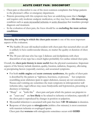
- 1. ACUTE CHEST PAIN / DISCOMFORT ̵ Chest pain or discomfort is one of the most common complaints that brings patients to the physician’s office or emergency department. ̵ Patients presenting with this symptom may have an underlying cause that is benign and requires only moderate analgesic medication, or they may have a life-threatening condition such as acute myocardial ischemia or aortic dissection that mandates prompt diagnosis and treatment. ̵ In the evaluation of chest pain, the focus should be on excluding the more serious conditions. History Assessing the setting in which the chest pain occurs is one of the most important aspects of the evaluation. The healthy 26-year-old medical resident with chest pain that occurred after on-call is unlikely to have cardiovascular disease, no matter the quality or duration of chest pain. The 58-year-old man who has type 2 diabetes and dyslipidemia with chest discomfort of any type has a much higher probability for cardiac-related chest pain. Overall, the chest pain history is more useful than the physical examination. Important aspects of the history include duration, quality, location, radiation, frequency, alleviating or precipitating factors (especially exercise), and associated symptoms. • For both stable angina and acute coronary syndromes, the quality of chest pain is described by the patient as “tightness, heaviness, or pressure,” but symptoms resembling acute abdomen (pain in upper abdomen, nausea) are not uncommon. Nausea and vomiting are sometimes the main symptoms in inferoposterior wall ischemia (also, vagal reflexes may cause bradycardia and hypotension, presenting as dizziness or fainting). • “Sharp” or “knife-like” chest pain and pain which the patient can pinpoint to an “exact area” are less likely to be related to ischemia or infarction, especially if the chest pain is reproduced by changes in position or palpation. • Myocardial infarction is associated with pain that lasts >20–30 minutes in duration. • Response of chest pain to nitroglycerin (within a few minutes) is most consistent with transient ischemia or esophageal spasm. Chest pain that worsens with nitroglycerin sometimes occurs with GERD
- 2. The response to nitroglycerin is not enough to confirm coronary disease as the cause of chest pain. • Acute coronary syndromes in women present with atypical symptoms: dyspnea, shortness of breath, fatigue. This may be due to the older age group in which myocardial ischemia and infarction occur in women. Physical Examination One of the most important parts in a chest pain examination is “initial impression.” Diaphoresis, tachypnea, and anxious expression should alert you to a potentially life- threatening process. Tachycardia and tachypnea are both nonspecific but occur in almost all cases of pulmonary embolism. • Check BP in both arms: a difference of >20 mm Hg systolic suggests aortic dissection and is present in ~70% of cases. • Hypotension may suggest massive pulmonary embolism or cardiac shock. • Fever may suggest pneumonia or mediastinitis (esophageal rupture) as the cause of chest pain. • Evidence of atherosclerosis (corneal lipid rings, narrowed retinal arteries, and pigment and hair changes in the legs) is commonly seen in patients with coronary syndromes. ̵ Inspect the chest wall for tender areas, respiratory motion, respiratory retractions, or accessory muscle use. If the tender area corresponds to the location of the patient’s pain and palpation exactly reproduces the pain, consider musculoskeletal chest pain as the cause of chest pain. ̵ Abnormal heart sounds and new murmurs are commonly found in certain chest pain syndromes. ➢ Wide physiologic splitting of the second heart sound (splitting wider with inspiration) can be found in right bundle branch block or in right ventricular infarction. ➢ New paradoxical splitting is most often due to left bundle branch block (LBBB), or anterior or lateral infarction. ➢ A new fourth heart sound can occur with angina or infarction. ➢ An S3 is more likely due to underlying heart failure. ➢ A new murmur may be significant: aortic regurgitation occurs in over half of patients with aortic dissection, while mitral regurgitation can occur in patients with angina or infarction and is due to papillary muscle dysfunction.
- 3. ̵ The lungs should be auscultated for crackles and asymmetrical breath sounds. Asymmetry of breath sounds may be found in patients with spontaneous pneumothorax. Absent lung sounds also may occur in pneumothorax and pleural effusions. ̵ The extremities should be examined for pulses, edema, calf tenderness, and signs of atherosclerotic vessel disease. ➢ Absence of pedal pulses may occur in aortic dissection. ➢ Any swelling of the legs, especially if unilateral, raises the odds of pulmonary embolism as the cause of chest pain. Testing All patients with chest pain should have a 12-lead electrocardiogram (ECG) since the ECG is the single most important test for the evaluation of the cause of chest pain. The ECG should be done immediately after initial stabilization and taking of vital signs. Most patients with myocardial infarction will have an abnormal initial ECG: • 50% with acute MI will have diagnostic findings (ST elevation or Q waves) • 35% will have findings consistent with ischemia (ST depression and/or T wave inversion) • In patients presenting with acute chest pain who have normal ECG, the chance of acute MI is much less than 10% (in some studies 1–2.6%). • An abnormal ECG can be seen in many non-cardiac conditions (pulmonary embolism, electrolyte abnormalities, aortic dissection). ̵ In interpreting the ECG, make every effort to obtain previous ECGs, so that abnormalities can be compared with those on the old tracing. Any ECG finding is assumed to be new unless proven otherwise by an old ECG (if one is available). Also, in patients with acute coronary syndromes, the ECG is the sole test required to select patients for emergency reperfusion. ̵ Serum cardiac biomarker determinations play a vital role in the evaluation of patients who present with acute chest pain and in the diagnosis of acute myocardial infarction. ̵ Serum markers such as aspartate transaminase, lactate dehydrogenase, and lactate dehydrogenase subforms no longer are used because they lack cardiac specificity and their delayed elevation precludes early diagnosis. ̵ Creatine kinase (CK) is found in striated muscle and tissues of the brain, kidney, lung, and GI tract. This widely available marker has low sensitivity and specificity for cardiac damage. Furthermore, CK levels may be elevated in a number of noncardiac
- 4. conditions, including trauma, seizures, renal insufficiency, hyperthermia, and hyperthyroidism. Currently, the CK marker largely has been replaced by cardiac troponins and CK-MB. CK-MB isoenzyme: CK-MB is cardiac specific and is useful for the early diagnosis of acute myocardial infarction. CK-MB typically is detectable in the serum 4–6 hours after the onset of ischemia, peaks in 12–24 hours, and normalizes in 2–3 days. Like the CK level, the peak CK-MB level does not predict infarct size; however, it can be used to detect early reinfarction. Serial CK-MB levels commonly are obtained at admission to the emergency department and are repeated in 6–12 hours. CK-MB subforms: CK-MB may be further characterized into subforms (or isoforms). CK-MB2 is found in myocardial tissue, and CK-MB1 is found in plasma. The CK-MB subform is not routinely used. Cardiac troponins: Troponins (T, I, C) are found in striated and cardiac muscle. Because the cardiac and skeletal muscle isoforms of troponin T and I differ, they are known as the “cardiac troponins.” They are the preferred markers for the diagnosis of myocardial injury. Troponin T and I generally have similar sensitivity and specificity for the detection of myocardial injury. Unlike troponin I levels, troponin T levels may be elevated in patients with renal disease, polymyositis, or dermatomyositis. The cardiac troponins typically are measured at emergency department admission and repeated in 6–12 hours. Patients with a normal CK-MB level but elevated troponin levels are considered to have sustained minor myocardial damage, or microinfarction, whereas patients with elevations of both CK-MB and troponins are considered to have had acute myocardial infarction. The cardiac troponins may remain elevated up to 2 weeks after symptom onset, which makes them useful as late markers of recent acute myocardial infarction. An elevated troponin T or I level is helpful in identifying patients at increased risk for death or the development of acute myocardial infarction. Increased risk is related to the high serum troponin levels. The troponins also can help identify low-risk patients who may be sent home with close follow-up.
- 5. Those with a normal or nearly normal ECG and a normal troponin I test 6 hours after admission had a very low risk of major cardiac events (0.3%) during the next 30 days. ̵ Myoglobin levels begin to rise as early as 1–4 hours after the onset of pain. Normal myoglobin at 4 hours has a very high negative predictive value. ̵ Chest x-ray should be obtained on patients with chest pain; it may show pneumothorax, pneumomediastinum (i.e., from esophageal rupture), pleural effusion, or infiltrates. Aortic dissection can cause widening of the mediastinum. Subtle findings such as loss of lung volume or unilateral decrease in vascular markings may suggest pulmonary embolism. ̵ Especially if a noncardiac diagnosis is suspected, arterial blood gases, BNP, and CT angiogram may be helpful for evaluating acute chest pain.
- 6. Causes of Chest Pain 1. Aortic Dissection ▪ Pain is sharp, tearing, and extremely severe; typically radiates to back; loss of pulses or aortic insufficiency often develop ▪ On chest x-ray, mediastinum is widened MI may occur if dissection extends into coronary artery ▪ Diagnosis confirmed by MRI, CT scan, or transesophageal echocardiogram 2. Pulmonary Embolism ▪ Dyspnea, tachycardia, and hypoxemia are prominent; pain is usually pleuritic, especially when pulmonary infarction develops. ▪ EKG is usually nonspecific but may show S wave in lead I, Q wave in lead III, or inverted T wave in lead III ▪ Diagnosis confirmed by CT angiogram 3. Pericarditis ▪ May be preceded by viral illness; pain is sharp, positional, pleuritic, and relieved by leaning forward. ▪ Pericardial rub often present ▪ Diffuse ST elevation occurs without evolution of Q waves ▪ CK level usually normal ▪ Responds to anti-inflammatory agents 4. Myocarditis ▪ May be preceded by viral illness; ▪ pain is generally vague and mild if present; ▪ total CK and MB fraction of CK (CK-MB) are often elevated; ▪ conduction abnormalities and Q waves may occur. 5. Musculoskeletal Disorders ▪ Most common cause of chest pain. ▪ Includes costochondritis, cervical osteoarthritis, radiculitis; ▪ pain is atypical, stabbing, localized, may be pleuritic; reproduced by motion or palpation; ▪ EKG changes absent. 6. GI Disorders ▪ Esophageal reflux is often made worse with recumbency or after meals, may be associated with regurgitation and relieved by antacids; episodes of spasm may be brought on by cold liquids, relieved by nitroglycerin, and may closely resemble angina or infarction; diagnosis may be confirmed by upper endoscopy or esophageal manometry.
- 7. ▪ Peptic ulcer disease, pancreatitis, and cholecystitis may occasionally mimic infarction; Abdominal tenderness is present, with radiation to back and elevated amylase in pancreatitis; Sonography can confirm cholecystitis. 7. Pneumothorax ▪ Onset abrupt with sharp pleuritic chest pain and dyspnea; ▪ breath sounds absent; ▪ chest x-ray confirms. 8. Pleuritis ▪ Pain is sharp and increases on inspiration; friction rub or dullness may be present; ▪ other respiratory symptoms and underlying pulmonary infection usually present.
- 8. Emergency department approach to chest pain ACS: acute coronary syndrome; ASA: aspirin; CXR: chest x-ray; ECG: electrocardiogram; HF: heart failure; PCI: percutaneous coronary intervention.