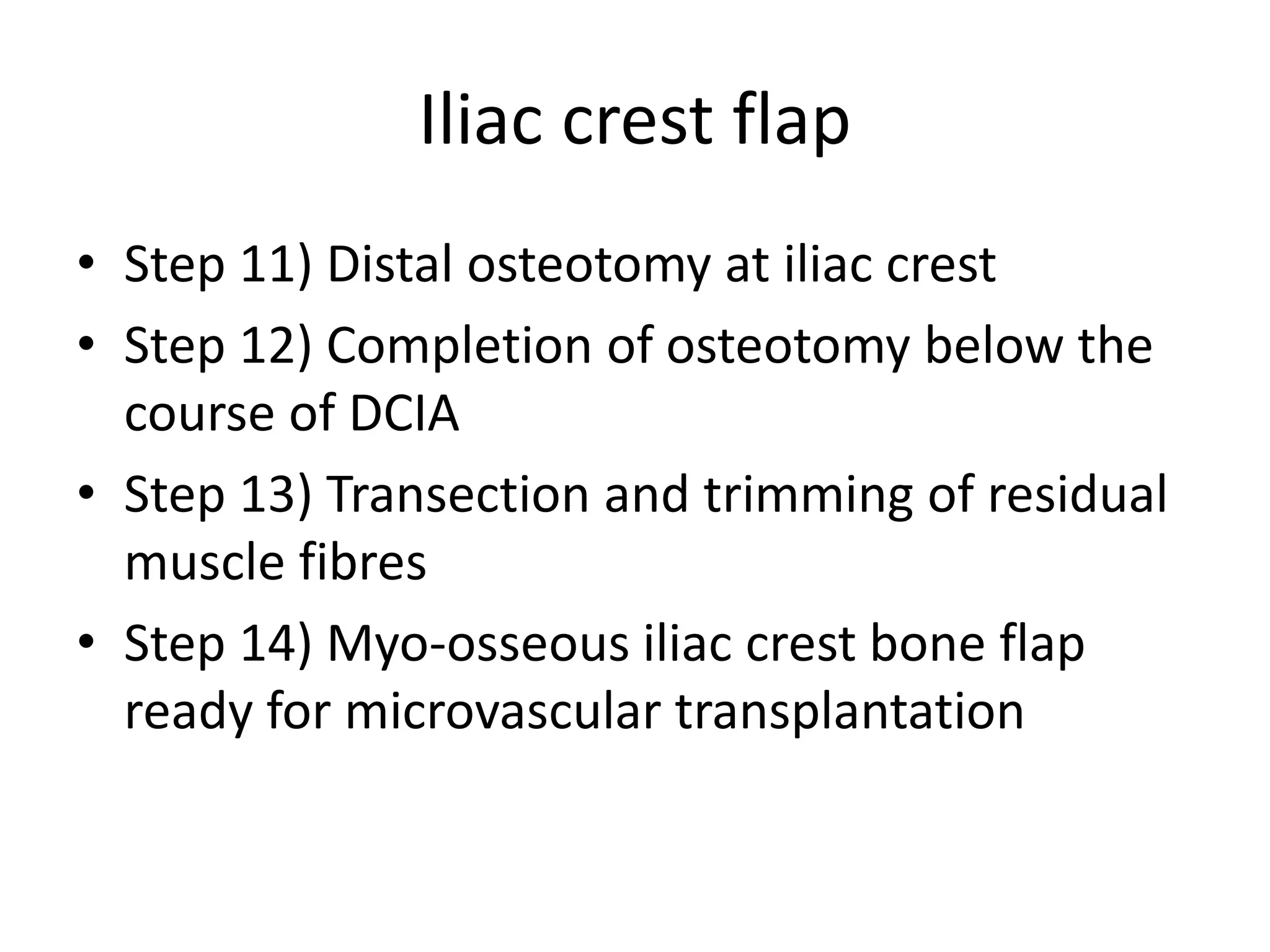
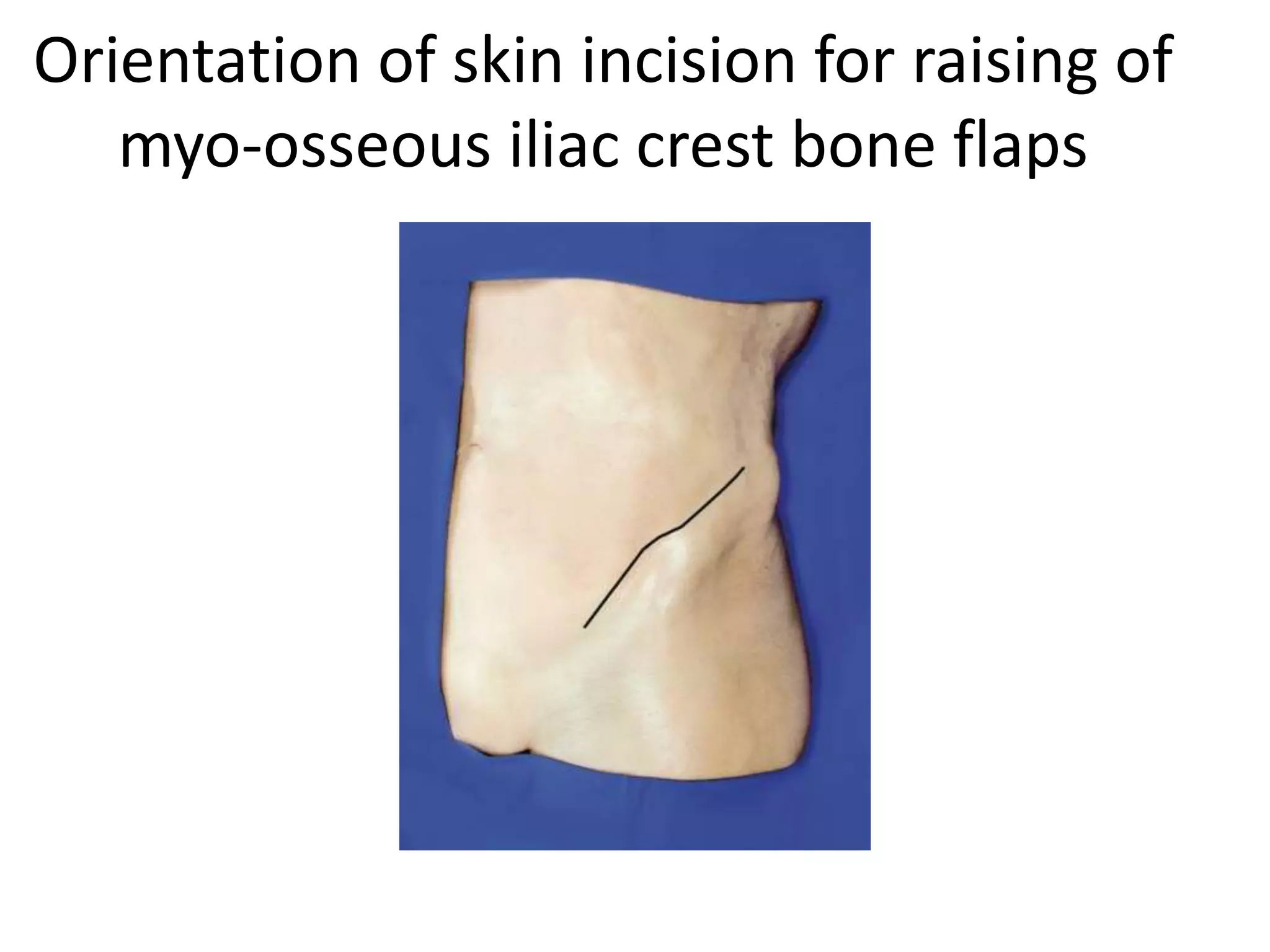
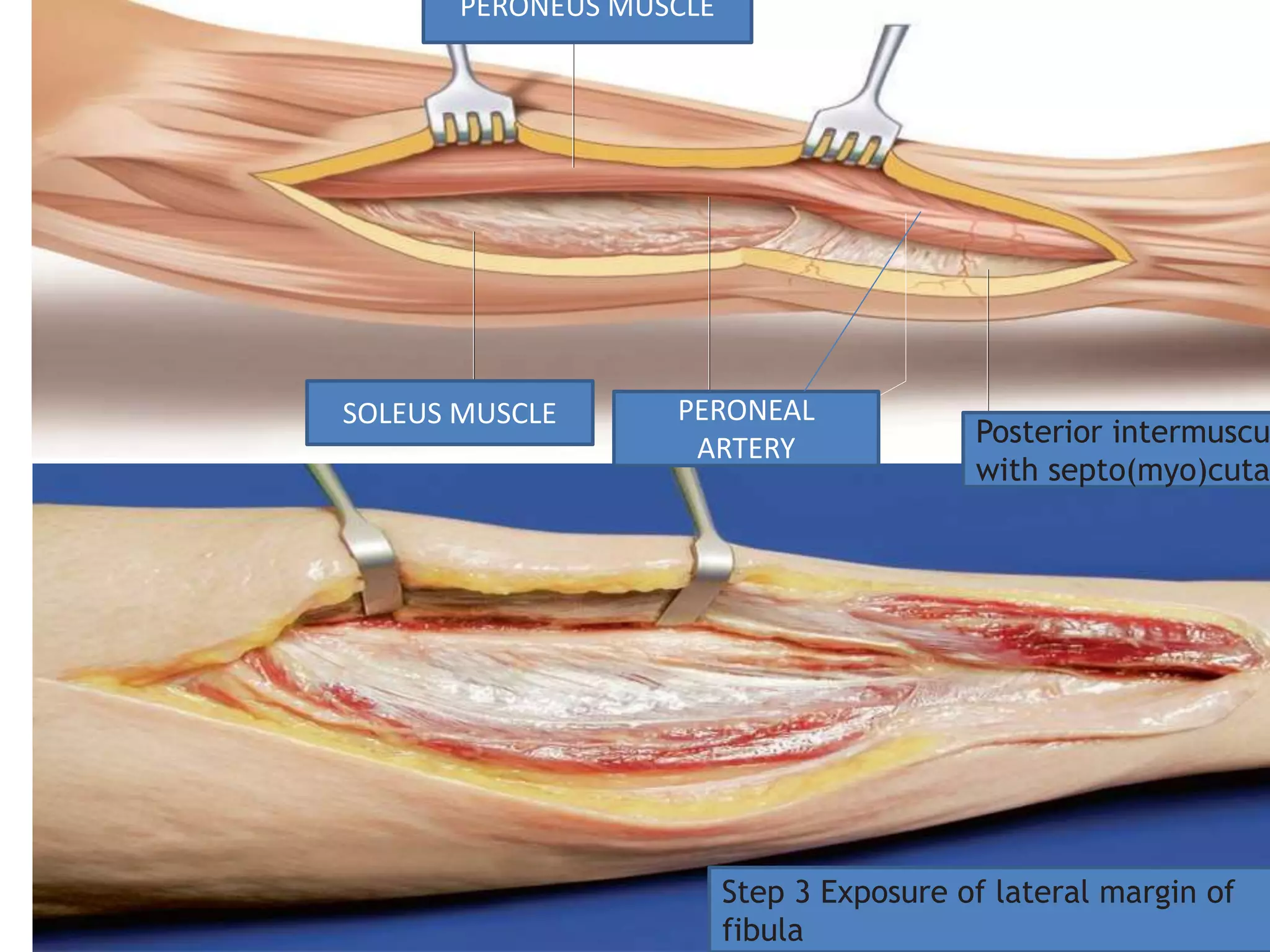
Flap techniques of DCIA,
FREE FIBULA AND RADIAL FOREARM
FREE FLAPS
Professor Dr Jameel Kifayatullah
Oral and Maxillofacial Surgeon
Khyber college of dentistry Peshawar
Iliac crest flap
• Step 1) Skin incision ,exposure of inguinal
ligament
• Step 2) identification of internal oblique
muscle
• Step 3) Exposure of vascular pedicle
• Step 4) Dissection of DCIA Towards the
anterior superior iliac spine
• Step 5)Skin incision along iliac crest
Iliac crest flap
• Step 6) Transection of Muscles at Lateral Rim
of iliac crest
• Step 7) Detachment of muscles from gluteal
surface of pelvis
• Step 8) Transection of oblique abdominal
muscles ,Exposure of iliacus muscle by
retracting the peritonium
• Step 9) Incision of iliacus muscle below DCIA
• Step 10) Transection of sartorius muscle at
ASIS
Iliac crest flap
• Step 11) Distal osteotomy at iliac crest
• Step 12) Completion of osteotomy below the
course of DCIA
• Step 13) Transection and trimming of residual
muscle fibres
• Step 14) Myo-osseous iliac crest bone flap
ready for microvascular transplantation
INGUINAL LIGAMAENT
Tuberculum pubicum Common femoral artery Profunda femoral
artery
ANTERIOR SUPERIOR
ILIAC SPINE
FEMORAL ARTERY
EXTERNAL ILIAC
ARTERY
DEEP CIRCUMFLEX
ILIAC ARTERY
ASCENDING
BRANCH
Orientation of skin incision for raising of
myo-osseous iliac crest bone flaps
Step 1 Skin incision, exposure of
inguinal ligament
Step 2 Identification of internal oblique muscle
Step 3 Exposure of vascular pedicle
Step 4 Dissection of DCIA towards the
anterior superior iliac spine (ASIS)
DEEP CIRCUMFL
ARTERY
LATERAL FEMORAL CUTANE
NERVE
ASCENDING BRANCH
ILIACUS MUSCLE
INTERNAL OBLIQUE
ABDOMINUS MUSCLE
EXTERNAL OBLIQUE
ABDOMINUS MUSCLE
ANSVERSE
DOMINUS
MUSCLE
MUSCLE
PSOAS
MUSCLE
INGUINAL
LIGAMENT
TRANSVERSUS
ABDOMINUS EXTERNAL OB
ABDOMINUS M
INTERNAL OBL
ABDOMINUS M
Deep circumflex iliac artery
LATERAL FEMORAL CUTANEOUS
NERVE CROSSING OVER DCIA
Step 5 Skin incision along iliac crest
Step 6 Transection of muscles at lateral
rim of iliac crest
Step 7 Detachment of muscles from
gluteal surface of pelvis
Step 8 Transection of oblique abdominal muscles,
exposure of iliacus muscle by retracting the peritoneum
Transverse abdominus muscle
Internal oblique
abdominis muscle
External oblique abdominus
muscle
Iliacus muscle
Deep circumflex iliac artery
Step 9 Incision of iliacus muscle below
DCIA
Step 10 Transection of sartorius
muscle at ASIS
Step 11 Distal osteotomy at iliac crest
Step 12 Completion of osteotomy
below the course of DCIA
Step 13 Transection and trimming of
residual muscle fibres
TRANSVERSE ABDOMINUS
MUSCLE
INTERNAL OBLIQUE
ABDOMINUS MUSCLE
EXTERNAL OBLIQUE
ABDOMINUS MUSCLE
DEEP CIRCUMFLEX ILIAC
ARTERY
ILIACUS MUSCLE
PERIPERITONIAL
FATTY TISSUE
Step 14 Myo-osseous iliac crest bone flap
ready for microvascular transplantation
ILIAC MUSCLE (CUT)
ILIAC BONE
MYO OSSEOUS FLAP
WITH PEDICLE
Flap perfusion is maintained until the recipient vessels are ready
for anastomoses
Fibular Flap raising technique
• Step 1) incision of skin and fascia
• Step 2) identification of perforator
• Step 3) Exposure of lateral margin of fibula
• Step 4) Detachment of soleus and Flexor
hallucis Longus Muscle
• Step 5) Opening of Deep Flexor
Space,Identification of Peroneal Branches
• Step 6) Exposure of Peroneal vessels
• Step 7) Dissection of vascula pedicle
Fibular Flap Raising technique
• Step 8) Distal Osteotomy
• Step 9) Proximal Osteotomy,Protection of Pedicle
• Step 10) Incision of Anterior Intermuscular
Septum
• Step 11) Exposure of interosseous membrane
• Step 12) Transection of interosseous membrane
• Step 13) Distal ligation of peroneal vessels
• Step 14) Division of Tibialis Posterior Muscle
Fibular Flap Raising technique
• Step 15) Incision of skin and fascia at Dorsal
periphery of Skin paddle
• Step 16) Complete elevation of skin paddle
including muscle cuff around perforator
• Step 17) Distal Transection of flexor hallucis
longus muscle
• Step 18) Proximal Transection of remaining
Flexor Hallucis muscle Fibers
• Step 19) Flap Raising Completed
• Doi: 10.1007./978-3-319-53670-5_9
Patient positioning and flap design
b
Patient positioning and flap design
a
Step 1 Incision of skin and fascia
Step 2 Identification of perforator
PERONEUS MUSCLE
SOLEUS MUSCLE PERONEAL
ARTERY Posterior intermuscu
with septo(myo)cuta
Step 3 Exposure of lateral margin of
fibula
Step 4 Detachment of soleus and
flexor hallucis longus muscle
Step 5 Opening of deep flexor space,
identification of peroneal branches
Step 6 Exposure of peroneal vessels
Step 7 Dissection of vascular pedicle
STEP 8 DISTAL OSTEOTOMY
Step 9 Proximal osteotomy, protection
of pedicle
Step 10 Incision of anterior
intermuscular septum
Step 11 Exposure of interosseous
membrane
Step 12 Transection of interosseous
membrane
Step 13 Distal ligation of peroneal
vessels
Step 14 Division of tibialis posterior
muscle
Step 15 Incision of skin and fascia at
dorsal periphery of skin paddle
Step 16 Complete elevation of skin paddle
including muscle cuff around perforator
Step 17 Distal transection of flexor
hallucis longus muscle
FLEXOR HALLUCIS LONGUS
MUSCLE(CUT)
Tibialis posterior muscle
covered by interosseous
membrane (cut)
PERONEUS
MUSCLE
EXTENSOR
MUSCLE
Posterior intermuscula
septum
PERONEAL ARTERY AND VENA
COMMITANTES
Step 18 Proximal transection of
remaining flexor hallucis muscle fibers
Step 19 Flap raising completed
Radial forearm Flap
• Step 1) ulnar skin incision and undermining of
forearm fascia
• Step 2) Incision of Fascia and exposure of
Flexor carpi ulnaris
• Step 3) distal skin incision ,Subfascial
dissection
• Step 4) Exposure of the flexor carpi radialis
tendon
• Step 5) identification of Radial vessels and
superficial radial nerve at distal flap margin
Radial forearm Flap
• Step 6) Ligation of radial vessels at distal flap
border
• Step 7) Radial skin incision
• Step 8) Dissecting the pedicle alonng
brachioradialis muscle
• Step 9)complete subfascial flap elevation
• Step 10) skin incision at proximal flap margin
and wavy incision fro exposure of pedicle
Radial forearm Flap
• Step 11 Exposure of additional superficial vein
(optional)
• Step 12 Exposure of vascular pedicle
• Step 13 Complete dissection of pedicle
• Step 14 Separation of residual attachments at
flap hilum
• Step 15 Flap raising completed with additional
superficial vein and nerve
Standard design of the radial forearm
flap
Step 1 Ulnar skin incision and
undermining of forearm fascia
Step 2 Incision of fascia and exposure
of flexor carpi ulnaris
Step 3 Distal skin incision and
subfascial dissection
Step 4 Exposure of the flexor carpi
radialis tendon
Step 5 Identification of radial vessels and
superficial radial nerve at distal flap margin
Step 6 Ligation of radial vessels at
distal flap border
Step 7 Radial skin incision
Step 8 Dissecting the pedicle along
brachioradialis muscle
Step 9 Complete subfascial flap
elevation
Step 10 Skin incision at proximal flap
margin and wavy incision for exposure
of pedicle
Step 11 Exposure of additional
superficial vein (optional)
Step 12 Exposure of vascular pedicle
Step 13 Complete dissection of pedicle
Step 14 Separation of residual
attachments at flap hilum
Step 15 Flap raising completed with
additional superficial vein and nerve

Flap techniques

  • 1.
    Flap techniques ofDCIA, FREE FIBULA AND RADIAL FOREARM FREE FLAPS Professor Dr Jameel Kifayatullah Oral and Maxillofacial Surgeon Khyber college of dentistry Peshawar
  • 2.
    Iliac crest flap •Step 1) Skin incision ,exposure of inguinal ligament • Step 2) identification of internal oblique muscle • Step 3) Exposure of vascular pedicle • Step 4) Dissection of DCIA Towards the anterior superior iliac spine • Step 5)Skin incision along iliac crest
  • 3.
    Iliac crest flap •Step 6) Transection of Muscles at Lateral Rim of iliac crest • Step 7) Detachment of muscles from gluteal surface of pelvis • Step 8) Transection of oblique abdominal muscles ,Exposure of iliacus muscle by retracting the peritonium • Step 9) Incision of iliacus muscle below DCIA • Step 10) Transection of sartorius muscle at ASIS
  • 4.
    Iliac crest flap •Step 11) Distal osteotomy at iliac crest • Step 12) Completion of osteotomy below the course of DCIA • Step 13) Transection and trimming of residual muscle fibres • Step 14) Myo-osseous iliac crest bone flap ready for microvascular transplantation
  • 5.
    INGUINAL LIGAMAENT Tuberculum pubicumCommon femoral artery Profunda femoral artery ANTERIOR SUPERIOR ILIAC SPINE FEMORAL ARTERY EXTERNAL ILIAC ARTERY DEEP CIRCUMFLEX ILIAC ARTERY ASCENDING BRANCH
  • 6.
    Orientation of skinincision for raising of myo-osseous iliac crest bone flaps
  • 7.
    Step 1 Skinincision, exposure of inguinal ligament
  • 8.
    Step 2 Identificationof internal oblique muscle
  • 9.
    Step 3 Exposureof vascular pedicle
  • 10.
    Step 4 Dissectionof DCIA towards the anterior superior iliac spine (ASIS)
  • 11.
    DEEP CIRCUMFL ARTERY LATERAL FEMORALCUTANE NERVE ASCENDING BRANCH ILIACUS MUSCLE INTERNAL OBLIQUE ABDOMINUS MUSCLE EXTERNAL OBLIQUE ABDOMINUS MUSCLE ANSVERSE DOMINUS MUSCLE MUSCLE PSOAS MUSCLE INGUINAL LIGAMENT TRANSVERSUS ABDOMINUS EXTERNAL OB ABDOMINUS M INTERNAL OBL ABDOMINUS M
  • 12.
    Deep circumflex iliacartery LATERAL FEMORAL CUTANEOUS NERVE CROSSING OVER DCIA
  • 13.
    Step 5 Skinincision along iliac crest
  • 14.
    Step 6 Transectionof muscles at lateral rim of iliac crest
  • 15.
    Step 7 Detachmentof muscles from gluteal surface of pelvis
  • 16.
    Step 8 Transectionof oblique abdominal muscles, exposure of iliacus muscle by retracting the peritoneum
  • 17.
    Transverse abdominus muscle Internaloblique abdominis muscle External oblique abdominus muscle Iliacus muscle Deep circumflex iliac artery
  • 18.
    Step 9 Incisionof iliacus muscle below DCIA
  • 19.
    Step 10 Transectionof sartorius muscle at ASIS
  • 20.
    Step 11 Distalosteotomy at iliac crest
  • 21.
    Step 12 Completionof osteotomy below the course of DCIA
  • 22.
    Step 13 Transectionand trimming of residual muscle fibres
  • 23.
    TRANSVERSE ABDOMINUS MUSCLE INTERNAL OBLIQUE ABDOMINUSMUSCLE EXTERNAL OBLIQUE ABDOMINUS MUSCLE DEEP CIRCUMFLEX ILIAC ARTERY ILIACUS MUSCLE PERIPERITONIAL FATTY TISSUE
  • 24.
    Step 14 Myo-osseousiliac crest bone flap ready for microvascular transplantation
  • 25.
    ILIAC MUSCLE (CUT) ILIACBONE MYO OSSEOUS FLAP WITH PEDICLE Flap perfusion is maintained until the recipient vessels are ready for anastomoses
  • 26.
    Fibular Flap raisingtechnique • Step 1) incision of skin and fascia • Step 2) identification of perforator • Step 3) Exposure of lateral margin of fibula • Step 4) Detachment of soleus and Flexor hallucis Longus Muscle • Step 5) Opening of Deep Flexor Space,Identification of Peroneal Branches • Step 6) Exposure of Peroneal vessels • Step 7) Dissection of vascula pedicle
  • 27.
    Fibular Flap Raisingtechnique • Step 8) Distal Osteotomy • Step 9) Proximal Osteotomy,Protection of Pedicle • Step 10) Incision of Anterior Intermuscular Septum • Step 11) Exposure of interosseous membrane • Step 12) Transection of interosseous membrane • Step 13) Distal ligation of peroneal vessels • Step 14) Division of Tibialis Posterior Muscle
  • 28.
    Fibular Flap Raisingtechnique • Step 15) Incision of skin and fascia at Dorsal periphery of Skin paddle • Step 16) Complete elevation of skin paddle including muscle cuff around perforator • Step 17) Distal Transection of flexor hallucis longus muscle • Step 18) Proximal Transection of remaining Flexor Hallucis muscle Fibers • Step 19) Flap Raising Completed • Doi: 10.1007./978-3-319-53670-5_9
  • 29.
  • 30.
  • 31.
    Step 1 Incisionof skin and fascia
  • 32.
    Step 2 Identificationof perforator
  • 33.
    PERONEUS MUSCLE SOLEUS MUSCLEPERONEAL ARTERY Posterior intermuscu with septo(myo)cuta Step 3 Exposure of lateral margin of fibula
  • 34.
    Step 4 Detachmentof soleus and flexor hallucis longus muscle
  • 35.
    Step 5 Openingof deep flexor space, identification of peroneal branches
  • 36.
    Step 6 Exposureof peroneal vessels
  • 37.
    Step 7 Dissectionof vascular pedicle
  • 38.
    STEP 8 DISTALOSTEOTOMY
  • 39.
    Step 9 Proximalosteotomy, protection of pedicle
  • 40.
    Step 10 Incisionof anterior intermuscular septum
  • 41.
    Step 11 Exposureof interosseous membrane
  • 42.
    Step 12 Transectionof interosseous membrane
  • 43.
    Step 13 Distalligation of peroneal vessels
  • 44.
    Step 14 Divisionof tibialis posterior muscle
  • 45.
    Step 15 Incisionof skin and fascia at dorsal periphery of skin paddle
  • 46.
    Step 16 Completeelevation of skin paddle including muscle cuff around perforator
  • 47.
    Step 17 Distaltransection of flexor hallucis longus muscle
  • 48.
    FLEXOR HALLUCIS LONGUS MUSCLE(CUT) Tibialisposterior muscle covered by interosseous membrane (cut) PERONEUS MUSCLE EXTENSOR MUSCLE Posterior intermuscula septum PERONEAL ARTERY AND VENA COMMITANTES
  • 49.
    Step 18 Proximaltransection of remaining flexor hallucis muscle fibers
  • 50.
    Step 19 Flapraising completed
  • 51.
    Radial forearm Flap •Step 1) ulnar skin incision and undermining of forearm fascia • Step 2) Incision of Fascia and exposure of Flexor carpi ulnaris • Step 3) distal skin incision ,Subfascial dissection • Step 4) Exposure of the flexor carpi radialis tendon • Step 5) identification of Radial vessels and superficial radial nerve at distal flap margin
  • 52.
    Radial forearm Flap •Step 6) Ligation of radial vessels at distal flap border • Step 7) Radial skin incision • Step 8) Dissecting the pedicle alonng brachioradialis muscle • Step 9)complete subfascial flap elevation • Step 10) skin incision at proximal flap margin and wavy incision fro exposure of pedicle
  • 53.
    Radial forearm Flap •Step 11 Exposure of additional superficial vein (optional) • Step 12 Exposure of vascular pedicle • Step 13 Complete dissection of pedicle • Step 14 Separation of residual attachments at flap hilum • Step 15 Flap raising completed with additional superficial vein and nerve
  • 54.
    Standard design ofthe radial forearm flap
  • 55.
    Step 1 Ulnarskin incision and undermining of forearm fascia
  • 56.
    Step 2 Incisionof fascia and exposure of flexor carpi ulnaris
  • 57.
    Step 3 Distalskin incision and subfascial dissection
  • 58.
    Step 4 Exposureof the flexor carpi radialis tendon
  • 59.
    Step 5 Identificationof radial vessels and superficial radial nerve at distal flap margin
  • 60.
    Step 6 Ligationof radial vessels at distal flap border
  • 61.
    Step 7 Radialskin incision
  • 62.
    Step 8 Dissectingthe pedicle along brachioradialis muscle
  • 63.
    Step 9 Completesubfascial flap elevation
  • 64.
    Step 10 Skinincision at proximal flap margin and wavy incision for exposure of pedicle
  • 65.
    Step 11 Exposureof additional superficial vein (optional)
  • 66.
    Step 12 Exposureof vascular pedicle
  • 67.
    Step 13 Completedissection of pedicle
  • 68.
    Step 14 Separationof residual attachments at flap hilum
  • 69.
    Step 15 Flapraising completed with additional superficial vein and nerve