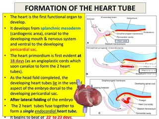
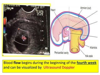
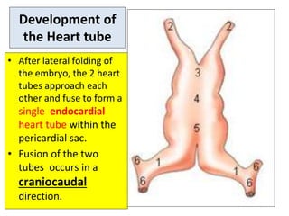
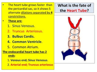
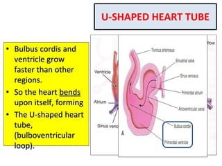
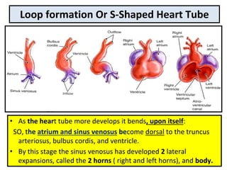
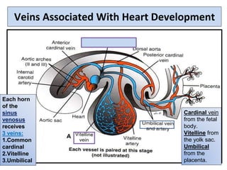
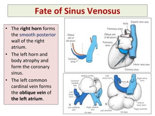
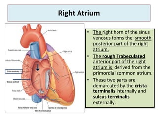
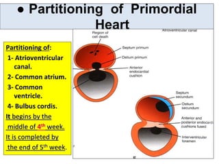
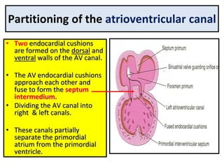
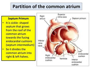
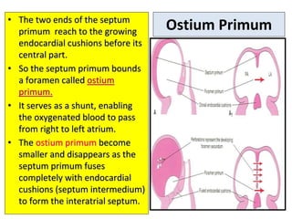
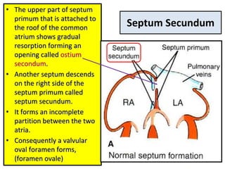
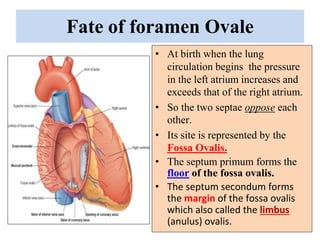
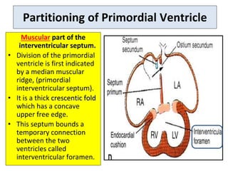
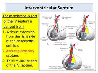
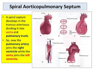
SlideShare a Scribd company logo
HEART DEVELOPMENT
Moderator-Dr. Sanjay Sharma
Presentor- Dr. Vishal Vaishnavi, Dr.
Dharmendra Singh
FORMATION OF THE HEART TUBE
• The heart is the first functional organ to
develop.
• It develops from splanchnic mesoderm
(cardiogenic area), cranial to the
developing mouth & nervous system
and ventral to the developing
pericardial sac.
• The heart primordium is first evident at
18 days (as an angioplastic cords which
soon canalize to form the 2 heart
tubes).
• As the head fold completed, the
developing heart tubes lie in the ventral
aspect of the embryo dorsal to the
developing pericardial sac.
• After lateral folding of the embryo
• The 2 heart tubes fuse together to
form a single endocardial heart tube.
• It begins to beat at 22 to 23 days.
Blood flow begins during the beginning of the fourth week
and can be visualized by Ultrasound Doppler
Development of
the Heart tube
• After lateral folding of
the embryo, the 2 heart
tubes approach each
other and fuse to form a
single endocardial
heart tube within the
pericardial sac.
• Fusion of the two
tubes occurs in a
craniocaudal
direction.
What is the fate of
the Heart Tube?
• The heart tube grows faster than
the pericardial sac, so it shows 5
alternate dilations separated by 4
constrictions.
• These are:
1. Sinus Venosus.
2. Truncus Arteriosus.
3. Bulbus Cordis.
4. Common Ventricle.
5. Common Atrium.
The endocardial heart tube has 2
ends:
1. Venous end; Sinus Venosus.
2. Arterial end; Truncus arteriosus
U-SHAPED HEART TUBE
• Bulbus cordis and
ventricle grow
faster than other
regions.
• So the heart bends
upon itself, forming
• The U-shaped heart
tube,
(bulboventricular
loop).
Loop formation Or S-Shaped Heart Tube
• As the heart tube more develops it bends, upon itself:
SO, the atrium and sinus venosus become dorsal to the truncus
arteriosus, bulbus cordis, and ventricle.
• By this stage the sinus venosus has developed 2 lateral
expansions, called the 2 horns ( right and left horns), and body.
Veins Associated With Heart Development
Each horn
of the
sinus
venosus
receives
3 veins:
1.Common
cardinal
2.Vitelline
3.Umbilical
Cardinal vein
from the fetal
body.
Vitelline from
the yolk sac.
Umbilical
from the
placenta.
Fate of Sinus Venosus
• The right horn forms
the smooth posterior
wall of the right
atrium.
• The left horn and
body atrophy and
form the coronary
sinus.
• The left common
cardinal vein forms
the oblique vein of
the left atrium.
Right Atrium
• The right horn of the sinus
venosus forms the smooth
posterior part of the right
atrium.
• The rough Trabeculated
anterior part of the right
atrium is derived from the
primordial common atrium.
• These two parts are
demarcated by the crista
terminalis internally and
sulcus terminalis
externally.
• Rough Trabeculated
part: derived from the
common primordial
atrium.
• The smooth part:
derived from the absorbed
Pulmonary Veins.
Left Atrium
● Partitioning of Primordial
Heart
Partitioning of:
1- Atrioventricular
canal.
2- Common atrium.
3- Common
ventricle.
4- Bulbus cordis.
It begins by the
middle of 4th week.
It is completed by
the end of 5th week.
Partitioning of the atrioventricular canal
• Two endocardial cushions
are formed on the dorsal and
ventral walls of the AV canal.
• The AV endocardial cushions
approach each other and
fuse to form the septum
intermedium.
• Dividing the AV canal into
right & left canals.
• These canals partially
separate the primordial
atrium from the primordial
ventricle.
Partition of the common atrium
Septum Primum
• It is sickle- shaped
septum that grows
from the roof of the
common atrium
towards the fusing
endocardial cushions
(septum intermedium)
• So it divides the
common atrium into
right & left halves.
Ostium Primum
• The two ends of the septum
primum reach to the growing
endocardial cushions before its
central part.
• So the septum primum bounds
a foramen called ostium
primum.
• It serves as a shunt, enabling
the oxygenated blood to pass
from right to left atrium.
• The ostium primum become
smaller and disappears as the
septum primum fuses
completely with endocardial
cushions (septum intermedium)
to form the interatrial septum.
Septum Secundum
• The upper part of septum
primum that is attached to
the roof of the common
atrium shows gradual
resorption forming an
opening called ostium
secondum.
• Another septum descends
on the right side of the
septum primum called
septum secundum.
• It forms an incomplete
partition between the two
atria.
• Consequently a valvular
oval foramen forms,
(foramen ovale)
• At birth when the lung
circulation begins the pressure
in the left atrium increases and
exceeds that of the right atrium.
• So the two septae oppose each
other.
• Its site is represented by the
Fossa Ovalis.
• The septum primum forms the
floor of the fossa ovalis.
• The septum secondum forms
the margin of the fossa ovalis
which also called the limbus
(anulus) ovalis.
Fate of foramen Ovale
Partitioning of Primordial Ventricle
Muscular part of the
interventricular septum.
• Division of the primordial
ventricle is first indicated
by a median muscular
ridge, (primordial
interventricular septum).
• It is a thick crescentic fold
which has a concave
upper free edge.
• This septum bounds a
temporary connection
between the two
ventricles called
interventricular foramen.
Interventricular Septum
The membranous part
of the IV septum is
derived from:
1- A tissue extension
from the right side
of the endocardial
cushion.
2- Aorticopulmonary
septum.
3- Thick muscular part
of the IV septum.
Spiral Aorticopulmonary Septum
• A spiral septum
develops in the
truncus arteriosus
dividing it into
aorta and
pulmonary trunk.
• So, now the
pulmonary artery
joins the right
ventricle while the
aorta joins the left
ventricle.
BULBUS CORDIS
• The bulbus cordis forms
the smooth upper part
of the two ventricles.
• In the right Ventricle:
• It forms the Conus
Arteriosus or
(Infundibulum) which
leads to the pulmonary
trunk.
• In the left ventricle:
• It forms the aortic
Vestibule which leads
to the aorta.
22
THANK
YOU

More Related Content

What's hot

Fundamentals of hfa perimetry
Fundamentals of hfa perimetryFundamentals of hfa perimetry
Fundamentals of hfa perimetry
Hossein Mirzaie
 
Extraocular muscles.pptx
Extraocular muscles.pptxExtraocular muscles.pptx
Extraocular muscles.pptx
shucayb1
 

What's hot (20)

Slit lamp biomicroscopy
Slit lamp biomicroscopySlit lamp biomicroscopy
Slit lamp biomicroscopy
 
FFA Dr Md Afzal Mahfuzullah
FFA Dr Md Afzal MahfuzullahFFA Dr Md Afzal Mahfuzullah
FFA Dr Md Afzal Mahfuzullah
 
Pal by nandini and jinal
Pal by nandini and jinalPal by nandini and jinal
Pal by nandini and jinal
 
Fundamentals of hfa perimetry
Fundamentals of hfa perimetryFundamentals of hfa perimetry
Fundamentals of hfa perimetry
 
Anatomy of lacrimal apparatus
Anatomy of lacrimal apparatusAnatomy of lacrimal apparatus
Anatomy of lacrimal apparatus
 
Fundus fluorescein angiography
Fundus fluorescein angiographyFundus fluorescein angiography
Fundus fluorescein angiography
 
Photochromatic lenses and tints www.eyenirvaan.com
Photochromatic lenses and tints   www.eyenirvaan.comPhotochromatic lenses and tints   www.eyenirvaan.com
Photochromatic lenses and tints www.eyenirvaan.com
 
Development of Retina
Development of Retina Development of Retina
Development of Retina
 
V isual field
V isual fieldV isual field
V isual field
 
Coronary CT
Coronary CTCoronary CT
Coronary CT
 
TRIAL CASE PPT
TRIAL CASE PPTTRIAL CASE PPT
TRIAL CASE PPT
 
Ferdous gonioscopy
Ferdous gonioscopy   Ferdous gonioscopy
Ferdous gonioscopy
 
Aqueous humor
Aqueous humorAqueous humor
Aqueous humor
 
FFA and ICG
FFA and ICGFFA and ICG
FFA and ICG
 
Lens power measurement
Lens power measurementLens power measurement
Lens power measurement
 
Log mar chart
Log mar chartLog mar chart
Log mar chart
 
Extraocular muscles.pptx
Extraocular muscles.pptxExtraocular muscles.pptx
Extraocular muscles.pptx
 
Anatomy of Orbit and its clinical importance
Anatomy of Orbit and its clinical importanceAnatomy of Orbit and its clinical importance
Anatomy of Orbit and its clinical importance
 
Perimetry
PerimetryPerimetry
Perimetry
 
Slit lamp biomicroscopy
Slit lamp biomicroscopySlit lamp biomicroscopy
Slit lamp biomicroscopy
 

Similar to Final heart development.pptx

Cardiovascularsystem 110514061056-phpapp02
Cardiovascularsystem 110514061056-phpapp02Cardiovascularsystem 110514061056-phpapp02
Cardiovascularsystem 110514061056-phpapp02
Ifrah Javaid
 
CVS Embryology (Amoud University)
CVS Embryology (Amoud University)CVS Embryology (Amoud University)
CVS Embryology (Amoud University)
moaminnca
 

Similar to Final heart development.pptx (20)

123.ppt
123.ppt123.ppt
123.ppt
 
development of heart.pptx
development of heart.pptxdevelopment of heart.pptx
development of heart.pptx
 
Embryology of heart and understand basic heart disease
Embryology of heart  and understand basic heart diseaseEmbryology of heart  and understand basic heart disease
Embryology of heart and understand basic heart disease
 
Final heart development.ppt
Final heart development.pptFinal heart development.ppt
Final heart development.ppt
 
Development of Cardiovascular System.pptx
Development of Cardiovascular System.pptxDevelopment of Cardiovascular System.pptx
Development of Cardiovascular System.pptx
 
Cardiovascularsystem 110514061056-phpapp02
Cardiovascularsystem 110514061056-phpapp02Cardiovascularsystem 110514061056-phpapp02
Cardiovascularsystem 110514061056-phpapp02
 
Development of heart.pptx
Development of heart.pptxDevelopment of heart.pptx
Development of heart.pptx
 
Embryology of heart and lung
Embryology of heart and lungEmbryology of heart and lung
Embryology of heart and lung
 
Heart Development.pptx
Heart Development.pptxHeart Development.pptx
Heart Development.pptx
 
Embryology of heart
Embryology of heartEmbryology of heart
Embryology of heart
 
Final heart development.pptx
Final heart development.pptxFinal heart development.pptx
Final heart development.pptx
 
Embryology of heart
Embryology of heartEmbryology of heart
Embryology of heart
 
CVS Embryology (Amoud University)
CVS Embryology (Amoud University)CVS Embryology (Amoud University)
CVS Embryology (Amoud University)
 
Cardiac Embryology basics.pptx
Cardiac Embryology basics.pptxCardiac Embryology basics.pptx
Cardiac Embryology basics.pptx
 
heart dev.pptx
heart dev.pptxheart dev.pptx
heart dev.pptx
 
embryology of heart
embryology of heartembryology of heart
embryology of heart
 
Embryology of the heart
Embryology of the heartEmbryology of the heart
Embryology of the heart
 
Embryonic normal development of heart
Embryonic normal development of heartEmbryonic normal development of heart
Embryonic normal development of heart
 
Development of heart
Development of heartDevelopment of heart
Development of heart
 
Heart development
Heart developmentHeart development
Heart development
 

More from SayanRDey (7)

Anatomy of lower limb
Anatomy  of lower  limbAnatomy  of lower  limb
Anatomy of lower limb
 
LIVER ABSCESS.pptx
LIVER ABSCESS.pptxLIVER ABSCESS.pptx
LIVER ABSCESS.pptx
 
abdomen_J.ppt
abdomen_J.pptabdomen_J.ppt
abdomen_J.ppt
 
Stomach
StomachStomach
Stomach
 
ulcer-190501174026.pdf
ulcer-190501174026.pdfulcer-190501174026.pdf
ulcer-190501174026.pdf
 
TPN.pptx
TPN.pptxTPN.pptx
TPN.pptx
 
salivary glands-csu.ppt
salivary glands-csu.pptsalivary glands-csu.ppt
salivary glands-csu.ppt
 

Recently uploaded

Mastering Wealth: A Path to Financial Freedom
Mastering Wealth: A Path to Financial FreedomMastering Wealth: A Path to Financial Freedom
Mastering Wealth: A Path to Financial Freedom
FatimaMary4
 

Recently uploaded (20)

The POPPY STUDY (Preconception to post-partum cardiovascular function in prim...
The POPPY STUDY (Preconception to post-partum cardiovascular function in prim...The POPPY STUDY (Preconception to post-partum cardiovascular function in prim...
The POPPY STUDY (Preconception to post-partum cardiovascular function in prim...
 
Multiple sclerosis diet.230524.ppt3.pptx
Multiple sclerosis diet.230524.ppt3.pptxMultiple sclerosis diet.230524.ppt3.pptx
Multiple sclerosis diet.230524.ppt3.pptx
 
Antiplatelets in IHD, Dose Duration, DAPT vs SAPT
Antiplatelets in IHD, Dose Duration, DAPT vs SAPTAntiplatelets in IHD, Dose Duration, DAPT vs SAPT
Antiplatelets in IHD, Dose Duration, DAPT vs SAPT
 
Temporal, Infratemporal & Pterygopalatine BY Dr.RIG.pptx
Temporal, Infratemporal & Pterygopalatine BY Dr.RIG.pptxTemporal, Infratemporal & Pterygopalatine BY Dr.RIG.pptx
Temporal, Infratemporal & Pterygopalatine BY Dr.RIG.pptx
 
Report Back from SGO 2024: What’s the Latest in Cervical Cancer?
Report Back from SGO 2024: What’s the Latest in Cervical Cancer?Report Back from SGO 2024: What’s the Latest in Cervical Cancer?
Report Back from SGO 2024: What’s the Latest in Cervical Cancer?
 
Anuman- An inference for helpful in diagnosis and treatment
Anuman- An inference for helpful in diagnosis and treatmentAnuman- An inference for helpful in diagnosis and treatment
Anuman- An inference for helpful in diagnosis and treatment
 
The History of Diagnostic Medical imaging
The History of Diagnostic Medical imagingThe History of Diagnostic Medical imaging
The History of Diagnostic Medical imaging
 
Compare home pulse pressure components collected directly from home
Compare home pulse pressure components collected directly from homeCompare home pulse pressure components collected directly from home
Compare home pulse pressure components collected directly from home
 
How to Give Better Lectures: Some Tips for Doctors
How to Give Better Lectures: Some Tips for DoctorsHow to Give Better Lectures: Some Tips for Doctors
How to Give Better Lectures: Some Tips for Doctors
 
ANATOMY OF THE LOWER URINARY TRACT AND MALE [Autosaved] [Autosaved].pptx
ANATOMY OF THE LOWER URINARY TRACT AND MALE [Autosaved] [Autosaved].pptxANATOMY OF THE LOWER URINARY TRACT AND MALE [Autosaved] [Autosaved].pptx
ANATOMY OF THE LOWER URINARY TRACT AND MALE [Autosaved] [Autosaved].pptx
 
HOT NEW PRODUCT! BIG SALES FAST SHIPPING NOW FROM CHINA!! EU KU DB BK substit...
HOT NEW PRODUCT! BIG SALES FAST SHIPPING NOW FROM CHINA!! EU KU DB BK substit...HOT NEW PRODUCT! BIG SALES FAST SHIPPING NOW FROM CHINA!! EU KU DB BK substit...
HOT NEW PRODUCT! BIG SALES FAST SHIPPING NOW FROM CHINA!! EU KU DB BK substit...
 
Aptopadesha Pramana / Pariksha: The Verbal Testimony
Aptopadesha Pramana / Pariksha: The Verbal TestimonyAptopadesha Pramana / Pariksha: The Verbal Testimony
Aptopadesha Pramana / Pariksha: The Verbal Testimony
 
Mastering Wealth: A Path to Financial Freedom
Mastering Wealth: A Path to Financial FreedomMastering Wealth: A Path to Financial Freedom
Mastering Wealth: A Path to Financial Freedom
 
Gauri Gawande(9) Constipation Final.pptx
Gauri Gawande(9) Constipation Final.pptxGauri Gawande(9) Constipation Final.pptx
Gauri Gawande(9) Constipation Final.pptx
 
linearity concept of significance, standard deviation, chi square test, stude...
linearity concept of significance, standard deviation, chi square test, stude...linearity concept of significance, standard deviation, chi square test, stude...
linearity concept of significance, standard deviation, chi square test, stude...
 
The hemodynamic and autonomic determinants of elevated blood pressure in obes...
The hemodynamic and autonomic determinants of elevated blood pressure in obes...The hemodynamic and autonomic determinants of elevated blood pressure in obes...
The hemodynamic and autonomic determinants of elevated blood pressure in obes...
 
TEST BANK For Wong’s Essentials of Pediatric Nursing, 11th Edition by Marilyn...
TEST BANK For Wong’s Essentials of Pediatric Nursing, 11th Edition by Marilyn...TEST BANK For Wong’s Essentials of Pediatric Nursing, 11th Edition by Marilyn...
TEST BANK For Wong’s Essentials of Pediatric Nursing, 11th Edition by Marilyn...
 
Young at heart: Cardiovascular health stations to empower healthy lifestyle b...
Young at heart: Cardiovascular health stations to empower healthy lifestyle b...Young at heart: Cardiovascular health stations to empower healthy lifestyle b...
Young at heart: Cardiovascular health stations to empower healthy lifestyle b...
 
Surat @ℂall @Girls ꧁❤8527049040❤꧂@ℂall @Girls Service Vip Top Model Safe
Surat @ℂall @Girls ꧁❤8527049040❤꧂@ℂall @Girls Service Vip Top Model SafeSurat @ℂall @Girls ꧁❤8527049040❤꧂@ℂall @Girls Service Vip Top Model Safe
Surat @ℂall @Girls ꧁❤8527049040❤꧂@ℂall @Girls Service Vip Top Model Safe
 
"Central Hypertension"‚ in China: Towards the nation-wide use of SphygmoCor t...
"Central Hypertension"‚ in China: Towards the nation-wide use of SphygmoCor t..."Central Hypertension"‚ in China: Towards the nation-wide use of SphygmoCor t...
"Central Hypertension"‚ in China: Towards the nation-wide use of SphygmoCor t...
 

Final heart development.pptx

  • 1. HEART DEVELOPMENT Moderator-Dr. Sanjay Sharma Presentor- Dr. Vishal Vaishnavi, Dr. Dharmendra Singh
  • 2. FORMATION OF THE HEART TUBE • The heart is the first functional organ to develop. • It develops from splanchnic mesoderm (cardiogenic area), cranial to the developing mouth & nervous system and ventral to the developing pericardial sac. • The heart primordium is first evident at 18 days (as an angioplastic cords which soon canalize to form the 2 heart tubes). • As the head fold completed, the developing heart tubes lie in the ventral aspect of the embryo dorsal to the developing pericardial sac. • After lateral folding of the embryo • The 2 heart tubes fuse together to form a single endocardial heart tube. • It begins to beat at 22 to 23 days.
  • 3. Blood flow begins during the beginning of the fourth week and can be visualized by Ultrasound Doppler
  • 4. Development of the Heart tube • After lateral folding of the embryo, the 2 heart tubes approach each other and fuse to form a single endocardial heart tube within the pericardial sac. • Fusion of the two tubes occurs in a craniocaudal direction.
  • 5. What is the fate of the Heart Tube? • The heart tube grows faster than the pericardial sac, so it shows 5 alternate dilations separated by 4 constrictions. • These are: 1. Sinus Venosus. 2. Truncus Arteriosus. 3. Bulbus Cordis. 4. Common Ventricle. 5. Common Atrium. The endocardial heart tube has 2 ends: 1. Venous end; Sinus Venosus. 2. Arterial end; Truncus arteriosus
  • 6. U-SHAPED HEART TUBE • Bulbus cordis and ventricle grow faster than other regions. • So the heart bends upon itself, forming • The U-shaped heart tube, (bulboventricular loop).
  • 7. Loop formation Or S-Shaped Heart Tube • As the heart tube more develops it bends, upon itself: SO, the atrium and sinus venosus become dorsal to the truncus arteriosus, bulbus cordis, and ventricle. • By this stage the sinus venosus has developed 2 lateral expansions, called the 2 horns ( right and left horns), and body.
  • 8. Veins Associated With Heart Development Each horn of the sinus venosus receives 3 veins: 1.Common cardinal 2.Vitelline 3.Umbilical Cardinal vein from the fetal body. Vitelline from the yolk sac. Umbilical from the placenta.
  • 9. Fate of Sinus Venosus • The right horn forms the smooth posterior wall of the right atrium. • The left horn and body atrophy and form the coronary sinus. • The left common cardinal vein forms the oblique vein of the left atrium.
  • 10. Right Atrium • The right horn of the sinus venosus forms the smooth posterior part of the right atrium. • The rough Trabeculated anterior part of the right atrium is derived from the primordial common atrium. • These two parts are demarcated by the crista terminalis internally and sulcus terminalis externally.
  • 11. • Rough Trabeculated part: derived from the common primordial atrium. • The smooth part: derived from the absorbed Pulmonary Veins. Left Atrium
  • 12. ● Partitioning of Primordial Heart Partitioning of: 1- Atrioventricular canal. 2- Common atrium. 3- Common ventricle. 4- Bulbus cordis. It begins by the middle of 4th week. It is completed by the end of 5th week.
  • 13. Partitioning of the atrioventricular canal • Two endocardial cushions are formed on the dorsal and ventral walls of the AV canal. • The AV endocardial cushions approach each other and fuse to form the septum intermedium. • Dividing the AV canal into right & left canals. • These canals partially separate the primordial atrium from the primordial ventricle.
  • 14. Partition of the common atrium Septum Primum • It is sickle- shaped septum that grows from the roof of the common atrium towards the fusing endocardial cushions (septum intermedium) • So it divides the common atrium into right & left halves.
  • 15. Ostium Primum • The two ends of the septum primum reach to the growing endocardial cushions before its central part. • So the septum primum bounds a foramen called ostium primum. • It serves as a shunt, enabling the oxygenated blood to pass from right to left atrium. • The ostium primum become smaller and disappears as the septum primum fuses completely with endocardial cushions (septum intermedium) to form the interatrial septum.
  • 16. Septum Secundum • The upper part of septum primum that is attached to the roof of the common atrium shows gradual resorption forming an opening called ostium secondum. • Another septum descends on the right side of the septum primum called septum secundum. • It forms an incomplete partition between the two atria. • Consequently a valvular oval foramen forms, (foramen ovale)
  • 17. • At birth when the lung circulation begins the pressure in the left atrium increases and exceeds that of the right atrium. • So the two septae oppose each other. • Its site is represented by the Fossa Ovalis. • The septum primum forms the floor of the fossa ovalis. • The septum secondum forms the margin of the fossa ovalis which also called the limbus (anulus) ovalis. Fate of foramen Ovale
  • 18. Partitioning of Primordial Ventricle Muscular part of the interventricular septum. • Division of the primordial ventricle is first indicated by a median muscular ridge, (primordial interventricular septum). • It is a thick crescentic fold which has a concave upper free edge. • This septum bounds a temporary connection between the two ventricles called interventricular foramen.
  • 19. Interventricular Septum The membranous part of the IV septum is derived from: 1- A tissue extension from the right side of the endocardial cushion. 2- Aorticopulmonary septum. 3- Thick muscular part of the IV septum.
  • 20. Spiral Aorticopulmonary Septum • A spiral septum develops in the truncus arteriosus dividing it into aorta and pulmonary trunk. • So, now the pulmonary artery joins the right ventricle while the aorta joins the left ventricle.
  • 21. BULBUS CORDIS • The bulbus cordis forms the smooth upper part of the two ventricles. • In the right Ventricle: • It forms the Conus Arteriosus or (Infundibulum) which leads to the pulmonary trunk. • In the left ventricle: • It forms the aortic Vestibule which leads to the aorta.