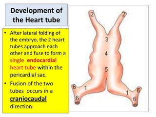
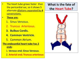
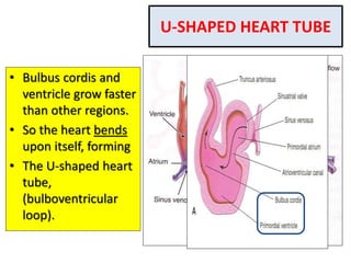
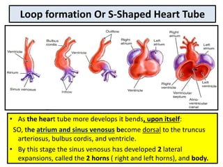
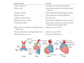
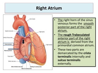
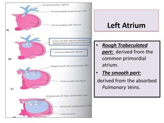
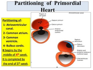
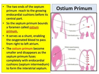
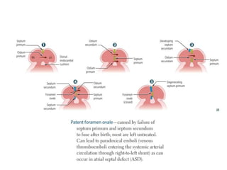
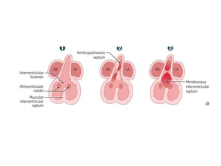
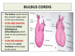
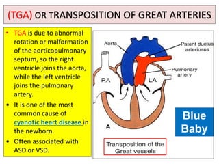
Objectives
• By the end of the lecture you should be able to:
• Describe the formation, site, union, division of the of the
heart tube.
• Describe the formation and fate of the sinus venosus.
• Describe the formation of the interatrial and the
interventricular septae.
• Describe the formation of the two atria and the two
ventricles.
• Describe the partitioning of the truncus arteriosus and
formation of the aorta and pulmonary trunk.
• List the most common cardiac anomalies.
Development of
the Heart tube
• After lateral folding of
the embryo, the 2 heart
tubes approach each
other and fuse to form a
single endocardial
heart tube within the
pericardial sac.
• Fusion of the two
tubes occurs in a
craniocaudal
direction.
What is the fate of
the Heart Tube?
• The heart tube grows faster than
the pericardial sac, so it shows 5
alternate dilations separated by 4
constrictions.
• These are:
1. Sinus Venosus.
2. Truncus Arteriosus.
3. Bulbus Cordis.
4. Common Ventricle.
5. Common Atrium.
The endocardial heart tube has 2
ends:
1. Venous end; Sinus Venosus.
2. Arterial end; Truncus arteriosus
U-SHAPED HEART TUBE
• Bulbus cordis and
ventricle grow faster
than other regions.
• So the heart bends
upon itself, forming
• The U-shaped heart
tube,
(bulboventricular
loop).
Loop formation Or S-Shaped Heart Tube
• As the heart tube more develops it bends, upon itself:
SO, the atrium and sinus venosus become dorsal to the truncus
arteriosus, bulbus cordis, and ventricle.
• By this stage the sinus venosus has developed 2 lateral
expansions, called the 2 horns ( right and left horns), and body.
Fate of Sinus Venosus
• The right horn forms
the smooth posterior
wall of the right
atrium.
• The left horn and
body atrophy and
form the coronary
sinus.
• The left common
cardinal vein forms
the oblique vein of
the left atrium.
Right Atrium
• The right horn of the sinus
venosus forms the smooth
posterior part of the right
atrium.
• The rough Trabeculated
anterior part of the right
atrium is derived from the
primordial common atrium.
• These two parts are
demarcated by the crista
terminalis internally and
sulcus terminalis
externally.
• Rough Trabeculated
part: derived from the
common primordial
atrium.
• The smooth part:
derived from the absorbed
Pulmonary Veins.
Left Atrium
Partitioning of Primordial
Heart
Partitioning of:
1- Atrioventricular
canal.
2- Common atrium.
3- Common
ventricle.
4- Bulbus cordis.
It begins by the
middle of 4th week.
It is completed by
the end of 5th week.
Partitioning of the atrioventricular canal
• Two endocardial cushions
are formed on the dorsal and
ventral walls of the AV canal.
• The AV endocardial cushions
approach each other and
fuse to form the septum
intermedium.
• Dividing the AV canal into
right & left canals.
• These canals partially
separate the primordial
atrium from the primordial
ventricle.
Partition of the common atrium
Septum Primum
• It is sickle- shaped
septum that grows
from the roof of the
common atrium
towards the fusing
endocardial cushions
(septum intermedium)
• So it divides the
common atrium into
right & left halves.
Ostium Primum
• The two ends of the septum
primum reach to the growing
endocardial cushions before its
central part.
• So the septum primum bounds
a foramen called ostium
primum.
• It serves as a shunt, enabling
the oxygenated blood to pass
from right to left atrium.
• The ostium primum become
smaller and disappears as the
septum primum fuses
completely with endocardial
cushions (septum intermedium)
to form the interatrial septum.
Septum Secundum
• The upper part of septum
primum that is attached to
the roof of the common
atrium shows gradual
resorption forming an
opening called ostium
secondum.
• Another septum descends
on the right side of the
septum primum called
septum secundum.
• It forms an incomplete
partition between the two
atria.
• Consequently a valvular
oval foramen forms,
(foramen ovale)
• At birth when the lung
circulation begins the pressure
in the left atrium increases and
exceeds that of the right atrium.
• So the two septae oppose each
other.
• Its site is represented by the
Fossa Ovalis.
• The septum primum forms the
floor of the fossa ovalis.
• The septum secondum forms
the margin of the fossa ovalis
which also called the limbus
(anulus) ovalis.
Fate of foramen Ovale
Partitioning of Primordial Ventricle
Muscular part of the
interventricular septum.
• Division of the primordial
ventricle is first indicated
by a median muscular
ridge, (primordial
interventricular septum).
• It is a thick crescentic fold
which has a concave
upper free edge.
• This septum bounds a
temporary connection
between the two
ventricles called
interventricular foramen.
Interventricular Septum
The membranous part
of the IV septum is
derived from:
1- A tissue extension
from the right side
of the endocardial
cushion.
2- Aorticopulmonary
septum.
3- Thick muscular part
of the IV septum.
Spiral Aorticopulmonary Septum
• A spiral septum
develops in the
truncus arteriosus
dividing it into
aorta and
pulmonary trunk.
• So, now the
pulmonary artery
joins the right
ventricle while the
aorta joins the left
ventricle.
BULBUS CORDIS
• The bulbus cordis forms
the smooth upper part
of the two ventricles.
• In the right Ventricle:
• It forms the Conus
Arteriosus or
(Infundibulum) which
leads to the pulmonary
trunk.
• In the left ventricle:
• It forms the aortic
Vestibule which leads
to the aorta.
MAJOR CARDIAC ANOALIES
Atrial Septal
Defects (ASD)
• Absence of septum
primum and septum
secundum, leads to
common atrium.
• Absence of Septum
Secundum
Excessive
resorption of
septum primum
(ASD)
Patent
foramen
ovale
• Roger’s disease
• Absence of the
membranous part
of interventricular
septum.
• Usually
accompanied by
other cardiac
defects.
TETRALOGY OF FALLOT
• Fallot’s Tetralogy:
• 1-VSD.
• 2- Pulmonary
stenosis.
• 3-Overriding of
the aorta
• 4- Right
ventricular
hypertrophy.
Blue
Baby
TETRALOGY
OF
FALLOT
Blue Baby
(TGA) OR TRANSPOSITION OF GREAT ARTERIES
• TGA is due to abnormal
rotation or malformation
of the aorticopulmonary
septum, so the right
ventricle joins the aorta,
while the left ventricle
joins the pulmonary
artery.
• It is one of the most
common cause of
cyanotic heart disease in
the newborn.
• Often associated with
ASD or VSD.
Blue
Baby
Persistent Truncus Arteriosus
 It is due to
failure of the
development of the
aorticopulmonary
(spiral) septum.
 It is usually
accompanied with
VSD.
31

Final heart development.pptx

  • 2.
    Objectives • By theend of the lecture you should be able to: • Describe the formation, site, union, division of the of the heart tube. • Describe the formation and fate of the sinus venosus. • Describe the formation of the interatrial and the interventricular septae. • Describe the formation of the two atria and the two ventricles. • Describe the partitioning of the truncus arteriosus and formation of the aorta and pulmonary trunk. • List the most common cardiac anomalies.
  • 3.
    Development of the Hearttube • After lateral folding of the embryo, the 2 heart tubes approach each other and fuse to form a single endocardial heart tube within the pericardial sac. • Fusion of the two tubes occurs in a craniocaudal direction.
  • 4.
    What is thefate of the Heart Tube? • The heart tube grows faster than the pericardial sac, so it shows 5 alternate dilations separated by 4 constrictions. • These are: 1. Sinus Venosus. 2. Truncus Arteriosus. 3. Bulbus Cordis. 4. Common Ventricle. 5. Common Atrium. The endocardial heart tube has 2 ends: 1. Venous end; Sinus Venosus. 2. Arterial end; Truncus arteriosus
  • 5.
    U-SHAPED HEART TUBE •Bulbus cordis and ventricle grow faster than other regions. • So the heart bends upon itself, forming • The U-shaped heart tube, (bulboventricular loop).
  • 6.
    Loop formation OrS-Shaped Heart Tube • As the heart tube more develops it bends, upon itself: SO, the atrium and sinus venosus become dorsal to the truncus arteriosus, bulbus cordis, and ventricle. • By this stage the sinus venosus has developed 2 lateral expansions, called the 2 horns ( right and left horns), and body.
  • 8.
    Fate of SinusVenosus • The right horn forms the smooth posterior wall of the right atrium. • The left horn and body atrophy and form the coronary sinus. • The left common cardinal vein forms the oblique vein of the left atrium.
  • 9.
    Right Atrium • Theright horn of the sinus venosus forms the smooth posterior part of the right atrium. • The rough Trabeculated anterior part of the right atrium is derived from the primordial common atrium. • These two parts are demarcated by the crista terminalis internally and sulcus terminalis externally.
  • 10.
    • Rough Trabeculated part:derived from the common primordial atrium. • The smooth part: derived from the absorbed Pulmonary Veins. Left Atrium
  • 11.
    Partitioning of Primordial Heart Partitioningof: 1- Atrioventricular canal. 2- Common atrium. 3- Common ventricle. 4- Bulbus cordis. It begins by the middle of 4th week. It is completed by the end of 5th week.
  • 12.
    Partitioning of theatrioventricular canal • Two endocardial cushions are formed on the dorsal and ventral walls of the AV canal. • The AV endocardial cushions approach each other and fuse to form the septum intermedium. • Dividing the AV canal into right & left canals. • These canals partially separate the primordial atrium from the primordial ventricle.
  • 13.
    Partition of thecommon atrium Septum Primum • It is sickle- shaped septum that grows from the roof of the common atrium towards the fusing endocardial cushions (septum intermedium) • So it divides the common atrium into right & left halves.
  • 14.
    Ostium Primum • Thetwo ends of the septum primum reach to the growing endocardial cushions before its central part. • So the septum primum bounds a foramen called ostium primum. • It serves as a shunt, enabling the oxygenated blood to pass from right to left atrium. • The ostium primum become smaller and disappears as the septum primum fuses completely with endocardial cushions (septum intermedium) to form the interatrial septum.
  • 15.
    Septum Secundum • Theupper part of septum primum that is attached to the roof of the common atrium shows gradual resorption forming an opening called ostium secondum. • Another septum descends on the right side of the septum primum called septum secundum. • It forms an incomplete partition between the two atria. • Consequently a valvular oval foramen forms, (foramen ovale)
  • 16.
    • At birthwhen the lung circulation begins the pressure in the left atrium increases and exceeds that of the right atrium. • So the two septae oppose each other. • Its site is represented by the Fossa Ovalis. • The septum primum forms the floor of the fossa ovalis. • The septum secondum forms the margin of the fossa ovalis which also called the limbus (anulus) ovalis. Fate of foramen Ovale
  • 18.
    Partitioning of PrimordialVentricle Muscular part of the interventricular septum. • Division of the primordial ventricle is first indicated by a median muscular ridge, (primordial interventricular septum). • It is a thick crescentic fold which has a concave upper free edge. • This septum bounds a temporary connection between the two ventricles called interventricular foramen.
  • 19.
    Interventricular Septum The membranouspart of the IV septum is derived from: 1- A tissue extension from the right side of the endocardial cushion. 2- Aorticopulmonary septum. 3- Thick muscular part of the IV septum.
  • 21.
    Spiral Aorticopulmonary Septum •A spiral septum develops in the truncus arteriosus dividing it into aorta and pulmonary trunk. • So, now the pulmonary artery joins the right ventricle while the aorta joins the left ventricle.
  • 22.
    BULBUS CORDIS • Thebulbus cordis forms the smooth upper part of the two ventricles. • In the right Ventricle: • It forms the Conus Arteriosus or (Infundibulum) which leads to the pulmonary trunk. • In the left ventricle: • It forms the aortic Vestibule which leads to the aorta.
  • 23.
  • 24.
    Atrial Septal Defects (ASD) •Absence of septum primum and septum secundum, leads to common atrium. • Absence of Septum Secundum
  • 25.
  • 26.
    • Roger’s disease •Absence of the membranous part of interventricular septum. • Usually accompanied by other cardiac defects.
  • 27.
    TETRALOGY OF FALLOT •Fallot’s Tetralogy: • 1-VSD. • 2- Pulmonary stenosis. • 3-Overriding of the aorta • 4- Right ventricular hypertrophy. Blue Baby
  • 28.
  • 29.
    (TGA) OR TRANSPOSITIONOF GREAT ARTERIES • TGA is due to abnormal rotation or malformation of the aorticopulmonary septum, so the right ventricle joins the aorta, while the left ventricle joins the pulmonary artery. • It is one of the most common cause of cyanotic heart disease in the newborn. • Often associated with ASD or VSD. Blue Baby
  • 30.
    Persistent Truncus Arteriosus It is due to failure of the development of the aorticopulmonary (spiral) septum.  It is usually accompanied with VSD.
  • 31.