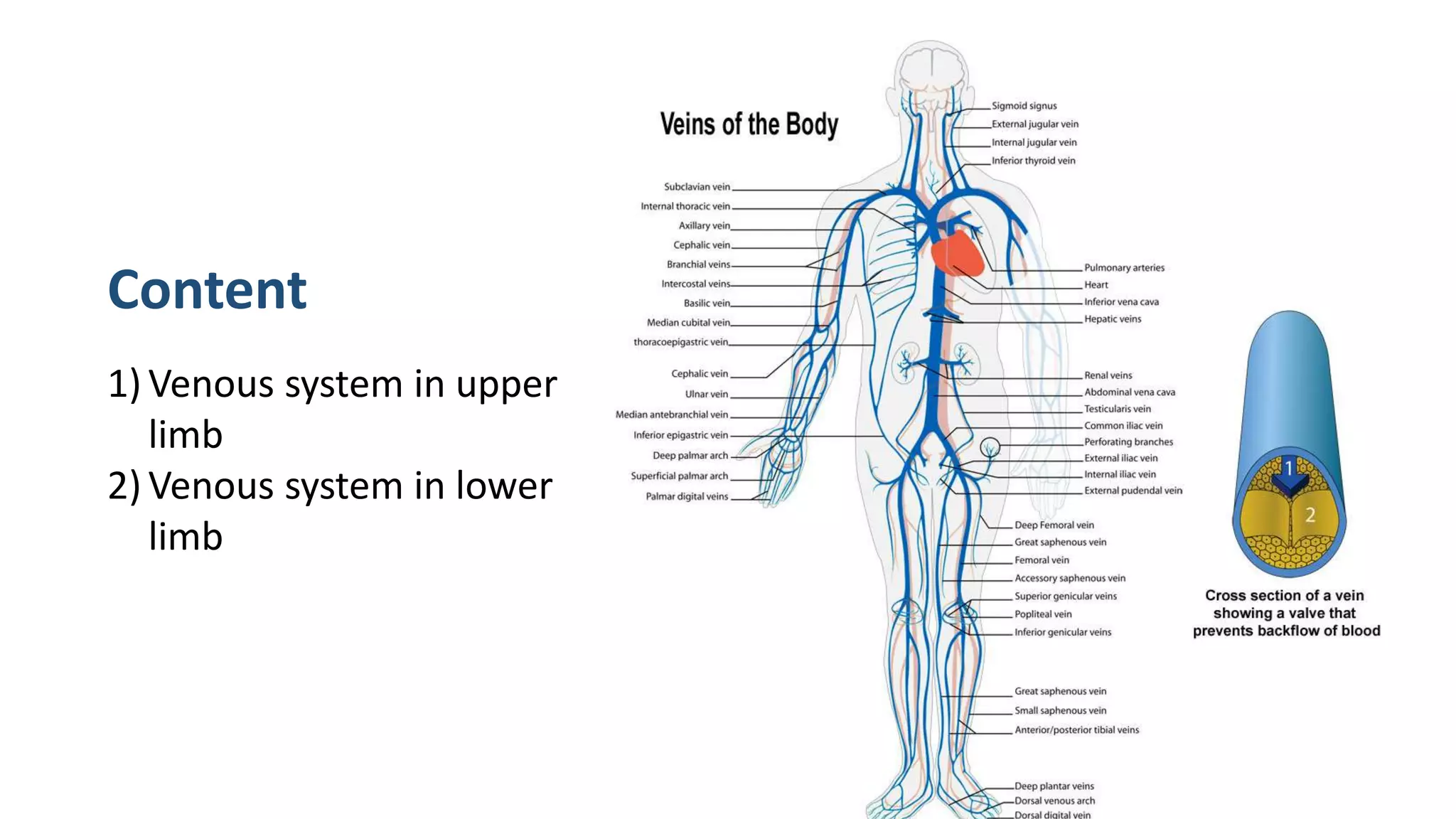
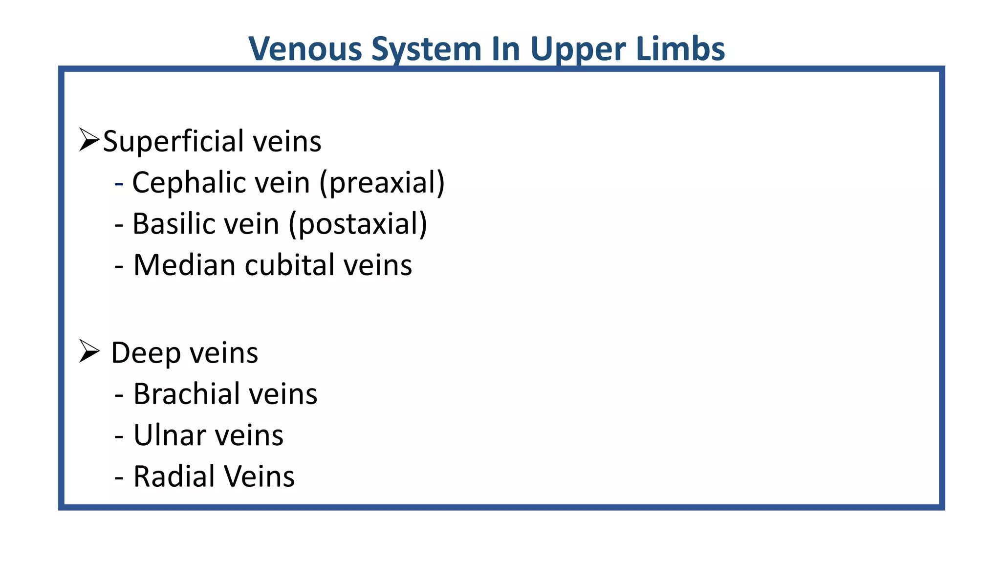
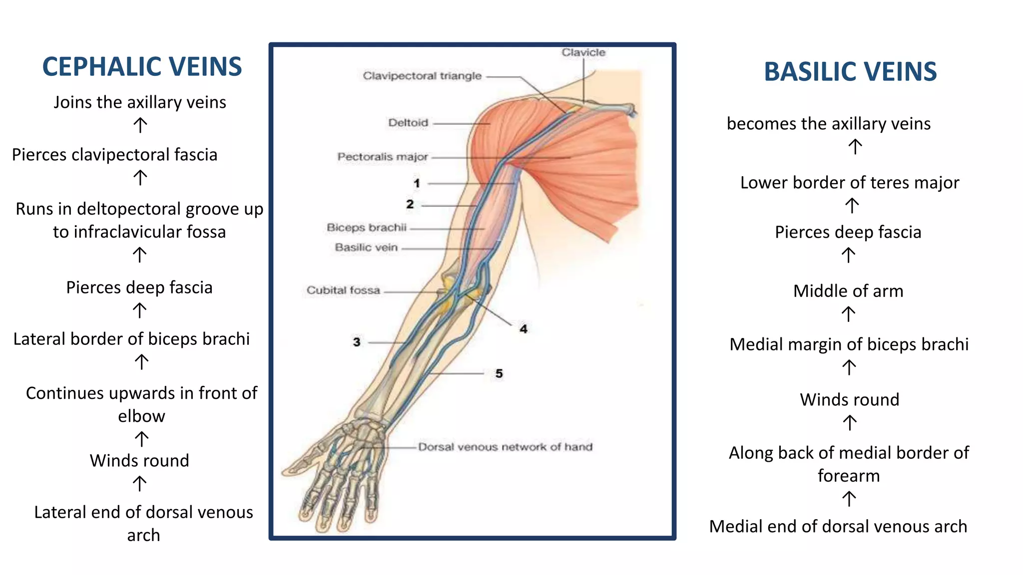
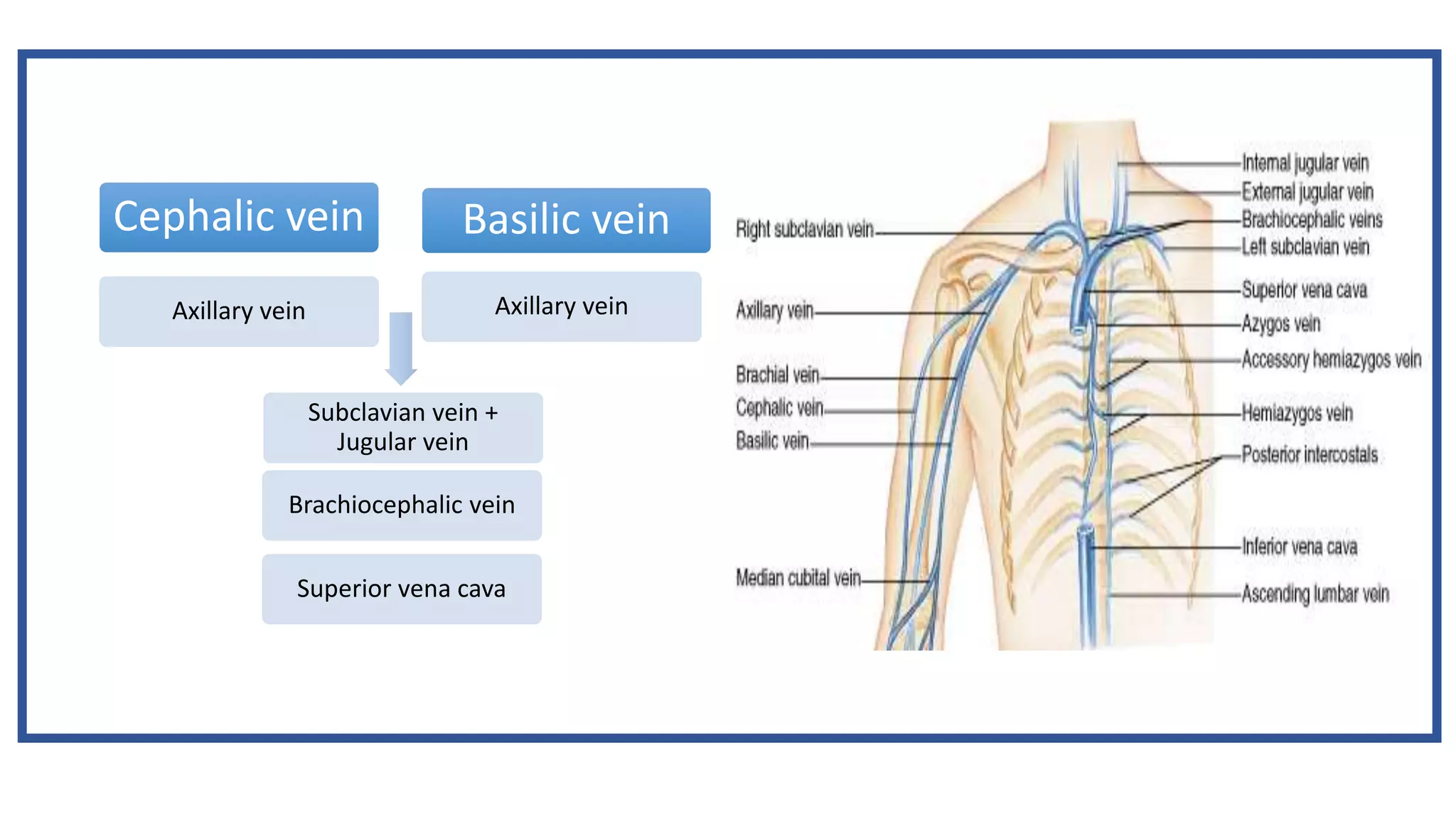
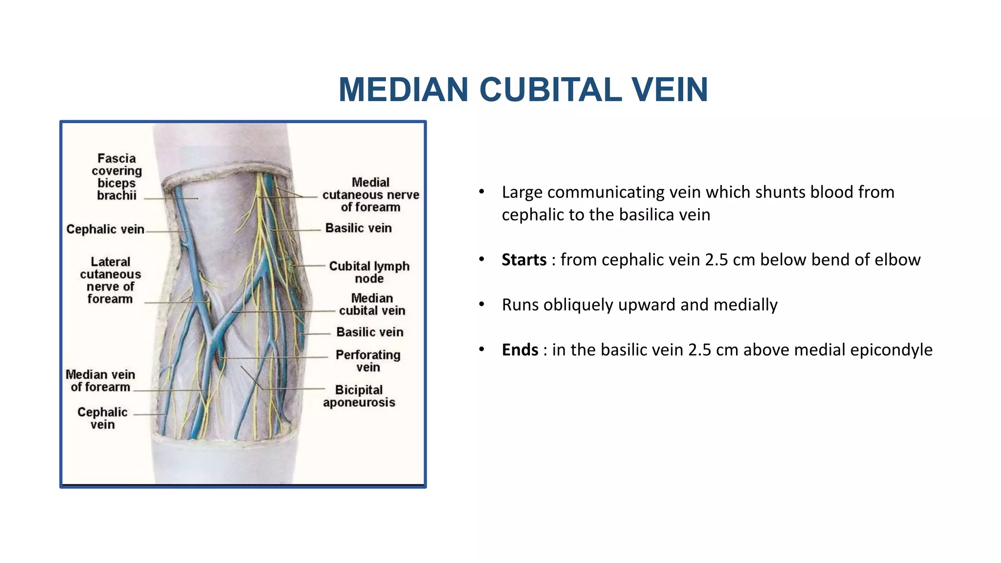
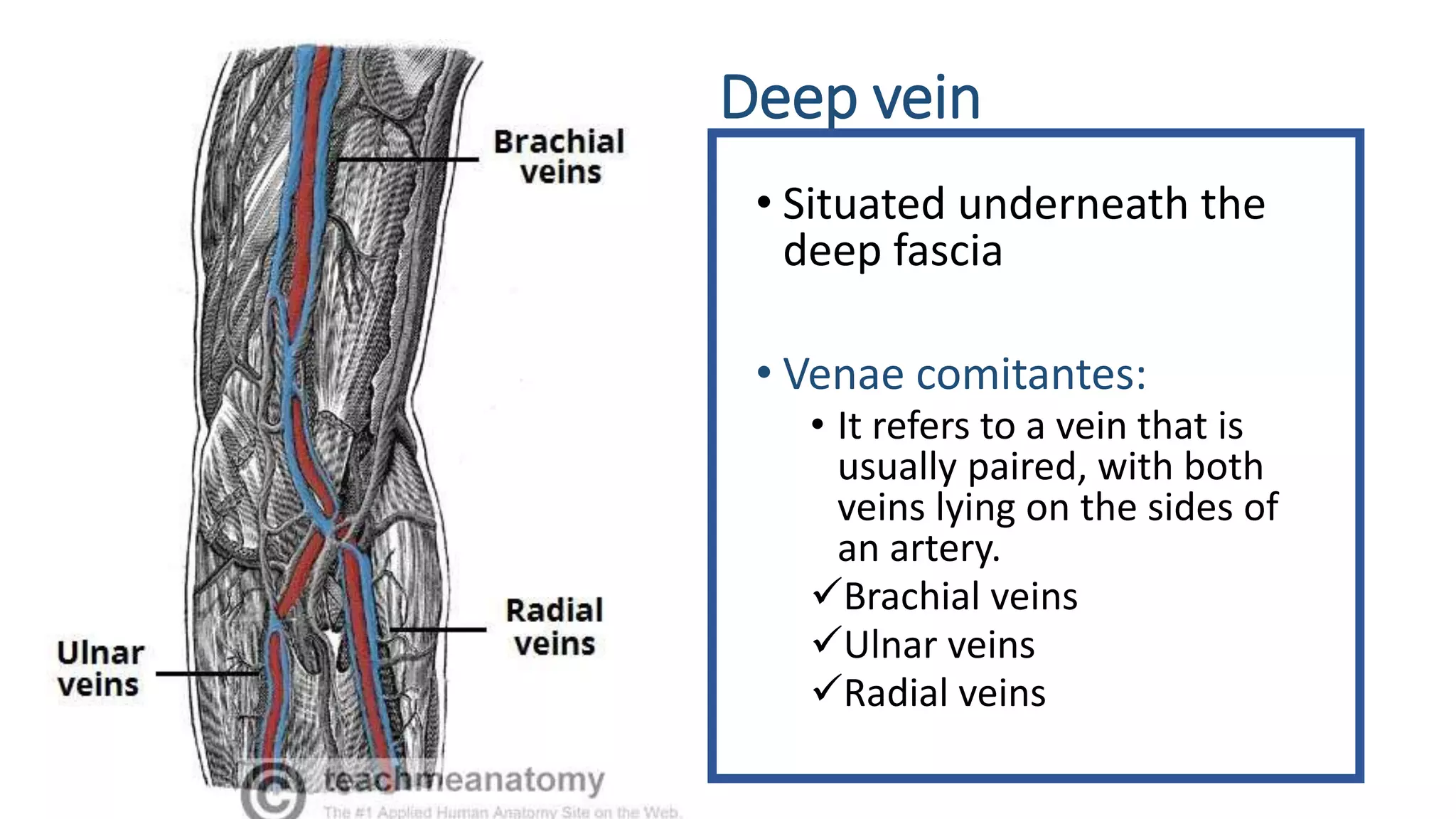
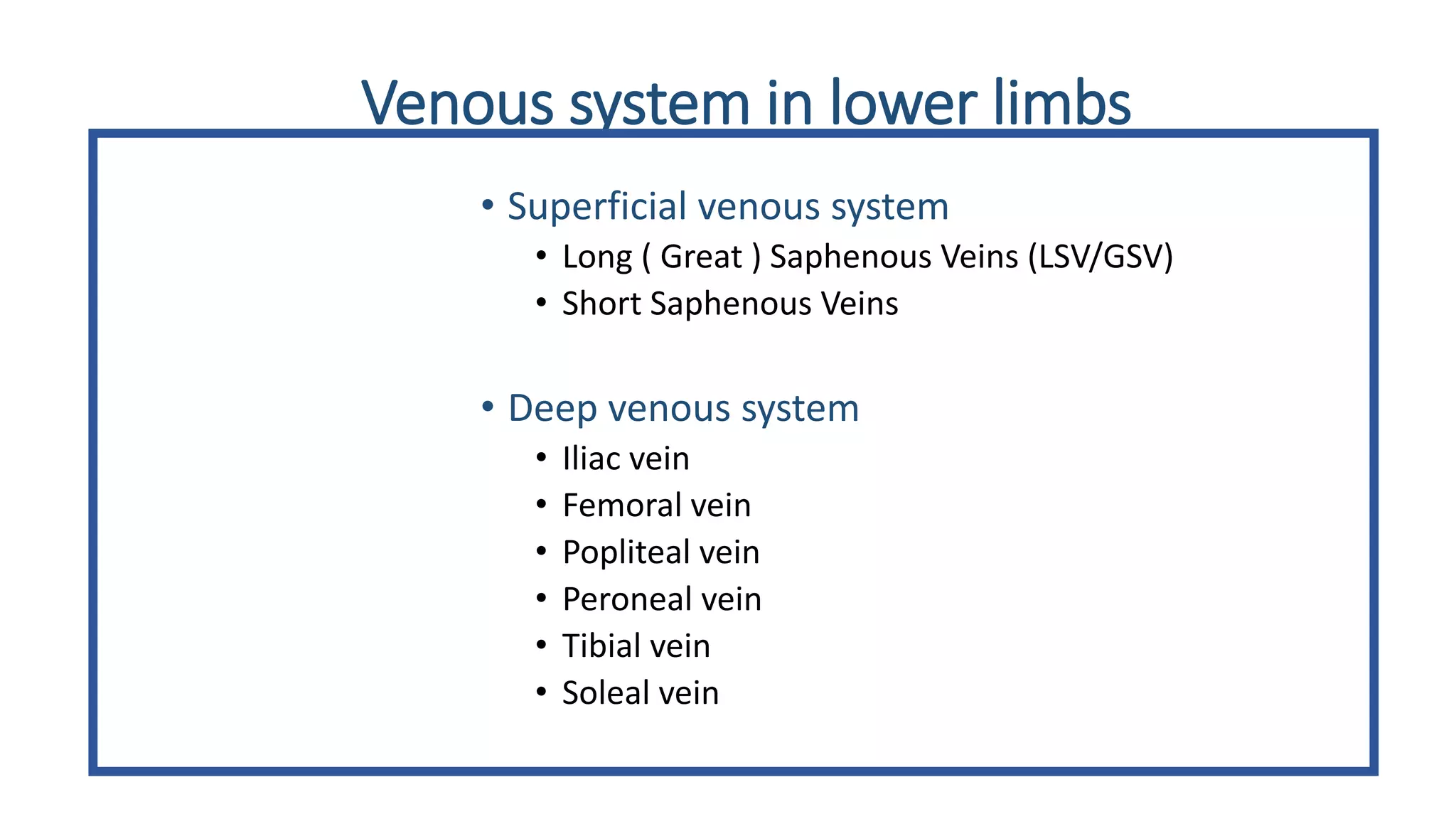
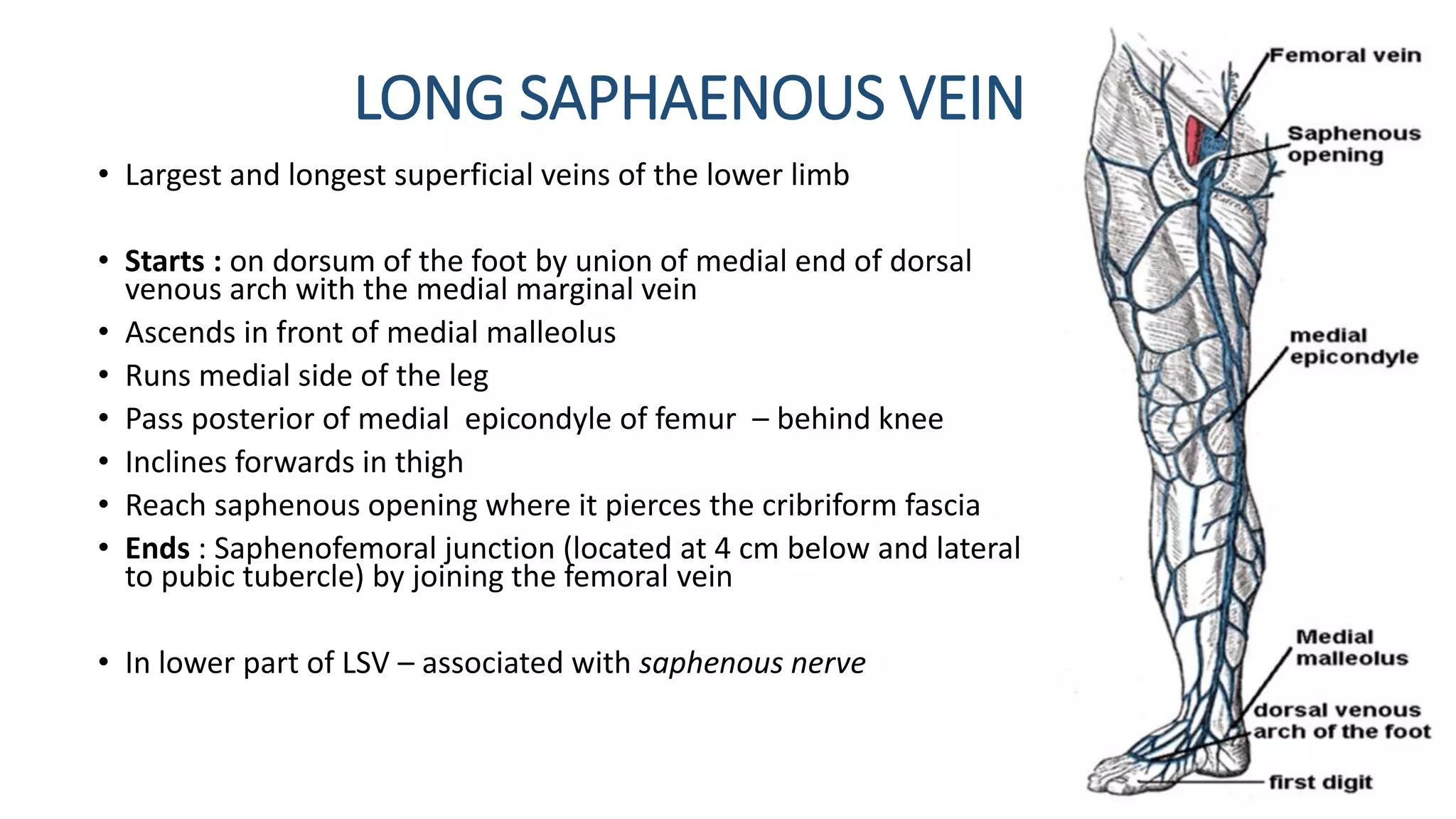
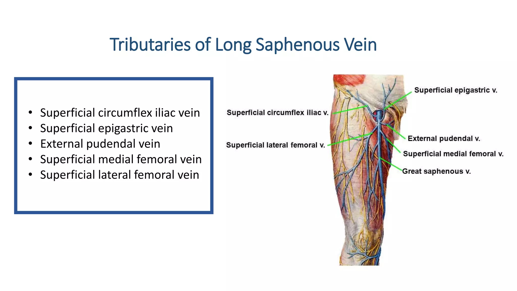
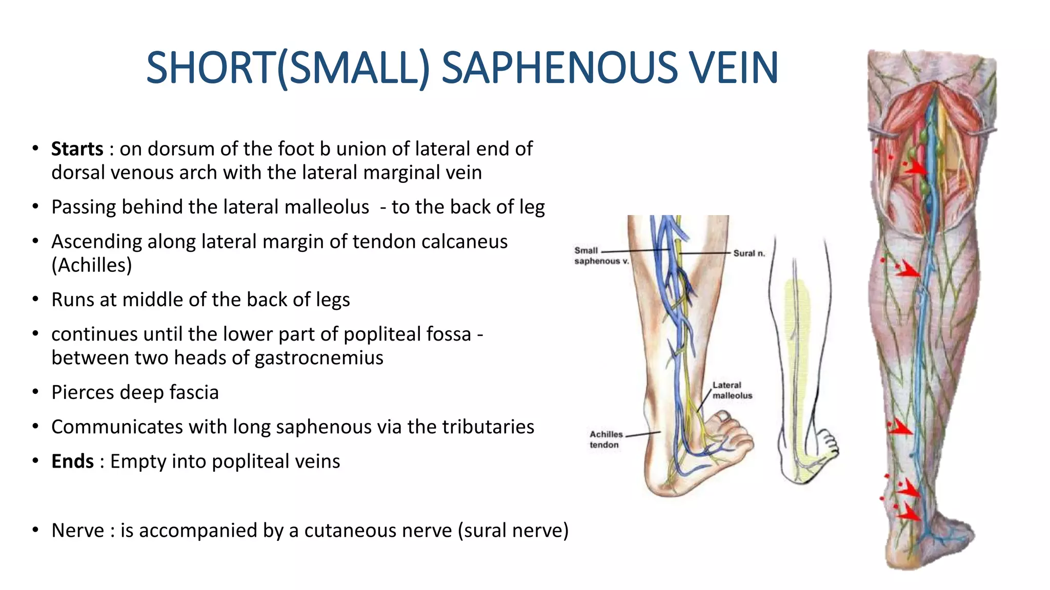
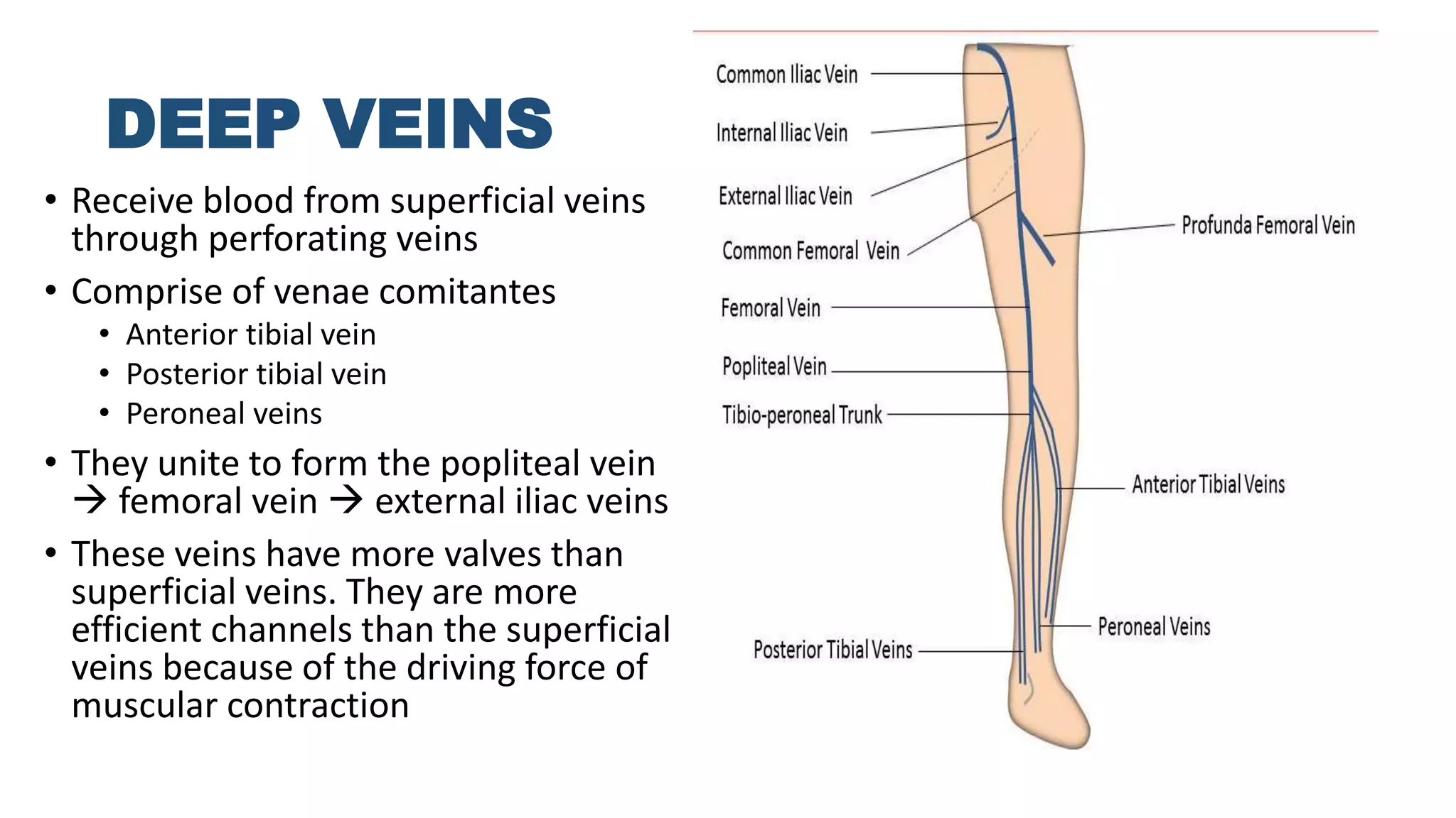
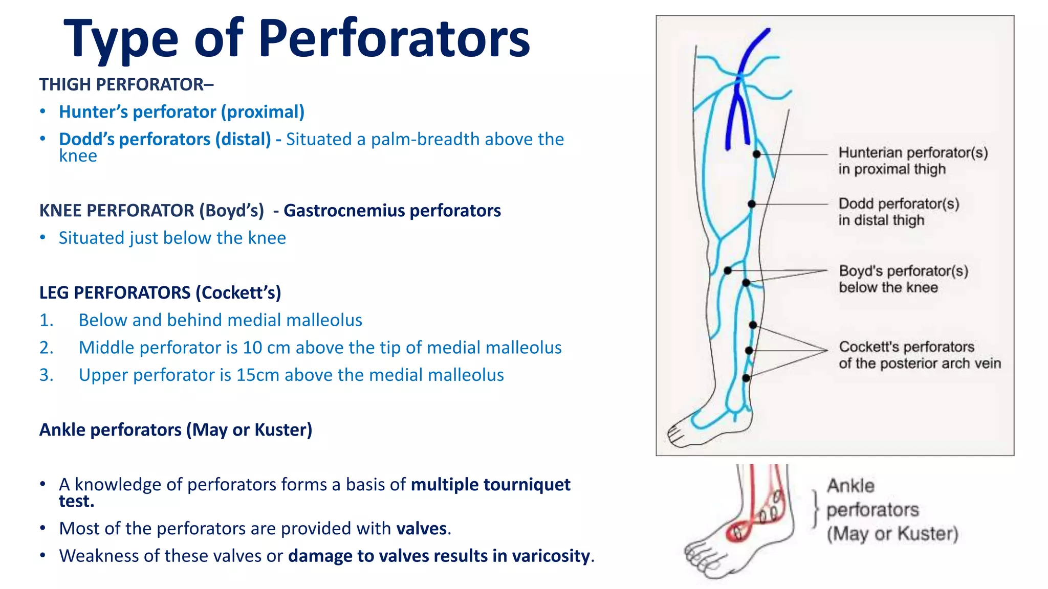
The document summarizes the anatomy of the venous system in the upper and lower limbs. In the upper limbs, it describes the superficial cephalic and basilic veins and their tributaries, as well as the deep brachial, ulnar, and radial veins. In the lower limbs, it outlines the long and short saphenous veins and their tributaries in the superficial system, and the deep femoral, popliteal, tibial and other veins. It also discusses perforating veins that connect the superficial and deep systems and their clinical significance.
The main artery of the lower limb is the femoral artery. It is a continuation of the external iliac artery (terminal branch of the abdominal aorta). The external iliac becomes the femoral artery when it crosses under the inguinal ligament and enters the femoral triangle.
In the femoral triangle, the profunda femoris artery arises from the posterolateral aspect of the femoral artery. It travels posteriorly and distally, giving off three main branches:
Perforating branches – Consists of three or four arteries that perforate the adductor magnus, contributing to the supply of the muscles in the medial and posterior thigh.
Lateral femoral circumflex artery – Wraps round the anterior, lateral side of the femur, supplying some of the muscles on the lateral aspect of the thigh.
Medial femoral circumflex artery – Wraps round the posterior side of the femur, supplying its neck and head. In a fracture of the femoral neck this artery can easily be damaged, and avascular necrosis of the femur head can occur.
In human anatomy, the thigh is the area between the hip (pelvis) and the knee. Anatomically, it is part of the lower limb. The single bone in the thigh is called the femur.
It is a power point presentation by Dr Zobayer Mahmud Khan while he was taking a lecture class of this topic in Sir Salimullah Medical College, Mitford, Dhaka. Here the venous drainage and the lymphatic drainage are stated clearly with many of the clinical events. Hope you can learn from here. Photographs used here are collected from The "Essential of human Anatomy (inferior extremity) by A.K. Datta".
Here is the powerpoint on relevent anatomy of multiple differentials for Inguinoscrtal swelling special for surgical diagnosis with very reliable References.
venous drainage of the upper limb, median vein of forearm, deep veins, basilic vein, cephalic vein, median cubital vein, superficial vein, dorsal venous arch,
Arterial Supply and Venous Drainage of Pelvis.pptxMathew Joseph
The rich vascular supply of the pelvis not only supports the structures contained within it, including the bladder, rectum, and reproductive organs, but also extends to the lower extremities. For a complete understanding of vascular anatomy as it pertains into the endovascular procedures of interventional radiology, it is useful to discuss the vascular structures in sections, from the bifurcation of the aorta and the inferior vena cava to the level of the common femoral arteries and veins. We will also review the anatomy of the iliac vessels, including their branches, common variants, and various collateral pathways
Honest Reviews of Tim Han LMA Course Program.pptxtimhan337
Personal development courses are widely available today, with each one promising life-changing outcomes. Tim Han’s Life Mastery Achievers (LMA) Course has drawn a lot of interest. In addition to offering my frank assessment of Success Insider’s LMA Course, this piece examines the course’s effects via a variety of Tim Han LMA course reviews and Success Insider comments.
Model Attribute Check Company Auto PropertyCeline George
In Odoo, the multi-company feature allows you to manage multiple companies within a single Odoo database instance. Each company can have its own configurations while still sharing common resources such as products, customers, and suppliers.
Welcome to TechSoup New Member Orientation and Q&A (May 2024).pdfTechSoup
In this webinar you will learn how your organization can access TechSoup's wide variety of product discount and donation programs. From hardware to software, we'll give you a tour of the tools available to help your nonprofit with productivity, collaboration, financial management, donor tracking, security, and more.
Biological screening of herbal drugs: Introduction and Need for
Phyto-Pharmacological Screening, New Strategies for evaluating
Natural Products, In vitro evaluation techniques for Antioxidants, Antimicrobial and Anticancer drugs. In vivo evaluation techniques
for Anti-inflammatory, Antiulcer, Anticancer, Wound healing, Antidiabetic, Hepatoprotective, Cardio protective, Diuretics and
Antifertility, Toxicity studies as per OECD guidelines
The French Revolution, which began in 1789, was a period of radical social and political upheaval in France. It marked the decline of absolute monarchies, the rise of secular and democratic republics, and the eventual rise of Napoleon Bonaparte. This revolutionary period is crucial in understanding the transition from feudalism to modernity in Europe.
For more information, visit-www.vavaclasses.com
Normal Labour/ Stages of Labour/ Mechanism of LabourWasim Ak
Normal labor is also termed spontaneous labor, defined as the natural physiological process through which the fetus, placenta, and membranes are expelled from the uterus through the birth canal at term (37 to 42 weeks
Operation “Blue Star” is the only event in the history of Independent India where the state went into war with its own people. Even after about 40 years it is not clear if it was culmination of states anger over people of the region, a political game of power or start of dictatorial chapter in the democratic setup.
The people of Punjab felt alienated from main stream due to denial of their just demands during a long democratic struggle since independence. As it happen all over the word, it led to militant struggle with great loss of lives of military, police and civilian personnel. Killing of Indira Gandhi and massacre of innocent Sikhs in Delhi and other India cities was also associated with this movement.
Unit 8 - Information and Communication Technology (Paper I).pdfThiyagu K
This slides describes the basic concepts of ICT, basics of Email, Emerging Technology and Digital Initiatives in Education. This presentations aligns with the UGC Paper I syllabus.
3. Venous System In Upper Limbs
Superficial veins
- Cephalic vein (preaxial)
- Basilic vein (postaxial)
- Median cubital veins
Deep veins
- Brachial veins
- Ulnar veins
- Radial Veins
4. Lateral end of dorsal venous
arch
Winds round
↑
Continues upwards in front of
elbow
↑
Lateral border of biceps brachi
↑
Pierces deep fascia
↑
Runs in deltopectoral groove up
to infraclavicular fossa
↑
Pierces clavipectoral fascia
↑
Joins the axillary veins
↑
CEPHALIC VEINS
Medial end of dorsal venous arch
Along back of medial border of
forearm
↑
Winds round
↑
Medial margin of biceps brachi
↑
Pierces deep fascia
↑
Middle of arm
↑
Lower border of teres major
↑
becomes the axillary veins
↑
BASILIC VEINS
6. MEDIAN CUBITAL VEIN
• Large communicating vein which shunts blood from
cephalic to the basilica vein
• Starts : from cephalic vein 2.5 cm below bend of elbow
• Runs obliquely upward and medially
• Ends : in the basilic vein 2.5 cm above medial epicondyle
7. Clinical and surgical correlation
1. These superficial veins are commonly used for
intravenous injection and for withdrawing blood
for transfusion or for testing.
2. Cannulation via basilic vein is more liable
compared to cephalic because basilic vein
becomes the axillary vein without angulation (will
not prone to obstruction)
3. Axillary vein thrombosis may occur due to
prolonged abduction of the arm above the head,
as in painting ceiling.
8. Deep vein
• Situated underneath the
deep fascia
• Venae comitantes:
• It refers to a vein that is
usually paired, with both
veins lying on the sides of
an artery.
Brachial veins
Ulnar veins
Radial veins
9. Venous system in lower limbs
• Superficial venous system
• Long ( Great ) Saphenous Veins (LSV/GSV)
• Short Saphenous Veins
• Deep venous system
• Iliac vein
• Femoral vein
• Popliteal vein
• Peroneal vein
• Tibial vein
• Soleal vein
10. LONG SAPHAENOUS VEIN
• Largest and longest superficial veins of the lower limb
• Starts : on dorsum of the foot by union of medial end of dorsal
venous arch with the medial marginal vein
• Ascends in front of medial malleolus
• Runs medial side of the leg
• Pass posterior of medial epicondyle of femur – behind knee
• Inclines forwards in thigh
• Reach saphenous opening where it pierces the cribriform fascia
• Ends : Saphenofemoral junction (located at 4 cm below and lateral
to pubic tubercle) by joining the femoral vein
• In lower part of LSV – associated with saphenous nerve
12. • Tributaries connect the long saphenous vein with short
saphenous veins
• Called as ‘Communicators’
13. SHORT(SMALL) SAPHENOUS VEIN
• Starts : on dorsum of the foot b union of lateral end of
dorsal venous arch with the lateral marginal vein
• Passing behind the lateral malleolus - to the back of leg
• Ascending along lateral margin of tendon calcaneus
(Achilles)
• Runs at middle of the back of legs
• continues until the lower part of popliteal fossa -
between two heads of gastrocnemius
• Pierces deep fascia
• Communicates with long saphenous via the tributaries
• Ends : Empty into popliteal veins
• Nerve : is accompanied by a cutaneous nerve (sural nerve)
14. DEEP VEINS
• Receive blood from superficial veins
through perforating veins
• Comprise of venae comitantes
• Anterior tibial vein
• Posterior tibial vein
• Peroneal veins
• They unite to form the popliteal vein
femoral vein external iliac veins
• These veins have more valves than
superficial veins. They are more
efficient channels than the superficial
veins because of the driving force of
muscular contraction
15.
16.
17. Salient features of deep vein:
• High pressure system, well-supported by powerful
muscles
• Powerful calf muscle contraction returns the blood
to the heart
• Connected to superficial veins by perforators
• Deep veins are also provided with valves except
soleal vein
18. Type of Perforators
THIGH PERFORATOR–
• Hunter’s perforator (proximal)
• Dodd’s perforators (distal) - Situated a palm-breadth above the
knee
KNEE PERFORATOR (Boyd’s) - Gastrocnemius perforators
• Situated just below the knee
LEG PERFORATORS (Cockett’s)
1. Below and behind medial malleolus
2. Middle perforator is 10 cm above the tip of medial malleolus
3. Upper perforator is 15cm above the medial malleolus
Ankle perforators (May or Kuster)
• A knowledge of perforators forms a basis of multiple tourniquet
test.
• Most of the perforators are provided with valves.
• Weakness of these valves or damage to valves results in varicosity.
19. References
• SRB’s Manual of Surgery, Bhat M, S. (2016). 5th ed. Jaypee
Brothers Medical Publishers
• Manipal Manual of Surgery, 4th Edition, K.Rajgopal Shenoy
• B D Chaurasia Human anatomy, 5th edition