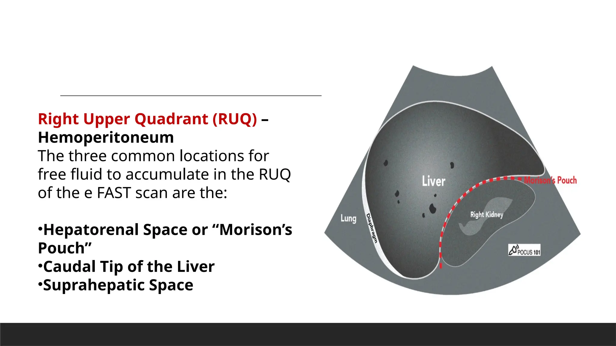
Focused Assessment
with Sonography for
Trauma (FAST) scan
D R .N I S H A N T M I S H R A
M B B S ( I M S , B A NA RA S H I N D U UN I V ERS I T Y, VA R A NA S I )
M D R A D I OD I AG N OS I S ( P G I , ROH TAK )
EX- MED I CA L OF F I C ER T EL EM ED I CI N E RRC B . H . U
S EN I OR RES I D EN T D OC TOR K. M ME D I C A L COL L EGE, M AT H UR A
CO N S U L ATA N T R A D I OLOGI ST R .K M I S S I ON H OS P I TA L , V RI N DAVA N
Focused Assessment with Sonography for
Trauma (FAST) scan is a point-of-care ultrasound (POCUS)
examination performed at the time of presentation of a
trauma patient.
Some studies have shown no significant difference in
diagnostic accuracy between radiologists and non-
radiologists .
Indications-
•Hemodynamically unstable trauma patient
•Abdominal and Thoracic Trauma: Blunt or Penetrating
•Previously stable trauma patient with acute worsening in clinical status
Limitations-
•Does not localize the injured abdominal organ
•Views may be limited in patients with subcutaneous emphysema
•Views may be limited in patients who have a hollow-viscus injury with free air in the abdomen
Patient Preparation
Patient lying supine with the exam table flat .
Machine Preparation
•Transducer: Curvilinear Probe can be used as well, however, the cardiac
views may be difficult to obtain given the large footprint of the
transducer.
•Preset: FAST exam or Abdominal Exam.
•Ultrasound Machine Placement: Place the machine on the patient’s
right side. This makes it possible to scan with your right hand and
manipulate ultrasound controls with your left hand.
1.Right Upper Quadrant View (RUQ)
2.Left Upper Quadrant View (LUQ)
3.Pelvic View
4.Cardiac View (Parasternal Long Axis
or Subxiphoid)
5.Lungs (Right and Left)
Recommended e FAST Exam
Sequence-
Step-By-Step e FAST Exam Protocol
The e FAST Exam serves to answer 4 questions:
1.Does my patient have free fluid in the Abdomen?
2.Does my patient have free fluid in the Thorax?
3.Does my patient have fluid in the Pericardium?
4.Does my patient have a Pneumothorax?
Step 1: e FAST Right Upper Quadrant View (RUQ)
Does my patient have free fluid in the abdomen or right thorax?
Since the liver is the most commonly injured organ in
blunt abdominal trauma, the right upper quadrant is
usually the most sensitive view of the eFAST exam.
•Orientate the probe indicator towards the patient’s head.
•Anchor your probe in the midaxillary line at the 10th intercostal space
RUQ Probe Position and Hand Placement
RUQ Normal View and Structures
•Using the liver as an acoustic window, identify the lung, liver, Morison’s Pouch,
diaphragm, and the long-axis of the right kidney.
•Morison’s Pouch is where you usually identify free fluid in the RUQ view.
•A Mirror Image Artifact is a normal finding signifying there is an aerated lung above the
diaphragm.
•You may have to slide up or down a rib space to identify the structures.
• These structures move as the diaphragm contracts and relaxes during the respiratory cycle. Consider asking
your patient to hold their breath to keep the desired organs from moving. Also, consider slightly rotating
the probe counterclockwise towards the bed so that the probe fits better between the rib spaces.
Step 2: eFAST Left Upper Quadrant View (LUQ)
Does my patient have free fluid in the abdomen or left thorax?
LUQ Probe Position and Hand Placement
•Grasp the linear probe between your thumb and first finger, like holding
a pencil.
•Orientate the probe indicator towards the patient’s head.
•Anchor your probe in the posterior axillary line around the 8th
intercostal space.
•You should have your “Knuckles to the bed” since the spleen is fairly
posterior.
Knuckles to the Bed” for the LUQ eFAST exam
view
LUQ Normal View and Structures-
•Using the spleen as an acoustic window, identify the spleen, perisplenic
space, diaphragm, and the long-axis view of the left kidney.
•Free fluid in the LUQ is most frequently seen in the peri splenic space (between the
spleen and the diaphragm). The reason is that there is a splenorenal ligament limiting
the ability of fluid to track in between the spleen and left kidney
•A Mirror Image Artifact is a normal finding, similar to the RUQ, signifying there is an
aerated lung above the diaphragm.
•You may have to move up or down a rib space to identify the structures.
•These structures move as the diaphragm contracts and relaxes during the respiratory
cycle. Consider asking your patient to hold their breath to keep the desired organs from
moving. Also, consider slightly rotating the probe clockwise towards the bed, so that the
probe fits better between the rib spaces.
Step 3: eFAST Pelvic View –
Does my patient have free fluid in the abdomen or pelvis?
When looking for free fluid in the pelvis it’s important to consider the sex of your patient,
as free fluid has a tendency to accumulate in different locations depending on the patient’s
gender.
Additionally, females may have a scant amount of normal physiologic fluid accumulation in
the pelvis (pouch of Douglas).
•Ideally, scan your patient with a full bladder (so the bladder can be used as an acoustic
window). Therefore, try to initiate the scan prior to foley catheter placement.
•Machine Depth: 10-15 cm
Pelvic Ultrasound – Longitudinal View
•Place the transducer with the indicator
pointing towards the patient’s head in the
patient’s midline, right above the pubic
symphysis.
•Rock the probe so that it points down
towards the pelvic cavity.
Make sure to point your probe into the pelvis!
One of the most important things to remember
when performing pelvic ultrasound in the eFAST
scan is that the bladder is directly posterior to the
pubic bone/symphysis. If you are unable to get
proper images, most likely your ultrasound probe
is placed too superiorly.
Male eFAST exam Pelvic View – Longitudinal Male eFAST exam Pelvic View – Longitudinal
•In males, identify the bladder (immediately posterior to the
symphysis), prostate/seminal vesicle, and rectovesical pouch in the longitudinal
view.
•The rectovesical pouch is where free fluid will accumulate in the male pelvis.
•In Females, identify the bladder, uterus, and Rectouterine Pouch (also called the Pouch of Douglas).
•The Pouch of Douglas is where free fluid will accumulate in the female pelvis.
Female eFAST exam Pelvic View – Longitudinal
•In all patients (male or female), observe
the lateral borders of the bladder to
identify free fluid by tilting/fanning the
probe left and right.
Pelvic Ultrasound – Transverse View
•Next, center the bladder and then rotate the
transducer 90 degrees counterclockwise. The
indicator should now point to the patient’s Right
side.
•Make sure to tilt the ultrasound probe so it scans
into the pelvic cavity.
Female eFAST exam Pelvic View – Transverse Male eFAST exam Pelvic View – Transverse
•Tilt/Fan the probe to examine the entire
pelvis from superior to inferior.
Transverse view of the eFAST Pelvic View
Step 4: e FAST Cardiac View
Does my patient have a pericardial effusion with cardiac tamponade?
The subxiphoid view is the preferred scan to evaluate the heart and
assess for any pericardial fluid. However, the subxiphoid view can
be difficult to obtain in some situations (e.g. patient body habitus
or abdominal pain with trauma). If the subxiphoid view cannot be
obtained, a parasternal long axis view is recommended.
•Hold the probe in the palm of your hand and use
an overhand grip.
•Point the probe indicator towards
the patient’s right with the ultrasound
machine depth set to around 15-20 cm.
•Using the liver as the acoustic
window, simultaneously press the probe into the
patient’s abdomen while tilting the tail of the
probe towards the patient’s feet
•Aim the ultrasound beam towards the patient’s
left shoulder.
Cardiac Subxiphoid View
Identify the liver, pericardium, right atrium, right ventricle, left atrium and left
ventricle
eFAST Subxiphoid View Structures
Cardiac Parasternal Long Axis
Consider obtaining the parasternal long-axis view of the heart in a patient when you are unable to
obtain the subxiphoid view. Otherwise, proceed to scanning the lungs.
•Grasp the linear probe between
your thumb and first finger,
like holding a pencil.
•Anchor your third and/or fourth
finger(s) in the 2nd or 3rd left
intercostal space, just lateral to
the sternum.
•Probe indicator towards the
patient’s left hip with the
machine depth set approximately 10-
15 cm e FAST Parasternal Long Axis View Probe Position
•e FAST Parasternal Long Axis View -
•Structures Identify the pericardium, mitral valve, aortic valve, right ventricle, descending aorta,
left atrium, and left ventricle
•You may have to move up or down a rib space to identify the structures
Step 5: eFAST Lung Views –
Does my patient have a pneumothorax?
The parietal pleura covers the surface of the thorax and the visceral
pleura covers the lungs.
The presence of these moving against one another produces lung
sliding on ultrasound.
Lung Probe Position and Hand Placement
•Point your indicator towards the patient’s head.
•Place your probe at the mid-clavicular line at the 2nd intercostal space of the right and left lungs
respectively. This point is the most sensitive spot for looking for pneumothorax in the supine patient.
•Anchor your probe in the space between two ribs and set the ultrasound machine depth to 3-5cm.
Identify Two Rib Shadows (Batwing Sign)
•The first lung ultrasound finding to confirm you are in the correct position is to look for the
two rib shadows or the “Batwing Sign.” This ensures that your probe is in between two ribs.
•The next finding you will want to look for is lung
sliding during respiration.
•Lung sliding is a normal finding where the visceral and
parietal pleura slide back and forth on one another as the
patient breathes. Some say this looks like tiny “ants
marching on a line.”
•“Ants Marching” sign is produced from the visceral and
parietal pleura moving against one another during
respiration.
•This is a simple finding but extremely useful since lung
sliding definitely means that the visceral and parietal pleura
are next to each other, effectively ruling out a pneumothorax.
Identify Lung Sliding
Identify Lung Sliding using M-Mode (Optional)
•If lung sliding is not readily apparent, it can be
further be evaluated using M-Mode. The goal of M-
Mode is to see if the patient has a normal seashore
sign.
•Place the ultrasound machine in M-Mode (M-Mode
for motion)
•Place the ultrasound doppler indicator/cursor over
the lung field (NOT over the rib).
•Look for the normal “Seashore Sign“: Sky =
Skin/Subcutaneous Tissue, Ocean= Muscle, Beach =
Lung sliding motion (sandy appearance).
•The Seashore Sign is a NORMAL finding.
•Contrast this with the Barcode sign (see in e FAST
pathology section below)
e FAST Ultrasound Pathology
Recall that fluid will appear black, or anechoic.
For the purposes of the eFAST exam we are looking for anechoic
(black) areas in the abdomen, chest, and heart that signify bleeding in
those potential spaces
.
For pneumothorax we will be evaluating the presence or absence of
lung sliding.
Hemoperitoneum – eFAST
The eFAST is moderately sensitive (approximately 80%) and highly specific (>90%) for
detecting free fluid from hemoperitoneum.
The general consensus is that there needs to be at least 200-250ml of blood before the
eFAST scan will appear positive.
Another important point to remember for the e FAST scan is that observing free fluid on
the eFAST scan does not localize the bleeding to a specific organ.
For example, if free fluid is noted in the pelvis, it could be originating from anywhere in the
abdomen, and does not localize the injury to the bladder.
A CT scan is needed to localize the origin of abdominal bleeding in a trauma patient.
Right Upper Quadrant (RUQ) –
Hemoperitoneum
The three common locations for
free fluid to accumulate in the RUQ
of the e FAST scan are the:
•Hepatorenal Space or “Morison’s
Pouch”
•Caudal Tip of the Liver
•Suprahepatic Space
Free Fluid at the Caudal Tip of the Liver Free Fluid in Morrison’s Pouch and Suprahepatic
Space
Left Upper Quadrant (LUQ) –
Hemoperitoneum
We will evaluate the LUQ in the eFAST for free
fluid in the following places:
•Perisplenic Space
•Spleen Tip
•Splenorenal Recess
POCUS 101 TIP: It is important to note that in the
LUQ the most common area to find fluid is in the
perisplenic space, NOT between the spleen and
the left kidney. This is because there is a
splenorenal ligament that attaches the spleen
and the left kidney preventing a significant
amount of fluid to accumulate there unless the
ligament is ruptured.
Free fluid in Perisplenic Space
Male Pelvis – Hemoperitoneum
In the male pelvis, you can find free fluid in the rectovesical pouch/space.
Hemothorax – eFAST
After evaluating the RUQ or LUQ, move the probe
superiorly one or two rib spaces to evaluate the
thorax for fluid accumulation.
A normal lung will have a Mirror Image Artifact
and you will be unable to see the spine going
above the diaphragm since all of the ultrasound
waves will be reflected back by the aerated lung.
Visualizing the patient’s spine above the
diaphragm implies that there is free fluid (e.g.
blood) in the thorax since ultrasound waves can
easily pass through the free fluid in the chest
cavity, allowing you to see the spine. This is
referred to as a Positive Spine Sign (click here for
a more in-depth explanation on the spine sign).
Absence of Spine. Normal Finding. Presence of Spine. Pathologic
Finding.
Pericardial Effusion and Tamponade – eFAST
•Anechoic free fluid can accumulate in the pericardial sac causing a pericardial effusion.
•Simply seeing a pericardial effusion not mean the patient has cardiac tamponade. Rather,
the fluid must be impairing cardiac filling for it to be considered tamponade.
•Consider tamponade when the following is observed:
• Right Atrial Systolic Collapse – the most sensitive (and earliest) echocardiographic
finding of tamponade (Perez-Casares, A., et al). Also referred to as the Trampoline
Sign.
• Right Ventricle Diastolic Collapse – the most specific echocardiographic finding for
tamponade (75-90%) (Armstrong, et al)
eFAST Subxiphoid View with Pericardial Effusion
eFAST Parasternal Long Axis view with Pericardial Effusion
and Tamponade (RV Diastolic Collapse)
Pneumothorax – eFAST
Here are three important steps to evaluating for pneumothorax when performing the eFAST scan:
First, if lung sliding is present, you can rule out pneumothorax with 100% accuracy at that
ultrasound point (Husain LF).
You can look for lung sliding with B-mode or M-mode:
Normal Lung Sliding with Seashore sign (M-mode)
Normal Lung Sliding (B-mode)
Second, if lung sliding is ABSENT, you should not automatically assume pneumothorax.
Recall other causes of reduced/absent lung sliding: severe consolidation, chemical pleurodesis,
acute infectious or inflammatory states, fibrotic lung diseases, acute respiratory distress
syndrome, or mainstem intubation.
Absence of Lung Sliding (B-mode) Absence of Lung Sliding – Barcode Sign (M-Mode)
For the purposes of the eFAST scan, it is highly likely that your patient has a pneumothorax if you
do not see lung sliding on B-mode or M-mode. If you want to confirm you can proceed to look
for the “Lung Point Sign” below.
Third, if a lung point is present, you can rule in pneumothorax with 100% accuracy (Chan S).
To confirm the presence of a pneumothorax, you should look for the “Lung Point Sign.“
The lung point is when you can see the transition between normal lung sliding and the absence
of lung sliding. This is the transition point between the collapsed lung and normal lung. If you
see this you can definitively rule in a pneumothorax. The Lung point sign also helps you quantify
how large a pneumothorax is.
If you think you may have found a lung point but are not sure, use M-Mode and place your
cursor at the intersection where you think lung sliding starts and stops. If you see a normal
seashore sign that turns into an abnormal barcode sign, then you have located the lung point
with M-Mode.
Lung Point Sign (B-mode) Lung Point (M-mode)
Pneumoperitoneum – eFAST
In the setting of trauma (especially penetrating trauma) you may encounter pneumoperitoneum,
or free air within the peritoneal cavity.
On abdominal ultrasound, the most common finding for pneumoperitoneum is the Enhanced
Peritoneal Stripe Sign (EPSS). This is when air within the peritoneal space rises and causes an
“echoing” of the usually single, hyperechoic peritoneal stripe that separates the abdominal wall
from underlying peritoneal fluid and fluid-filled organs (Indiran).
If there is a large amount of pneumoperitoneum, your image of abdominal organs will be
obscured by air wherever you place your probe.
POCUS 101 tip: if you can’t get any good abdominal views despite having your probe in the correct
position, have a high suspicion for pneumoperitoneum.
Enhanced peritoneal stripe sign (EPSS) seen anterior to the liver in both images (straight
arrows), indicating the abnormal presence of air between the liver and the anterior
abdominal wall (Indiran).
e FAST Algorithm and Summary
•Remember that the POCUS eFAST ultrasound exam is most beneficial in hemodynamically
unstable patients who are unable to go to the CT scanner. A positive eFAST scan can help the
surgeon identify the general region of bleeding (i.e. abdomen vs heart vs lungs) to plan their
surgical approach.
•A negative initial eFAST exam in patients with a highly-suspicious mechanism of injury may
benefit from a CT scan or serial eFAST exams, especially in the context of a worsening clinical
status (e.g. worsening vitals, hemodynamic instability, worsening pain, or worsening abdominal
exam) as patients can also have a delayed presentation.
The chief aim of the study, in a trauma patient, is to identify
intraperitoneal free fluid (assumed to be haemoperitoneum in the context of
trauma) allowing for an immediate transfer to operating theatre, CT or other. Solid
organ injury is seldom identified, and when present may warrant further
investigation.
Many papers have been published detailing the pros and cons of this
investigation 1,2
. FAST scanning has a reported sensitivity of ~90% (range 75-100%)
and a specificity of ~95% (range 88-100%) for detecting intraperitoneal free fluid 4
.
Sensitivity for detecting solid organ injuries is much lower.
Most studies in the emergency medicine literature dictate that peritoneal free fluid will
not be identified by ultrasonography until more than 500 mL is present. Therefore, a
negative exam will not preclude a bleed which will eventually become
significant. Moreover, mesenteric vascular injuries, solid organ injuries, hollow viscus
injuries, and diaphragmatic injuries may not result in free intraperitoneal fluid, and thus
may not be detected 10
.
It has replaced diagnostic peritoneal lavage as the preferred initial method for
assessment of haemoperitoneum.
In several recent studies, the sensitivity and specificity of thoracic ultrasonography use
for the detection of pneumothorax after blunt injury was 86-98% and 97-100%,
respectively, outperforming the supine chest x-ray
The original “FAST Exam” consisted of 3 views: the right upper quadrant, the left upper
quadrant, and the pelvis to rule out bleeding in the abdomen from trauma. It is also
commonly referred to as the “FAST Scan“
The eFAST exam incorporates the evaluation of the lungs and heart in addition to
the abdomen.
E FAST is efficient way to be able to use Point of Care Ultrasound (POCUS) to:
•Perform the complete eFAST Ultrasound Exam Protocol in 5 simple Steps
•Evaluate a patient with suspected intra-abdominal or intrathoracic free fluid collection
•Evaluate a patient for suspected cardiac tamponade
•Evaluate a patient for a suspected pneumothorax
Technique
•patient in supine position
•3.5-5.0 MHz convex transducer
•five regions may be scanned
•Pericardial view :
commonly referred to as the subcostal or subxiphoid view
• to examine the pericardium, the liver in the epigastric region is most commonly used as a
sonographic window to the heart
• the potential space between the visceral and parietal pericardium is examined for a
pericardial effusion
• if anatomical factors preclude epigastric probe placement, parasternal or apical four-
chamber views may be used
•Right flank view
• commonly referred to as the perihepatic view, Morison pouch view or right upper quadrant
view
• four potential spaces are sequentially examined for the accumulation of free fluid
• the hepatorenal interface (Morison pouch) is first identified, with subsequent assessment
of the more cephalad subphrenic and pleural spaces
• visualisation of the inferior pole of the kidney, which is a continuation of the right paracolic
gutter, defines the caudad extent of an adequate view
•Left flank view
• commonly referred to as the perisplenic or left upper quadrant view
• four potential spaces are sequentially examined in an analogous fashion to the right flank,
albeit the splenorenal interface is assessed on the left
•Pelvic view
• commonly referred to as the suprapubic view, this space is the most dependent peritoneal
space in the supine trauma patient
• a transverse sweep, using the bladder as a sonographic window, the pouch of Douglas or
rectovesical space is explored for free fluid
An extended FAST or "eFAST" scan is now standard of care, and is performed by
incorporating two views assessing the anterior thorax 7
:
•anterior pleural views
• the anterior pleura is assessed for the presence or absence of lung sliding as a sensitive,
but non-specific, indicator of a traumatic pneumothorax
• the probe is placed in a sagittal orientation in the midclavicular line between the clavicle
and diaphragm
• anterior and lateral interrogation of interspaces 5-8 bilaterally is recommended
Causes of false negatives
•obesity: severely limits assessment of the peritoneal cavity
•subcutaneous emphysema
•posterior acoustic enhancement caused by the fluid-filled bladder can result in free fluid
being missed in the pelvic view
Causes of false positives
•epicardial fat pads, the descending aorta, and pericardial cysts have been mistakenly
identified as an effusion
•pre-existing ascites, pleural, and pericardial effusions due to medical conditions
•seminal vesicles mistaken for pelvic free fluid in the young male patient

Focused Assessment with Sonography for Trauma (FAST.pptx

  • 1.
    Focused Assessment with Sonographyfor Trauma (FAST) scan D R .N I S H A N T M I S H R A M B B S ( I M S , B A NA RA S H I N D U UN I V ERS I T Y, VA R A NA S I ) M D R A D I OD I AG N OS I S ( P G I , ROH TAK ) EX- MED I CA L OF F I C ER T EL EM ED I CI N E RRC B . H . U S EN I OR RES I D EN T D OC TOR K. M ME D I C A L COL L EGE, M AT H UR A CO N S U L ATA N T R A D I OLOGI ST R .K M I S S I ON H OS P I TA L , V RI N DAVA N
  • 2.
    Focused Assessment withSonography for Trauma (FAST) scan is a point-of-care ultrasound (POCUS) examination performed at the time of presentation of a trauma patient. Some studies have shown no significant difference in diagnostic accuracy between radiologists and non- radiologists .
  • 3.
    Indications- •Hemodynamically unstable traumapatient •Abdominal and Thoracic Trauma: Blunt or Penetrating •Previously stable trauma patient with acute worsening in clinical status Limitations- •Does not localize the injured abdominal organ •Views may be limited in patients with subcutaneous emphysema •Views may be limited in patients who have a hollow-viscus injury with free air in the abdomen
  • 4.
    Patient Preparation Patient lyingsupine with the exam table flat .
  • 5.
    Machine Preparation •Transducer: CurvilinearProbe can be used as well, however, the cardiac views may be difficult to obtain given the large footprint of the transducer. •Preset: FAST exam or Abdominal Exam. •Ultrasound Machine Placement: Place the machine on the patient’s right side. This makes it possible to scan with your right hand and manipulate ultrasound controls with your left hand.
  • 6.
    1.Right Upper QuadrantView (RUQ) 2.Left Upper Quadrant View (LUQ) 3.Pelvic View 4.Cardiac View (Parasternal Long Axis or Subxiphoid) 5.Lungs (Right and Left) Recommended e FAST Exam Sequence-
  • 7.
    Step-By-Step e FASTExam Protocol The e FAST Exam serves to answer 4 questions: 1.Does my patient have free fluid in the Abdomen? 2.Does my patient have free fluid in the Thorax? 3.Does my patient have fluid in the Pericardium? 4.Does my patient have a Pneumothorax?
  • 8.
    Step 1: eFAST Right Upper Quadrant View (RUQ) Does my patient have free fluid in the abdomen or right thorax? Since the liver is the most commonly injured organ in blunt abdominal trauma, the right upper quadrant is usually the most sensitive view of the eFAST exam.
  • 9.
    •Orientate the probeindicator towards the patient’s head. •Anchor your probe in the midaxillary line at the 10th intercostal space RUQ Probe Position and Hand Placement
  • 10.
    RUQ Normal Viewand Structures •Using the liver as an acoustic window, identify the lung, liver, Morison’s Pouch, diaphragm, and the long-axis of the right kidney. •Morison’s Pouch is where you usually identify free fluid in the RUQ view. •A Mirror Image Artifact is a normal finding signifying there is an aerated lung above the diaphragm. •You may have to slide up or down a rib space to identify the structures. • These structures move as the diaphragm contracts and relaxes during the respiratory cycle. Consider asking your patient to hold their breath to keep the desired organs from moving. Also, consider slightly rotating the probe counterclockwise towards the bed so that the probe fits better between the rib spaces.
  • 12.
    Step 2: eFASTLeft Upper Quadrant View (LUQ) Does my patient have free fluid in the abdomen or left thorax? LUQ Probe Position and Hand Placement •Grasp the linear probe between your thumb and first finger, like holding a pencil. •Orientate the probe indicator towards the patient’s head. •Anchor your probe in the posterior axillary line around the 8th intercostal space. •You should have your “Knuckles to the bed” since the spleen is fairly posterior.
  • 13.
    Knuckles to theBed” for the LUQ eFAST exam view
  • 14.
    LUQ Normal Viewand Structures- •Using the spleen as an acoustic window, identify the spleen, perisplenic space, diaphragm, and the long-axis view of the left kidney. •Free fluid in the LUQ is most frequently seen in the peri splenic space (between the spleen and the diaphragm). The reason is that there is a splenorenal ligament limiting the ability of fluid to track in between the spleen and left kidney •A Mirror Image Artifact is a normal finding, similar to the RUQ, signifying there is an aerated lung above the diaphragm. •You may have to move up or down a rib space to identify the structures. •These structures move as the diaphragm contracts and relaxes during the respiratory cycle. Consider asking your patient to hold their breath to keep the desired organs from moving. Also, consider slightly rotating the probe clockwise towards the bed, so that the probe fits better between the rib spaces.
  • 16.
    Step 3: eFASTPelvic View – Does my patient have free fluid in the abdomen or pelvis? When looking for free fluid in the pelvis it’s important to consider the sex of your patient, as free fluid has a tendency to accumulate in different locations depending on the patient’s gender. Additionally, females may have a scant amount of normal physiologic fluid accumulation in the pelvis (pouch of Douglas). •Ideally, scan your patient with a full bladder (so the bladder can be used as an acoustic window). Therefore, try to initiate the scan prior to foley catheter placement. •Machine Depth: 10-15 cm
  • 17.
    Pelvic Ultrasound –Longitudinal View •Place the transducer with the indicator pointing towards the patient’s head in the patient’s midline, right above the pubic symphysis. •Rock the probe so that it points down towards the pelvic cavity.
  • 18.
    Make sure topoint your probe into the pelvis! One of the most important things to remember when performing pelvic ultrasound in the eFAST scan is that the bladder is directly posterior to the pubic bone/symphysis. If you are unable to get proper images, most likely your ultrasound probe is placed too superiorly.
  • 19.
    Male eFAST examPelvic View – Longitudinal Male eFAST exam Pelvic View – Longitudinal •In males, identify the bladder (immediately posterior to the symphysis), prostate/seminal vesicle, and rectovesical pouch in the longitudinal view. •The rectovesical pouch is where free fluid will accumulate in the male pelvis.
  • 20.
    •In Females, identifythe bladder, uterus, and Rectouterine Pouch (also called the Pouch of Douglas). •The Pouch of Douglas is where free fluid will accumulate in the female pelvis. Female eFAST exam Pelvic View – Longitudinal
  • 21.
    •In all patients(male or female), observe the lateral borders of the bladder to identify free fluid by tilting/fanning the probe left and right.
  • 22.
    Pelvic Ultrasound –Transverse View •Next, center the bladder and then rotate the transducer 90 degrees counterclockwise. The indicator should now point to the patient’s Right side. •Make sure to tilt the ultrasound probe so it scans into the pelvic cavity.
  • 23.
    Female eFAST examPelvic View – Transverse Male eFAST exam Pelvic View – Transverse
  • 24.
    •Tilt/Fan the probeto examine the entire pelvis from superior to inferior. Transverse view of the eFAST Pelvic View
  • 25.
    Step 4: eFAST Cardiac View Does my patient have a pericardial effusion with cardiac tamponade? The subxiphoid view is the preferred scan to evaluate the heart and assess for any pericardial fluid. However, the subxiphoid view can be difficult to obtain in some situations (e.g. patient body habitus or abdominal pain with trauma). If the subxiphoid view cannot be obtained, a parasternal long axis view is recommended.
  • 26.
    •Hold the probein the palm of your hand and use an overhand grip. •Point the probe indicator towards the patient’s right with the ultrasound machine depth set to around 15-20 cm. •Using the liver as the acoustic window, simultaneously press the probe into the patient’s abdomen while tilting the tail of the probe towards the patient’s feet •Aim the ultrasound beam towards the patient’s left shoulder. Cardiac Subxiphoid View
  • 27.
    Identify the liver,pericardium, right atrium, right ventricle, left atrium and left ventricle eFAST Subxiphoid View Structures
  • 28.
    Cardiac Parasternal LongAxis Consider obtaining the parasternal long-axis view of the heart in a patient when you are unable to obtain the subxiphoid view. Otherwise, proceed to scanning the lungs. •Grasp the linear probe between your thumb and first finger, like holding a pencil. •Anchor your third and/or fourth finger(s) in the 2nd or 3rd left intercostal space, just lateral to the sternum. •Probe indicator towards the patient’s left hip with the machine depth set approximately 10- 15 cm e FAST Parasternal Long Axis View Probe Position
  • 29.
    •e FAST ParasternalLong Axis View - •Structures Identify the pericardium, mitral valve, aortic valve, right ventricle, descending aorta, left atrium, and left ventricle •You may have to move up or down a rib space to identify the structures
  • 30.
    Step 5: eFASTLung Views – Does my patient have a pneumothorax? The parietal pleura covers the surface of the thorax and the visceral pleura covers the lungs. The presence of these moving against one another produces lung sliding on ultrasound.
  • 31.
    Lung Probe Positionand Hand Placement •Point your indicator towards the patient’s head. •Place your probe at the mid-clavicular line at the 2nd intercostal space of the right and left lungs respectively. This point is the most sensitive spot for looking for pneumothorax in the supine patient. •Anchor your probe in the space between two ribs and set the ultrasound machine depth to 3-5cm.
  • 32.
    Identify Two RibShadows (Batwing Sign) •The first lung ultrasound finding to confirm you are in the correct position is to look for the two rib shadows or the “Batwing Sign.” This ensures that your probe is in between two ribs.
  • 33.
    •The next findingyou will want to look for is lung sliding during respiration. •Lung sliding is a normal finding where the visceral and parietal pleura slide back and forth on one another as the patient breathes. Some say this looks like tiny “ants marching on a line.” •“Ants Marching” sign is produced from the visceral and parietal pleura moving against one another during respiration. •This is a simple finding but extremely useful since lung sliding definitely means that the visceral and parietal pleura are next to each other, effectively ruling out a pneumothorax. Identify Lung Sliding
  • 34.
    Identify Lung Slidingusing M-Mode (Optional) •If lung sliding is not readily apparent, it can be further be evaluated using M-Mode. The goal of M- Mode is to see if the patient has a normal seashore sign. •Place the ultrasound machine in M-Mode (M-Mode for motion) •Place the ultrasound doppler indicator/cursor over the lung field (NOT over the rib). •Look for the normal “Seashore Sign“: Sky = Skin/Subcutaneous Tissue, Ocean= Muscle, Beach = Lung sliding motion (sandy appearance). •The Seashore Sign is a NORMAL finding. •Contrast this with the Barcode sign (see in e FAST pathology section below)
  • 35.
    e FAST UltrasoundPathology Recall that fluid will appear black, or anechoic. For the purposes of the eFAST exam we are looking for anechoic (black) areas in the abdomen, chest, and heart that signify bleeding in those potential spaces . For pneumothorax we will be evaluating the presence or absence of lung sliding.
  • 36.
    Hemoperitoneum – eFAST TheeFAST is moderately sensitive (approximately 80%) and highly specific (>90%) for detecting free fluid from hemoperitoneum. The general consensus is that there needs to be at least 200-250ml of blood before the eFAST scan will appear positive. Another important point to remember for the e FAST scan is that observing free fluid on the eFAST scan does not localize the bleeding to a specific organ. For example, if free fluid is noted in the pelvis, it could be originating from anywhere in the abdomen, and does not localize the injury to the bladder. A CT scan is needed to localize the origin of abdominal bleeding in a trauma patient.
  • 37.
    Right Upper Quadrant(RUQ) – Hemoperitoneum The three common locations for free fluid to accumulate in the RUQ of the e FAST scan are the: •Hepatorenal Space or “Morison’s Pouch” •Caudal Tip of the Liver •Suprahepatic Space
  • 38.
    Free Fluid atthe Caudal Tip of the Liver Free Fluid in Morrison’s Pouch and Suprahepatic Space
  • 39.
    Left Upper Quadrant(LUQ) – Hemoperitoneum We will evaluate the LUQ in the eFAST for free fluid in the following places: •Perisplenic Space •Spleen Tip •Splenorenal Recess POCUS 101 TIP: It is important to note that in the LUQ the most common area to find fluid is in the perisplenic space, NOT between the spleen and the left kidney. This is because there is a splenorenal ligament that attaches the spleen and the left kidney preventing a significant amount of fluid to accumulate there unless the ligament is ruptured.
  • 40.
    Free fluid inPerisplenic Space
  • 41.
    Male Pelvis –Hemoperitoneum In the male pelvis, you can find free fluid in the rectovesical pouch/space.
  • 42.
    Hemothorax – eFAST Afterevaluating the RUQ or LUQ, move the probe superiorly one or two rib spaces to evaluate the thorax for fluid accumulation. A normal lung will have a Mirror Image Artifact and you will be unable to see the spine going above the diaphragm since all of the ultrasound waves will be reflected back by the aerated lung. Visualizing the patient’s spine above the diaphragm implies that there is free fluid (e.g. blood) in the thorax since ultrasound waves can easily pass through the free fluid in the chest cavity, allowing you to see the spine. This is referred to as a Positive Spine Sign (click here for a more in-depth explanation on the spine sign).
  • 43.
    Absence of Spine.Normal Finding. Presence of Spine. Pathologic Finding.
  • 44.
    Pericardial Effusion andTamponade – eFAST •Anechoic free fluid can accumulate in the pericardial sac causing a pericardial effusion. •Simply seeing a pericardial effusion not mean the patient has cardiac tamponade. Rather, the fluid must be impairing cardiac filling for it to be considered tamponade. •Consider tamponade when the following is observed: • Right Atrial Systolic Collapse – the most sensitive (and earliest) echocardiographic finding of tamponade (Perez-Casares, A., et al). Also referred to as the Trampoline Sign. • Right Ventricle Diastolic Collapse – the most specific echocardiographic finding for tamponade (75-90%) (Armstrong, et al)
  • 45.
    eFAST Subxiphoid Viewwith Pericardial Effusion eFAST Parasternal Long Axis view with Pericardial Effusion and Tamponade (RV Diastolic Collapse)
  • 46.
    Pneumothorax – eFAST Hereare three important steps to evaluating for pneumothorax when performing the eFAST scan: First, if lung sliding is present, you can rule out pneumothorax with 100% accuracy at that ultrasound point (Husain LF). You can look for lung sliding with B-mode or M-mode: Normal Lung Sliding with Seashore sign (M-mode) Normal Lung Sliding (B-mode)
  • 47.
    Second, if lungsliding is ABSENT, you should not automatically assume pneumothorax. Recall other causes of reduced/absent lung sliding: severe consolidation, chemical pleurodesis, acute infectious or inflammatory states, fibrotic lung diseases, acute respiratory distress syndrome, or mainstem intubation. Absence of Lung Sliding (B-mode) Absence of Lung Sliding – Barcode Sign (M-Mode)
  • 48.
    For the purposesof the eFAST scan, it is highly likely that your patient has a pneumothorax if you do not see lung sliding on B-mode or M-mode. If you want to confirm you can proceed to look for the “Lung Point Sign” below. Third, if a lung point is present, you can rule in pneumothorax with 100% accuracy (Chan S). To confirm the presence of a pneumothorax, you should look for the “Lung Point Sign.“ The lung point is when you can see the transition between normal lung sliding and the absence of lung sliding. This is the transition point between the collapsed lung and normal lung. If you see this you can definitively rule in a pneumothorax. The Lung point sign also helps you quantify how large a pneumothorax is. If you think you may have found a lung point but are not sure, use M-Mode and place your cursor at the intersection where you think lung sliding starts and stops. If you see a normal seashore sign that turns into an abnormal barcode sign, then you have located the lung point with M-Mode.
  • 49.
    Lung Point Sign(B-mode) Lung Point (M-mode)
  • 50.
    Pneumoperitoneum – eFAST Inthe setting of trauma (especially penetrating trauma) you may encounter pneumoperitoneum, or free air within the peritoneal cavity. On abdominal ultrasound, the most common finding for pneumoperitoneum is the Enhanced Peritoneal Stripe Sign (EPSS). This is when air within the peritoneal space rises and causes an “echoing” of the usually single, hyperechoic peritoneal stripe that separates the abdominal wall from underlying peritoneal fluid and fluid-filled organs (Indiran). If there is a large amount of pneumoperitoneum, your image of abdominal organs will be obscured by air wherever you place your probe. POCUS 101 tip: if you can’t get any good abdominal views despite having your probe in the correct position, have a high suspicion for pneumoperitoneum.
  • 51.
    Enhanced peritoneal stripesign (EPSS) seen anterior to the liver in both images (straight arrows), indicating the abnormal presence of air between the liver and the anterior abdominal wall (Indiran).
  • 52.
    e FAST Algorithmand Summary •Remember that the POCUS eFAST ultrasound exam is most beneficial in hemodynamically unstable patients who are unable to go to the CT scanner. A positive eFAST scan can help the surgeon identify the general region of bleeding (i.e. abdomen vs heart vs lungs) to plan their surgical approach. •A negative initial eFAST exam in patients with a highly-suspicious mechanism of injury may benefit from a CT scan or serial eFAST exams, especially in the context of a worsening clinical status (e.g. worsening vitals, hemodynamic instability, worsening pain, or worsening abdominal exam) as patients can also have a delayed presentation.
  • 54.
    The chief aimof the study, in a trauma patient, is to identify intraperitoneal free fluid (assumed to be haemoperitoneum in the context of trauma) allowing for an immediate transfer to operating theatre, CT or other. Solid organ injury is seldom identified, and when present may warrant further investigation. Many papers have been published detailing the pros and cons of this investigation 1,2 . FAST scanning has a reported sensitivity of ~90% (range 75-100%) and a specificity of ~95% (range 88-100%) for detecting intraperitoneal free fluid 4 . Sensitivity for detecting solid organ injuries is much lower.
  • 55.
    Most studies inthe emergency medicine literature dictate that peritoneal free fluid will not be identified by ultrasonography until more than 500 mL is present. Therefore, a negative exam will not preclude a bleed which will eventually become significant. Moreover, mesenteric vascular injuries, solid organ injuries, hollow viscus injuries, and diaphragmatic injuries may not result in free intraperitoneal fluid, and thus may not be detected 10 . It has replaced diagnostic peritoneal lavage as the preferred initial method for assessment of haemoperitoneum. In several recent studies, the sensitivity and specificity of thoracic ultrasonography use for the detection of pneumothorax after blunt injury was 86-98% and 97-100%, respectively, outperforming the supine chest x-ray
  • 56.
    The original “FASTExam” consisted of 3 views: the right upper quadrant, the left upper quadrant, and the pelvis to rule out bleeding in the abdomen from trauma. It is also commonly referred to as the “FAST Scan“ The eFAST exam incorporates the evaluation of the lungs and heart in addition to the abdomen. E FAST is efficient way to be able to use Point of Care Ultrasound (POCUS) to: •Perform the complete eFAST Ultrasound Exam Protocol in 5 simple Steps •Evaluate a patient with suspected intra-abdominal or intrathoracic free fluid collection •Evaluate a patient for suspected cardiac tamponade •Evaluate a patient for a suspected pneumothorax
  • 57.
    Technique •patient in supineposition •3.5-5.0 MHz convex transducer •five regions may be scanned
  • 58.
    •Pericardial view : commonlyreferred to as the subcostal or subxiphoid view • to examine the pericardium, the liver in the epigastric region is most commonly used as a sonographic window to the heart • the potential space between the visceral and parietal pericardium is examined for a pericardial effusion • if anatomical factors preclude epigastric probe placement, parasternal or apical four- chamber views may be used
  • 60.
    •Right flank view •commonly referred to as the perihepatic view, Morison pouch view or right upper quadrant view • four potential spaces are sequentially examined for the accumulation of free fluid • the hepatorenal interface (Morison pouch) is first identified, with subsequent assessment of the more cephalad subphrenic and pleural spaces • visualisation of the inferior pole of the kidney, which is a continuation of the right paracolic gutter, defines the caudad extent of an adequate view
  • 62.
    •Left flank view •commonly referred to as the perisplenic or left upper quadrant view • four potential spaces are sequentially examined in an analogous fashion to the right flank, albeit the splenorenal interface is assessed on the left
  • 63.
    •Pelvic view • commonlyreferred to as the suprapubic view, this space is the most dependent peritoneal space in the supine trauma patient • a transverse sweep, using the bladder as a sonographic window, the pouch of Douglas or rectovesical space is explored for free fluid
  • 64.
    An extended FASTor "eFAST" scan is now standard of care, and is performed by incorporating two views assessing the anterior thorax 7 : •anterior pleural views • the anterior pleura is assessed for the presence or absence of lung sliding as a sensitive, but non-specific, indicator of a traumatic pneumothorax • the probe is placed in a sagittal orientation in the midclavicular line between the clavicle and diaphragm • anterior and lateral interrogation of interspaces 5-8 bilaterally is recommended
  • 65.
    Causes of falsenegatives •obesity: severely limits assessment of the peritoneal cavity •subcutaneous emphysema •posterior acoustic enhancement caused by the fluid-filled bladder can result in free fluid being missed in the pelvic view
  • 66.
    Causes of falsepositives •epicardial fat pads, the descending aorta, and pericardial cysts have been mistakenly identified as an effusion •pre-existing ascites, pleural, and pericardial effusions due to medical conditions •seminal vesicles mistaken for pelvic free fluid in the young male patient