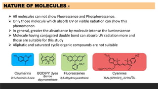
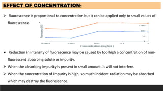
Factors Affecting Fluorescence and Phosphorescence
IMA
Instrumental method of analysis
B pharmacy
Semester VII
as per PCI Syllabus
nature of molecule
adsorption
light
oxygen
photodecamposition
temperature
This document discusses fluorimetry and phosphorimetry. It defines them as measurement techniques, with fluorimetry measuring fluorescence intensity at a particular wavelength, and phosphorimetry measuring phosphorescence in conjunction with pulsed radiation. It describes the principles behind photoluminescence, including fluorescence and phosphorescence. Factors affecting these processes and instrumentation used are summarized, including light sources, filters, monochromators, and detectors. Applications in pharmaceutical, clinical, environmental, and entertainment fields are also briefly outlined.
This document discusses the instrumentation of UV spectrophotometry. It describes the key components which include sources of UV radiation like hydrogen discharge lamps, xenon discharge lamps, and mercury arc lamps. It also discusses monochromators like gratings to produce monochromatic light, and sample holders/cuvettes to hold liquid samples. Common detectors mentioned are barrier layer cells, phototubes, and photomultiplier tubes. Finally, it explains the basic setup of single beam and double beam UV spectrophotometers used for analysis.
The document describes the principles and components of flame photometry. Flame photometry measures the intensity of light emitted from metal atoms excited by the heat of a flame. When a solution is sprayed into the flame, the solvent evaporates and the metal atoms are excited and emit light of characteristic wavelengths. A mirror collects the light, which is separated into its wavelengths by a prism or grating. A photodetector measures the light intensities, which correspond to concentrations of metals in the original solution. Common applications include analyzing body fluids, soils, and water.
Flame photometry is a technique that uses the intensity of light emitted from a flame to determine the concentration of certain metal ions in a sample. When a sample is introduced into the flame, the metal ions are atomized and excited. As they return to the ground state, they emit light of characteristic wavelengths. The intensity of light emitted can then be measured to determine the concentration of the metal ions. Flame photometry is used to analyze samples for concentrations of ions like sodium, potassium, calcium, and lithium. It has applications in analyzing body fluids and determining metal concentrations in materials like cement.
Introduction and Principle of IR spectroscopyRajaram Kshetri
This document provides an introduction to infrared (IR) spectrophotometry. It discusses how IR spectroscopy analyzes molecular vibrations when molecules absorb IR radiation that matches their natural vibrational frequencies. The document outlines the principle of IR spectroscopy and describes the different types of molecular vibrations observed in IR spectra, including stretching and bending vibrations. It also discusses the criteria for a molecule to absorb IR radiation, such as having a change in dipole moment when vibrations occur.
This document discusses fragmentation techniques in mass spectroscopy. It begins by defining mass spectroscopy as a technique used to determine molecular weight of compounds. The key points made are:
1) Molecular ions formed during ionization undergo fragmentation into smaller daughter ions.
2) Fragmentation occurs through simple cleavage like homolytic, heterolytic, or semi-heterolytic cleavage or through rearrangement reactions like elimination, ortho, or McLafferty rearrangements.
3) Specific fragmentation patterns can provide information about functional groups present like aldehydes producing m/z 44 ions or ketones producing m/z 58 ions.
The document discusses atomic absorption spectroscopy. It begins with an introduction describing how atomic absorption spectroscopy measures the concentration of an element by measuring the amount of light absorbed at a characteristic wavelength when it passes through atoms of that element. It then describes the principle, instrumentation, applications, and sources of interference in atomic absorption spectroscopy. The key sources of interference discussed are non-spectral interferences such as matrix, chemical, and ionization interferences and spectral interferences such as background absorption.
The document discusses infrared (IR) spectroscopy, which analyzes the interaction of infrared radiation with matter. IR spectroscopy can provide information about a compound's chemical structure and molecular structure by measuring its absorption of IR radiation. It is widely used to analyze organic materials and some inorganic molecules. The document then describes various components of IR instrumentation, including IR radiation sources like the Nernst glower and globar, monochromators that separate wavelengths, sample cells and techniques, and detectors like thermocouples, bolometers, and thermistors that measure the radiation absorbed.
Spin-spin coupling occurs between neighboring NMR-active nuclei and causes splitting of NMR spectra. The splitting pattern is related to the number of equivalent hydrogen atoms near the nuclei. The distance between peaks in a split signal is the coupling constant (J) measured in Hertz. Factors like number of bonds between nuclei, bond angles, and molecular rigidity can affect the coupling constant value. Complex coupling results when a set of hydrogen is coupled to two or more nonequivalent neighbors, producing more complex splitting patterns.
spectrofluorometer is the instrument for recording fluorescence emission and absorption spectra When a beam of light is incident on certain substances they emit visible light or radiations. This is known as fluorescence. Fluorescence starts immediately after the absorption of light and stops as soon as the incident light is cut off. The substances showing this phenomenon are known as flourescent substances.
This document provides an overview of flame emission spectroscopy and atomic absorption spectroscopy. It discusses the theory, principles, instrumentation, interferences and applications of both techniques. Flame emission spectroscopy measures the light emitted from excited atoms in a flame, while atomic absorption spectroscopy measures the absorption of light by ground state atoms. Both can be used to analyze metals but atomic absorption spectroscopy provides better precision and is applicable to more elements.
The document discusses ultraviolet-visible spectroscopy and its applications in analyzing molecular structure. It describes the four types of electronic transitions that can occur in molecules when exposed to UV-VIS light: sigma-to-sigma, n-to-sigma, n-to-pi, and pi-to-pi transitions. Beer's Law and Lambert's Law are introduced, relating absorbance to concentration, path length, and molar absorptivity. Deviations from Beer's Law can occur due to chemical changes in the sample or limitations of the instrumentation. Chromophores and auxochromes are defined as groups that impart or shift color to compounds.
Factors affecting IR absorption frequency Vrushali Tambe
1. Many factors affect the absorption frequency in IR spectroscopy, including reduced mass, bond strength, hydrogen bonding, electronic effects, and molecular structure.
2. Coupling between vibrations and Fermi resonance can cause frequency shifts and intensity changes. Hydrogen bonding causes broad bands while strong bonds absorb at higher frequencies.
3. Electronic effects like induction, mesomerism, and conjugation influence frequency by altering bond strength. Ring size, hybridization, and physical state also impact the absorption frequency.
Infrared spectroscopy (IR spectroscopy) is the spectroscopy that deals with the infrared
region of the electromagnetic spectrum, that is light with a longer wavelength and
lower frequency than visible light.
Infrared Spectroscopy is the analysis of infrared light interacting with a molecule.
FT-IR spectroscopy uses a Michelson interferometer to measure the absorption of infrared light by molecules. The key components are a source, beam splitter, two mirrors, sample, detector, and computer. Infrared light from the source is split at the beam splitter, reflected by the mirrors, and recombined to generate an interferogram, which is Fourier transformed by the computer into an infrared absorption spectrum. FT-IR spectroscopy can be used to determine molecular structure in gases, explore interstellar composition, perform quantitative analysis, and identify functional groups and bonds based on their characteristic vibrational frequencies.
This document provides an overview of infrared (IR) spectroscopy. It discusses the principle behind IR spectroscopy, the different modes of molecular vibration, instrumentation including sources, detectors and monochromators. It also covers sample handling techniques, factors that affect vibrational frequencies and applications of IR spectroscopy such as structure elucidation.
Flame photometry is a technique that uses a flame to atomize samples and a spectrophotometer to measure the intensity of light emitted by the atoms. It works by introducing a liquid sample containing metal ions into a flame, which excites the metal atoms causing them to emit light of characteristic wavelengths. This allows for qualitative and quantitative analysis of metals in samples. The key components of a flame photometry instrument are the sample delivery system, burner and flame, monochromator, detector, and readout system. Common interferences include spectral and chemical overlap between elements.
Nuclear magnetic resonance spectroscopy involves subjecting atomic nuclei to magnetic fields and measuring the electromagnetic radiation absorbed and emitted. Fourier transform NMR provides increased sensitivity by combining multiple free induction decay signals measured in the time domain. A Fourier transform converts these signals to an NMR spectrum in the frequency domain. The Michelson interferometer induces interference of light waves by splitting and recombining beams that traveled different path lengths, allowing observation of interference patterns related to the wavelength of light.
The coupling constant is the distance between peaks in a multiplet in NMR spectroscopy. It is measured in Hertz and does not depend on external magnetic field strength. The value of the coupling constant provides information to distinguish multiplets and can indicate structural features like cis/trans isomers. Coupling occurs between protons close in space, known as geminal, vicinal, and sometimes long-range coupling over several bonds. The coupling constant is affected by factors like bond angle, dihedral angle, and electronegativity of substituents.
Mass spectrometry is a technique that ionizes chemical species and sorts the ions based on their mass-to-charge ratio. It can be used to determine molecular masses and elucidate molecular structures of organic compounds. There are several types of ions produced including molecular ions, fragment ions, and isotope ions. Compounds undergo various fragmentation modes like homolytic cleavage, heterolytic cleavage, retro-Diels-Alder reactions, hydrogen transfers and McLafferty rearrangements. Mass spectrometry has applications in fields like drug development, environmental analysis, and clinical diagnosis.
Instrumentation of uv visible spectroscopyZainab&Sons
UV-visible spectroscopy uses light in the UV and visible ranges. It works by passing light through a sample and measuring how much light is absorbed. Key components are a light source, monochromator, sample cell, detector, and recorder. For UV light a hydrogen lamp is used as the source and quartz is used for the cell and prism. It can be used to identify functional groups and conjugation, detect impurities, and determine molecular structure and in quantitative analysis. Applications include qualitative and quantitative analysis of organic compounds.
This document discusses factors that affect fluorescence and phosphorescence. It defines fluorescence and phosphorescence as types of molecular luminescence that are excited by photon absorption. The main difference is that fluorescence involves no change in electron spin, while phosphorescence does involve a change. Several factors can influence emission, including molecular structure and rigidity, temperature, solvent properties, pH, dissolved oxygen, concentration, and the presence of heavy atoms. More rigid and planar structures favor fluorescence and phosphorescence. Higher temperatures, viscosities, and oxygen levels decrease emission, while appropriate solvent polarity and pH can increase it.
UV-visible spectroscopy involves measuring the absorption of light in the UV and visible light ranges. It is useful for determining conjugation and distinguishing between conjugated and non-conjugated compounds. It has applications in identifying unknown compounds, determining the extent of conjugation, and elucidating the structures of molecules like vitamins. It can also provide information about configuration, hydrogen bonding, molecular weight, and detect impurities. The Woodward-Fieser rules allow calculating the expected absorption maxima for certain functional groups.
Mass spectrometry and ionization techniquesSurbhi Narang
Mass spectrometry is a technique that identifies chemicals based on their mass and charge. It works by ionizing chemical compounds and separating the resulting ions based on their mass-to-charge ratio. The document discusses the key components and principles of mass spectrometry including various ionization methods, mass analyzers, and applications such as sequencing proteins, determining molecular weights, and drug discovery.
fluorimetry and phosphorimetry m pharmacy notes scop sataranikhil salunkhe
This document provides an introduction to fluorimetry and phosphorimetry. It defines fluorescence as the emission of visible light when certain substances are exposed to light, while phosphorescence is the continued emission of light even after the light source is removed. The principle involves excitation of electrons from the highest occupied to the lowest unoccupied molecular orbital. Factors that affect fluorescence and phosphorescence include concentration, oxygen levels, pH, temperature, substituents, scatter, and adsorption. Applications include determination of various compounds in samples like urine, serum, food, and more.
1. Fluorescence is the emission of light from a substance that has absorbed light or other electromagnetic radiation. It occurs in certain biological molecules like fireflies, corals, and genetically engineered fish.
2. Fluorescence results from electrons absorbing energy and getting excited to higher energy molecular orbitals, then dropping down and emitting photons of lower energy. The Jablonski diagram illustrates this process.
3. Many factors influence fluorescence, including molecular structure, temperature, solvent, pH, and structural rigidity. Fluorescent dyes like FITC and cyanine dyes are used in applications like labeling and fluorescence resonance energy transfer.
Introduction of Fluorescent Probes and DyesBOC Sciences
Fluorescence spectroscopy and fluorescence imaging are essential tools for scientific research today. Fluorescent probes/dyes are highly luminescent materials used in fluorescence spectrum analysis and biological imaging. Fluorescent probes/dyes can be widely used in research fields such as fluorescence immunity, cell staining, nucleic acid detection, in vivo imaging and environmental monitoring. For more information, please visit https://probes.bocsci.com.
This document provides an overview of spectrofluorimetry and fluorescence. It begins by explaining the principles behind fluorescence - how absorption of UV or visible light causes electrons to transition to an excited state and then emit light as they fall back down. It then discusses fluorescence and fluorimetry in more detail. The rest of the document covers the Jablonski diagram, factors that affect fluorescence, types of quenching, instrumentation used including light sources, filters, sample cells and detectors, and applications of fluorescence including pharmaceutical analysis.
Spectroscopic methods in inorganic Chemistry: Fluorescence spectroscopy Chris Sonntag
Fluorescence spectroscopy involves absorption of UV or visible light by a molecule, promoting electrons to an excited state. The molecule then relaxes and emits light of a longer wavelength. It has many applications, including determination of organic and some inorganic substances at low concentrations in areas like food analysis, pharmaceuticals, and clinical samples. Factors like conjugation, substituents, temperature, and oxygen presence can influence fluorescence intensity. It is a sensitive and specific technique compared to absorption spectroscopy.
This document discusses fluorometry and its application in analyzing cephalosporin antibiotics. It begins with defining types of luminescence including fluorescence and providing an overview of the principles of fluorometry. It then discusses structural factors that affect fluorescence and advantages of fluorometry such as high sensitivity. The document provides examples of using fluorometry to analyze specific cephalosporins like cefoxitin and cefuroxime. It also briefly discusses other methods for analyzing cephalosporins and concludes by stating fluorometry is well suited for trace analysis of pharmaceutical compounds.
Phenols are aromatic compounds that contain a hydroxyl group attached to a benzene ring. Some key points:
- Phenol is a weak acid due to delocalization of charge in the phenoxide ion. Electron withdrawing groups increase acidity while electron donating groups decrease acidity.
- Phenol and its derivatives like cresol and resorcinol have many uses including as precursors to drugs, plastics, and dyes. They are also used as disinfectants, preservatives, and wood preservatives.
- Qualitative tests for phenols include turning blue litmus red due to acidity, and forming colored complexes with ferric chloride and bromine water.
This document describes a spectrofluorometric method for determining two cephalosporin drugs, cefadroxile and cefuroxime sodium, in pharmaceutical formulations. The method involves reacting the drugs with 1,2-naphthoquinone-4-sulfonate under alkaline conditions to form fluorescent derivatives, extracting them with chloroform, and measuring fluorescence intensity. The method was optimized and validated, demonstrating good linearity, accuracy, precision and sensitivity for quantifying the drugs within certain concentration ranges in samples. The effects of various parameters on the analysis were also examined.
Fluorescence spectroscopy is a technique that uses fluorescence from molecules to analyze samples. Certain molecules emit light at longer wavelengths after absorbing ultraviolet or visible light (fluorescence). This technique is highly sensitive and can detect fluorescent compounds even when present at low concentrations. It has various applications like determining drugs in formulations, studying drug-protein binding, and bioanalysis. Factors like temperature, pH, concentration, and molecular structure can influence fluorescence intensity. Fluorometers contain a light source, wavelength selection devices, and photodetectors to measure fluorescence from samples.
Fluorescence is the phenomenon whereby a molecule, after absorption radiation, emits radiation of a longer wavelength.
A compound absorbs radiation in the UV-rgion and emits visible light.
Absorption of uv/visible radiation causes transition of electrons from ground state (low energy) to excited state (high energy).
This increase in wavelength is known as the Stokes shift.
This document summarizes a study that investigated the potential for photochemical reactions of two thiazide compounds, bendroflumethiazide and hydroflumethiazide, when exposed to sunlight-range illumination. Spectroscopy analysis revealed that both compounds strongly absorb UV light in specific wavelength ranges. When solutions of the compounds were illuminated over time, their absorption spectra changed, indicating photochemical reactions were occurring. However, the reaction kinetics could not be definitively determined due to interference from photoproducts also absorbing at the same wavelengths. Further separation and analysis of photoproducts using HPLC will be needed to fully characterize the photochemical reactions.
This document discusses factors that affect fluorimetry and quenching. It lists several factors that can influence fluorescence, including the nature of molecules, substituents, concentration, adsorption, light, oxygen, pH, temperature, and viscosity. It also describes different types of quenching such as self-quenching, chemical quenching, static quenching, and collisional quenching. Chemical quenching can occur due to changes in pH, presence of oxygen, or heavy metals. Static quenching involves complex formation between the fluorophore and quencher. Collisional quenching occurs through interactions between an excited fluorophore and quencher molecule.
Fluorescent dyes are molecules that absorb light at one wavelength and emit it at a longer wavelength. They are useful for labeling and studying biomolecules. Some common fluorescent dyes include fluorescein, rhodamine, and GFP. Fluorescent dyes have many applications, such as cancer research where they allow over 1500 protein spots to be detected from microdissected tissue, physiological sensing inside deep tissue, monitoring acidified organelles during autophagy, and assessing contamination during drilling operations. The Jablonski diagram illustrates the excited states involved in the fluorescent process.
this presentation describes the basics of photosynthesis. it includes Significance of photosynthesis, Photosynthetic apparatus, Absorption & action spectra, Absorption & action spectra, Factors affecting photosynthesis, Photosynthetic apparatus, Position of photosynthetic pigments, Photosynthetic pigments, Functions of carotenoids, Phycobilins, Principle /Blackman’s law of limiting factors.
1) The absorption of light by organic compounds involves the promotion of electrons from ground state to excited state molecular orbitals. Sigma electrons undergo σ-σ* transitions at shorter wavelengths while pi and non-bonding electrons undergo π-π* and n-π* transitions at longer wavelengths.
2) Chromophores are functional groups responsible for electronic transitions, imparting color. Auxochromes enhance absorption by chromophores through resonance. Conjugation and pH can shift absorption to longer wavelengths while dilution, solvents, and temperature can affect absorption spectra.
3) Spectrophotometry is widely used for quantitative analysis due to its sensitivity, selectivity, accuracy and ease. Both absorbing and non-absorbing
This document discusses factors that affect the position and intensity of UV bands in spectroscopy, including conjugation, steric effects, pH, and solvent polarity. It explains that increasing conjugation causes a bathochromic shift to longer wavelengths. Steric hindrance and higher pH can decrease intensity by disrupting conjugation. More polar solvents can shift bands by stabilizing or destabilizing molecular orbitals.
This document discusses the use of fluorescence dyes to stain cells and make their structures visible under a microscope. It explains that while early dyes stained broad cellular components, fluorescence dyes have increased specificity by binding to particular proteins or molecules. Common fluorescence dyes excite at specific wavelengths and emit light of longer wavelengths, allowing structures to be seen against a dark background. The document outlines the basic components of a fluorescence microscope and how it works, and notes some applications of fluorescence staining like detecting vitamins, drugs, and lipofuscin deposits in aging cells.
Fluorescence spectroscopy involves three main processes: excitation, where a molecule absorbs a photon and reaches an excited state; internal conversion and vibrational relaxation in the excited state; and fluorescence emission, where the molecule returns to the ground state and emits a photon. It has many applications including structural elucidation of molecules, monitoring molecular interactions and conformational changes, and tracking ions and biomolecules in cells. Specifically, intrinsic protein fluorescence relies on tryptophan residues, while extrinsic labels are often used for non-fluorescent compounds. Fluorescence resonance energy transfer (FRET) also allows measuring distances between fluorophores to study biomolecular interactions and conformational dynamics.
Similar to Factors affecting fluorescence and phosphorescence.pptx (20)
Describing and Interpreting an Immersive Learning Case with the Immersion Cub...Leonel Morgado
Current descriptions of immersive learning cases are often difficult or impossible to compare. This is due to a myriad of different options on what details to include, which aspects are relevant, and on the descriptive approaches employed. Also, these aspects often combine very specific details with more general guidelines or indicate intents and rationales without clarifying their implementation. In this paper we provide a method to describe immersive learning cases that is structured to enable comparisons, yet flexible enough to allow researchers and practitioners to decide which aspects to include. This method leverages a taxonomy that classifies educational aspects at three levels (uses, practices, and strategies) and then utilizes two frameworks, the Immersive Learning Brain and the Immersion Cube, to enable a structured description and interpretation of immersive learning cases. The method is then demonstrated on a published immersive learning case on training for wind turbine maintenance using virtual reality. Applying the method results in a structured artifact, the Immersive Learning Case Sheet, that tags the case with its proximal uses, practices, and strategies, and refines the free text case description to ensure that matching details are included. This contribution is thus a case description method in support of future comparative research of immersive learning cases. We then discuss how the resulting description and interpretation can be leveraged to change immersion learning cases, by enriching them (considering low-effort changes or additions) or innovating (exploring more challenging avenues of transformation). The method holds significant promise to support better-grounded research in immersive learning.
Sexuality - Issues, Attitude and Behaviour - Applied Social Psychology - Psyc...PsychoTech Services
A proprietary approach developed by bringing together the best of learning theories from Psychology, design principles from the world of visualization, and pedagogical methods from over a decade of training experience, that enables you to: Learn better, faster!
The cost of acquiring information by natural selectionCarl Bergstrom
This is a short talk that I gave at the Banff International Research Station workshop on Modeling and Theory in Population Biology. The idea is to try to understand how the burden of natural selection relates to the amount of information that selection puts into the genome.
It's based on the first part of this research paper:
The cost of information acquisition by natural selection
Ryan Seamus McGee, Olivia Kosterlitz, Artem Kaznatcheev, Benjamin Kerr, Carl T. Bergstrom
bioRxiv 2022.07.02.498577; doi: https://doi.org/10.1101/2022.07.02.498577
Current Ms word generated power point presentation covers major details about the micronuclei test. It's significance and assays to conduct it. It is used to detect the micronuclei formation inside the cells of nearly every multicellular organism. It's formation takes place during chromosomal sepration at metaphase.
ESA/ACT Science Coffee: Diego Blas - Gravitational wave detection with orbita...Advanced-Concepts-Team
Presentation in the Science Coffee of the Advanced Concepts Team of the European Space Agency on the 07.06.2024.
Speaker: Diego Blas (IFAE/ICREA)
Title: Gravitational wave detection with orbital motion of Moon and artificial
Abstract:
In this talk I will describe some recent ideas to find gravitational waves from supermassive black holes or of primordial origin by studying their secular effect on the orbital motion of the Moon or satellites that are laser ranged.
(June 12, 2024) Webinar: Development of PET theranostics targeting the molecu...Scintica Instrumentation
Targeting Hsp90 and its pathogen Orthologs with Tethered Inhibitors as a Diagnostic and Therapeutic Strategy for cancer and infectious diseases with Dr. Timothy Haystead.
Authoring a personal GPT for your research and practice: How we created the Q...Leonel Morgado
Thematic analysis in qualitative research is a time-consuming and systematic task, typically done using teams. Team members must ground their activities on common understandings of the major concepts underlying the thematic analysis, and define criteria for its development. However, conceptual misunderstandings, equivocations, and lack of adherence to criteria are challenges to the quality and speed of this process. Given the distributed and uncertain nature of this process, we wondered if the tasks in thematic analysis could be supported by readily available artificial intelligence chatbots. Our early efforts point to potential benefits: not just saving time in the coding process but better adherence to criteria and grounding, by increasing triangulation between humans and artificial intelligence. This tutorial will provide a description and demonstration of the process we followed, as two academic researchers, to develop a custom ChatGPT to assist with qualitative coding in the thematic data analysis process of immersive learning accounts in a survey of the academic literature: QUAL-E Immersive Learning Thematic Analysis Helper. In the hands-on time, participants will try out QUAL-E and develop their ideas for their own qualitative coding ChatGPT. Participants that have the paid ChatGPT Plus subscription can create a draft of their assistants. The organizers will provide course materials and slide deck that participants will be able to utilize to continue development of their custom GPT. The paid subscription to ChatGPT Plus is not required to participate in this workshop, just for trying out personal GPTs during it.
The debris of the ‘last major merger’ is dynamically youngSérgio Sacani
The Milky Way’s (MW) inner stellar halo contains an [Fe/H]-rich component with highly eccentric orbits, often referred to as the
‘last major merger.’ Hypotheses for the origin of this component include Gaia-Sausage/Enceladus (GSE), where the progenitor
collided with the MW proto-disc 8–11 Gyr ago, and the Virgo Radial Merger (VRM), where the progenitor collided with the
MW disc within the last 3 Gyr. These two scenarios make different predictions about observable structure in local phase space,
because the morphology of debris depends on how long it has had to phase mix. The recently identified phase-space folds in Gaia
DR3 have positive caustic velocities, making them fundamentally different than the phase-mixed chevrons found in simulations
at late times. Roughly 20 per cent of the stars in the prograde local stellar halo are associated with the observed caustics. Based
on a simple phase-mixing model, the observed number of caustics are consistent with a merger that occurred 1–2 Gyr ago.
We also compare the observed phase-space distribution to FIRE-2 Latte simulations of GSE-like mergers, using a quantitative
measurement of phase mixing (2D causticality). The observed local phase-space distribution best matches the simulated data
1–2 Gyr after collision, and certainly not later than 3 Gyr. This is further evidence that the progenitor of the ‘last major merger’
did not collide with the MW proto-disc at early times, as is thought for the GSE, but instead collided with the MW disc within
the last few Gyr, consistent with the body of work surrounding the VRM.
Factors affecting fluorescence and phosphorescence.pptx
1. By-
Mr. Samarth H. Giri
Ashokrao Mane College Of Pharmacy
TOPIC –
Factors affecting
Fluorescence and
Phosphorescence,
Applications of
Fluorimetry.
2. Factors Affecting Fluorescence and Phosphorescence -
1) Nature of Molecule
2) Nature of Substituent
3) Effect of Concentration
4) Adsorption
5) Light / Radiation
6) Oxygen
7) pH
8) Photodecomposition
9) Temperature
10)Viscosity
3. NATURE OF MOLECULES -
All molecules can not show Fluorescence and Phosphorescence.
Only those molecule which absorb UV or visible radiation can show this
phenomenon.
In general, greater the absorbance by molecule intense the luminescence
Molecule having conjugated double bond can absorb UV radiation more and
those are suitable for this study
Aliphatic and saturated cyclic organic compounds are not suitable
4. NATURE OF SUBSTITUENT -
Substituents often exhibit a marked effect on the fluorescence and phosphorescence of
molecules.
Different groups show different types of effect on fluorescence and phosphorescence.
Electron donating groups like -NH₂ and OH often enhance fluorescence.
Electron withdrawing groups like -COOH, -NO₂, -N=N- and halides decrease or destroy
fluorescence.
Groups like -SO3H, NH4 and alkyl groups do not have much effect on both
phosphorescence and fluorescence.
5. EFFECT OF CONCENTRATION-
fluorescence is proportional to concentration but it can be applied only to small values of
fluorescence.
Reduction in intensity of fluorescence may be caused by too high a concentration of non-
fluorescent absorbing solute or impurity.
When the absorbing impurity is present in small amount, it will not interfere.
When the concentration of impurity is high, so much incident radiation may be absorbed
which may destroy the fluorescence.
6. ADSORPTION-
Adsorption of substance on the wall of the sample cells may cause a serious problem.
This happens when the concentration of substance solution is strong hence the strong
stock solution must be diluted.
The method require a very dilute solution (10-100 times) weaker than those used in
absorption study.
RADIATION-
Monochromatic light is essential for the excitation and fluorescence.
7. OXYGEN-
The presence of oxygen may interfere by direct oxidation of the fluorescent substance to
non-fluorescent products or by quenching of fluorescence.
Eg-Anthracene.
TEMPERATURE , VISCOSITY-
QUENCHING-
Quenching is the reduction of fluorescence intensity by the presence substance in the sample
other than the fluorescent analyte.
8. pH-
Alteration of the pH of the solution will have a significant effect on fluorescence ,if absorption
curve of the solute is changed.
Eg- Aniline
9. APPLICATIONS OF FLUORIMETRY-
1) Determination of vitamin B1 (Thiamine):Thiamine is non-fluorescent but its oxidation
product thiochrome have fluoresces with blue colour. The property is used for the
determination of thiamine in the food samples like meat, cereal etc.
2) Determination of vitamin B2 (Riboflavin):Determination of vitamin B2 is done by a
fluorescence method because the fluorescent power depends upon the exact conditions
and upon the nature and amount of impurities.
3) Organic analysis: To carry out qualitative as well as quantitative analysis of aromatic
compounds present in cigarette smoke, air-pollutant, concentrate and automobile exhaust.
10. 4) Analysis of pharmaceuticals : The substances such as allyl morphine, PAS, chloroquine,
folic acid, menadione, phenobarbitone, procaine, thymol can be analysed by coupling with
suitable reagents.
5) Determination of indoles, phenols, phenothiazines , Napthols , proteins plant pigment and
steroids .
6) Detection of impurities at nanogram quantities.