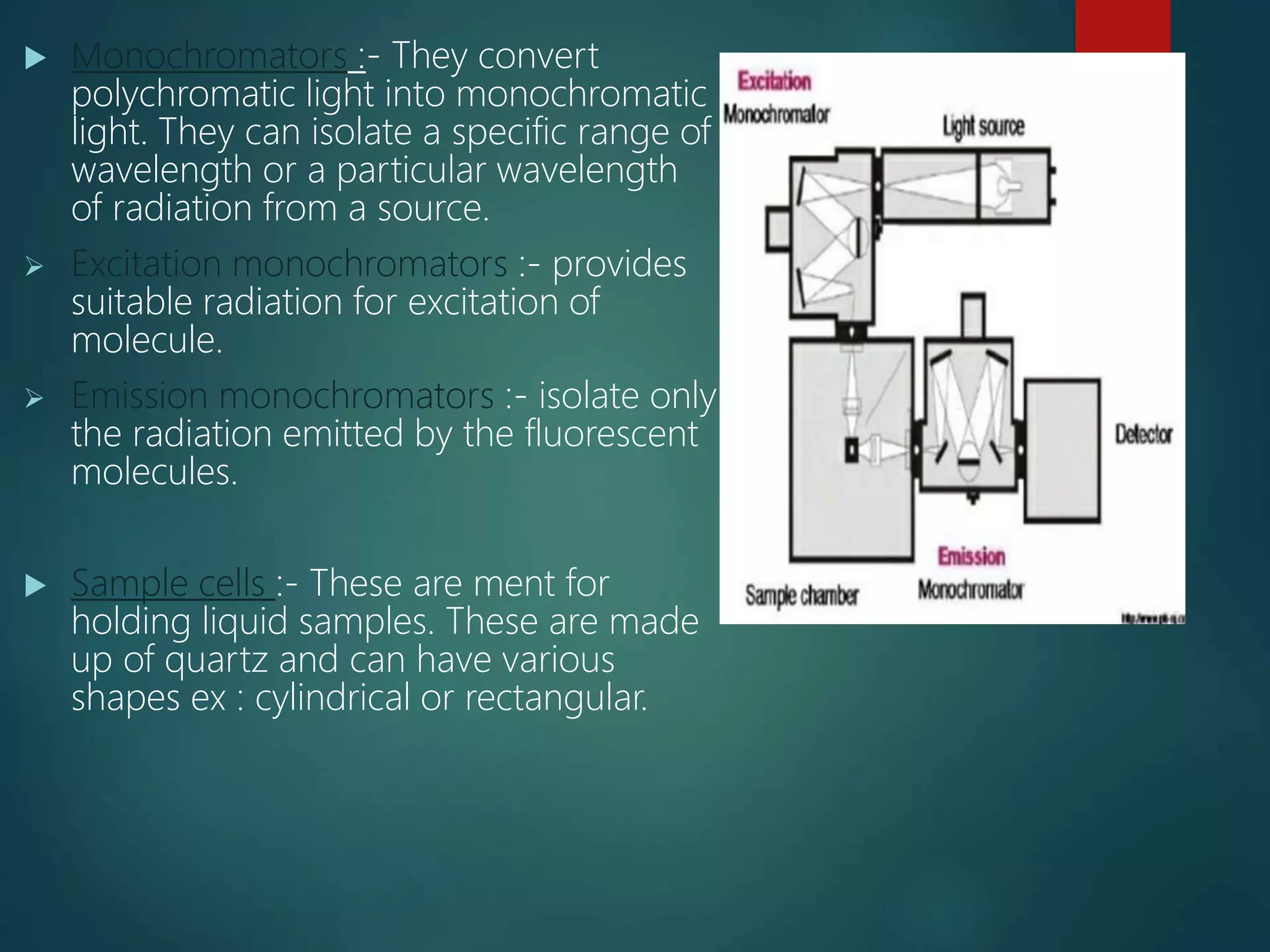
spectrofluorometer is the instrument for recording fluorescence emission and absorption spectra When a beam of light is incident on certain substances they emit visible light or radiations. This is known as fluorescence. Fluorescence starts immediately after the absorption of light and stops as soon as the incident light is cut off. The substances showing this phenomenon are known as flourescent substances.
In this slide contains principle of IR spectroscopy and sampling techniques.
Presented by: R.Banuteja (Department of pharmaceutical analysis).
RIPER, anantpur.
PRINCIPLES of FT-NMR & 13C NMR
Fourier Transform
FOURIER TRANSFORM NMR SPECTROSCOPY
THEORY OF FT-NMR
13C NMR SPECTROSCOPY
Principle
Why C13-NMR is required though we have H1-NMR?
CHARACTERISTIC FEATURES OF 13 C NMR
Chemical Shifts
NUCLEAR OVERHAUSER ENHANCEMENT
Short-Comings of 13C-NMR Spectra
a substance can absorb any visible light or external radiation and then again emit it. this called fluorescence and the process of reduction in fluorescence intensity is called quenching. this presentation is all about quenching of fluorescence.
Fluorimetry, principle, Concept of singlet,doublet,and triplet electronic sta...Vandana Devesh Sharma
Content-Principle
concept of singlet, doublet and triplet electronic stages,
Internal and external conversions,
Factors affecting fluorescence,
quenching,
Instrumentation and
applications
Types of luminescence including
bioluminescence,
chemiluminescence,
Fluorescence, and
phosphorescence
These various forms of luminescence differ in their method of emitting light.
Bioluminescence
Chemiluminescence
Fluorescence is the emission of light by a substance that has absorbed light or other electromagnetic radiation.
In most cases, the emitted light has a longer wavelength, and therefore a lower photon energy, than the absorbed radiation
In fluorescence, absorption and emission light takes place in very short time (10-12 or 10-9 seconds)
Fluorescence starts immediately after the absorption of light and stops as soon as the incident light is cut off
Eg -The fluorescent clothes, shoes
Fluorescence is the emission of light by a substance that has absorbed light or other electromagnetic radiation.
In most cases, the emitted light has a longer wavelength, and therefore a lower photon energy, than the absorbed radiation
In fluorescence, absorption and emission light takes place in very short time (10-12 or 10-9 seconds) Fluorimetry
An analytical technique for identifying and characterizing minute amounts of substance by excitation of the substance with a beam of ultraviolet/Visible light and detection and measurement of the characteristic wavelength of fluorescent light emitted.Excited – State Processes in molecules
the presentation gives knowledge about principle or fluorometry, factors that affect fluorescence including quenching instruments used in fluorometry, and the applications of fluorometry. added references in the end for more knowledge.
Principle
Interferences
Instrumentation and
Applications
The principle of flame photometer
is based on the measurement of the emitted light intensity when a metal is introduced into the flame.
The wavelength of the colour gives information about the element and
the colour of the flame gives information about the amount of the element present in the sample.
Flame photometry is one of the branches of atomic absorption spectroscopy.
It is also known as flame emission spectroscopy.
Currently, it has become a necessary tool in the field of analytical chemistry. Used to
Determine the concentration of certain metal ions like
potassium,lithium, calcium, cesium etc. In flame photometer spectra the metal ions are used in the form of atoms.
(IUPAC) Committee on Spectroscopic Nomenclature has named this technique as flame atomic emission spectrometry (FAES). Principle of Flame photometer
The compounds of the alkali and alkaline earth metals (Group II) dissociate into atoms when introduced into the flame.
Some of these atoms further get excited to even higher levels. But these atoms are not stable at higher levels.
Hence, these atoms emit radiations when returning back to the ground state.
These radiations generally lie in the visible region of the spectrum.
Each of the alkali and alkaline earth metals has a specific wavelength. Instrumentation-Source of flame, Nebuliser, Monochromator(Prism monochromator, Grating monochromators)DETECTOR (
The radiation emitted by the elements is mostly in the visible region and measured by photo detector. Hence conventional detectors like photo voltaic cell or photo tubes or photomultiplier tube is used), READ OUT DEVICE
[The signal from the detector is shown as a response in the digital read out device. The readings are displayed in an arbitrary scale (% Flame Intensity).], working of flame photometer, Advantages and disadvantage of flame photometer, Errors /interference in Flame Photometry-Flame Temperature, chemical interference, Radiation interference
Application of flame photometry
fluorometry is used in pharmaceutical fields.An analytic method for detecting and measuring fluorescence in compounds that uses ultraviolet light stimulating the compounds, causing them to emit visible light. An important topic studied in instrumental analysis.
Fluorimetry is a technique used in analytical chemistry and biochemistry to measure the concentration of a substance in a sample by analyzing the fluorescence it emits when exposed to specific wavelengths of light. This technique is based on the principle of fluorescence, which is the emission of light (or photons) by a molecule when it absorbs photons at a shorter wavelength.
Here's how fluorimetry works:
Excitation: A sample is exposed to a specific wavelength of light, known as the excitation wavelength, which is typically in the ultraviolet or visible range. This excitation light is absorbed by the molecules of interest in the sample, causing them to move to higher energy states.
Emission: After absorbing the excitation light, the molecules return to their ground state by releasing energy in the form of fluorescent light at longer wavelengths. The emitted light is typically at a longer wavelength than the excitation light, and it is specific to the particular molecule or compound being analyzed.
Detection: A detector, such as a photomultiplier tube or a photodiode, is used to measure the intensity of the emitted fluorescent light. The detector is sensitive to the specific wavelength of light emitted by the target molecules.
Data Analysis: The intensity of the emitted fluorescent light is correlated with the concentration of the substance in the sample. By comparing the intensity of the emitted light to a calibration curve or standard, the concentration of the substance can be determined.
Fluorimetry has various applications in chemistry and biology. It is commonly used for quantifying the concentration of fluorescent dyes, proteins, nucleic acids (e.g., DNA and RNA), and other biomolecules. It is also employed in environmental analysis, drug discovery, and medical diagnostics.
One of the advantages of fluorimetry is its high sensitivity, which allows for the detection of very low concentrations of analytes. Additionally, it offers high selectivity because the emitted fluorescence is specific to the target molecule.
Overall, fluorimetry is a valuable analytical tool that helps researchers and scientists measure and analyze a wide range of substances with high precision and sensitivity
A fluorometer or fluorimeter is a device used to measure parameters of fluorescence: its intensity and wavelength distribution of emission spectrum after excitation by a certain spectrum of light. These parameters are used to identify the presence and the number of specific molecules in a medium.
Fluorescence is the phenomenon whereby a molecule, after absorption radiation, emits radiation of a longer wavelength.
A compound absorbs radiation in the UV-rgion and emits visible light.
Absorption of uv/visible radiation causes transition of electrons from ground state (low energy) to excited state (high energy).
This increase in wavelength is known as the Stokes shift.
Luminescence is the emission of light by a substance. It occurs when an electron returns to the electronic ground state from an excited state and loses its excess energy as a photon.
It is of 3 types.
Fluorescence spectroscopy.
Phosphorescence spectroscopy.
Chemiluminescence spectroscopy
Fluorescence spectroscopy. : When a beam of light is incident on certain substances they emit visible light or radiations. This is known as fluorescence. Fluorescence starts immediately after the absorption of light and stops as soon as the incident light is cut off. The substances showing this phenomenon are known as flourescent substances
Phosphorescence spectroscopy: When light radiation is incident on certain substances they emit light continuously even after the incident light is cut off.
This type of delayed fluorescence is called phosphorescence.
Substances showing phosphorescence are phosphorescent substances.
Chemiluminescence (also chemoluminescence) is the emission of light (luminescence) as the result of a chemical reaction. There may also be limited emission of heat
Fluorescence
Phosphorescence
Radiation less processes
Vibration relaxation
Internal conversion
External conversion
Intersystem crossing
Jablonski diagram is a graphical representation of the various transitions(electronic states, vibrational levels) that can occur after a molecule has been excited photochemically.
When a molecule is raised from its ground state to a higher state using light, photochemistry occurs.
The molecule in the excited state has a shorter lifetime and significantly more energy than the ground state from which it was formed.
As a result, molecules in the excited state are much more reactive.
A photochemical or photophysical process deactivates an excited state.
Therefore, the fate of the excited molecules is described by using the Jablonski diagram, which only focuses on the photophysical process occurring during the excitation and deactivation process.
Radiative transitions involve the absorption of a photon, if the transition occurs to a higher energy level, or the emission of a photon, for a transition to a lower level.
Nonradiative transitions arise through several different mechanisms, all differently labeled in the diagram. Relaxation of the excited state to its lowest vibrational level is called vibrational relaxation. This process involves the dissipation of energy from the molecule to its surroundings, and thus it cannot occur for isolated molecules. A second type of nonradiative transition is internal conversion (IC), which occurs when a vibrational state of an electronically excited state can couple to a vibrational state of a lower electronic state.
A third type is intersystem crossing (ISC); this is a transition to a state with a different spin multiplicity. In molecules with large spin-orbit coupling, intersystem crossing is much more important than in molecules that exhibit only small spin-orbit coupling. ISC can
A brief information about the SCOP protein database used in bioinformatics.
The Structural Classification of Proteins (SCOP) database is a comprehensive and authoritative resource for the structural and evolutionary relationships of proteins. It provides a detailed and curated classification of protein structures, grouping them into families, superfamilies, and folds based on their structural and sequence similarities.
This presentation explores a brief idea about the structural and functional attributes of nucleotides, the structure and function of genetic materials along with the impact of UV rays and pH upon them.
Multi-source connectivity as the driver of solar wind variability in the heli...Sérgio Sacani
The ambient solar wind that flls the heliosphere originates from multiple
sources in the solar corona and is highly structured. It is often described
as high-speed, relatively homogeneous, plasma streams from coronal
holes and slow-speed, highly variable, streams whose source regions are
under debate. A key goal of ESA/NASA’s Solar Orbiter mission is to identify
solar wind sources and understand what drives the complexity seen in the
heliosphere. By combining magnetic feld modelling and spectroscopic
techniques with high-resolution observations and measurements, we show
that the solar wind variability detected in situ by Solar Orbiter in March
2022 is driven by spatio-temporal changes in the magnetic connectivity to
multiple sources in the solar atmosphere. The magnetic feld footpoints
connected to the spacecraft moved from the boundaries of a coronal hole
to one active region (12961) and then across to another region (12957). This
is refected in the in situ measurements, which show the transition from fast
to highly Alfvénic then to slow solar wind that is disrupted by the arrival of
a coronal mass ejection. Our results describe solar wind variability at 0.5 au
but are applicable to near-Earth observatories.
Seminar of U.V. Spectroscopy by SAMIR PANDASAMIR PANDA
Spectroscopy is a branch of science dealing the study of interaction of electromagnetic radiation with matter.
Ultraviolet-visible spectroscopy refers to absorption spectroscopy or reflect spectroscopy in the UV-VIS spectral region.
Ultraviolet-visible spectroscopy is an analytical method that can measure the amount of light received by the analyte.
Professional air quality monitoring systems provide immediate, on-site data for analysis, compliance, and decision-making.
Monitor common gases, weather parameters, particulates.
THE IMPORTANCE OF MARTIAN ATMOSPHERE SAMPLE RETURN.Sérgio Sacani
The return of a sample of near-surface atmosphere from Mars would facilitate answers to several first-order science questions surrounding the formation and evolution of the planet. One of the important aspects of terrestrial planet formation in general is the role that primary atmospheres played in influencing the chemistry and structure of the planets and their antecedents. Studies of the martian atmosphere can be used to investigate the role of a primary atmosphere in its history. Atmosphere samples would also inform our understanding of the near-surface chemistry of the planet, and ultimately the prospects for life. High-precision isotopic analyses of constituent gases are needed to address these questions, requiring that the analyses are made on returned samples rather than in situ.
Earliest Galaxies in the JADES Origins Field: Luminosity Function and Cosmic ...Sérgio Sacani
We characterize the earliest galaxy population in the JADES Origins Field (JOF), the deepest
imaging field observed with JWST. We make use of the ancillary Hubble optical images (5 filters
spanning 0.4−0.9µm) and novel JWST images with 14 filters spanning 0.8−5µm, including 7 mediumband filters, and reaching total exposure times of up to 46 hours per filter. We combine all our data
at > 2.3µm to construct an ultradeep image, reaching as deep as ≈ 31.4 AB mag in the stack and
30.3-31.0 AB mag (5σ, r = 0.1” circular aperture) in individual filters. We measure photometric
redshifts and use robust selection criteria to identify a sample of eight galaxy candidates at redshifts
z = 11.5 − 15. These objects show compact half-light radii of R1/2 ∼ 50 − 200pc, stellar masses of
M⋆ ∼ 107−108M⊙, and star-formation rates of SFR ∼ 0.1−1 M⊙ yr−1
. Our search finds no candidates
at 15 < z < 20, placing upper limits at these redshifts. We develop a forward modeling approach to
infer the properties of the evolving luminosity function without binning in redshift or luminosity that
marginalizes over the photometric redshift uncertainty of our candidate galaxies and incorporates the
impact of non-detections. We find a z = 12 luminosity function in good agreement with prior results,
and that the luminosity function normalization and UV luminosity density decline by a factor of ∼ 2.5
from z = 12 to z = 14. We discuss the possible implications of our results in the context of theoretical
models for evolution of the dark matter halo mass function.
2. introduction
Absorption of uv/visible radiation causes transition of electrons
from ground state (low energy) to exited state is not stable,
excess energy is lost.
Molecule contains electron, electron and non bonding (n)
electron.
1) The electrons may be present in bonding molecular orbital. It
is called as highest occupied molecular orbital (HOMO). It has
lest energy and more stable.
2) When the molecules absorbs radiant energy from a light
source, the bonding electrons may be promoted to
antibonding molecular orbital (LUMO. It has more energy and
hence less stable.
2
3. principle
The process of promotion of electrons from H0MO to LUMO
with absorption of energy is called as excitation.
Singlet state :- a state in which all the electron in a molecule
are paired ↓↑
Doublet state :- a state in which un paired electrons is present
↓ or ↑
Triplet state :- a state in which unpaired electrons of same spin
present ↑↑
Singlet excited state :- a state in which electrons are unpaired
but of opposite spin like ↑ ↓ (unpaired and opposite spin)
3
4. When light of appropriate wavelength is
absorbed by a molecule the electrons are
promoted from singlet ground state to
singlet excited state. Once the molecule is
in this excited state to relaxation can occur
via several process. For ex by emission of
radiation. The process can be the following
1) Collisional deactivation
2) Fluorescence
3) Phosphorescence
4
5. 1) Collisional de activation :- in which entire energy lost due to
collision de activation and no radiation emitted.
2) Fluorescence :- excited singlet state is highly unstable.
Relaxation of electrons from exited singlet ground state
with emission of light.
3) Phosphorescence :- At favorable condition like low
temperature and absence of oxygen there is transition from
excited singlet state to triplet state which is called as inner
system crossing. The emission of radiation when electrons
undergo transition from triplet state to singlet ground state
is called as phosphorescence.
5
7. Advantages
More sensitive when
compared to other absorption
techniques. Concentration as
low as цg/ml or ng/ml can be
determined.
Precision upto 1% can be
achieved easily.
As both excitation and
emission wave lengths are
characteristic it is more specific
than absorption methods.
Disadvantage
The susceptibility to
environmental condition and
virtual impossibility of
predicting whether a
compound will fluorescence.
The other major problem is
quenching, whereby the
energy transferred to the other
molecules.
7
8. Factors effecting fluorescence intensity
1) Concentration
2) Quantum yield of fluorescence
3) Intensity of incident light
4) Adsorption
5) Oxygen
6) PH
7) Temperature and viscosity
8) Photodecomposition
9) Quenchers
10) Scatter
8
9. Concentration :- Fluorescence intensity is proportional to
concentration of substance only when the absorbance is less
than 0.02 .
Quantum yield of fluorescence Ø :- Number of photons
emitted / Number of photons absorbed. It is always less than
1 since some energy is lost by radiation less pathways (
Collisional, Intersystem crossing, Vibrational relaxation)
Intensity of incident light :- Increase in the intensity light on
the sample fluorescence intensity also increases.
Adsorption :- Adsorption of sample solution in the container
may leads to a serious problem.
Oxygen :- Oxidation of fluorescent species to a non
fluorescent species, quenches fluorescent substance
9
10. Ph :- Alteration of ph of a solution will have significant effect on
fluorescence. For ex Aniline in alkali medium gives visible
fluorescence and in acid gives invisible region.
Temperature and viscosity:- temperature increases can increase the
collisional de activation and reduce fluorescent intensity.
If viscosity of solution is more the frequency of collisions are
reduced and increase in fluorescent intensity.
Photochemical decomposition:- Absorption of intense radiation
leads to photochemical decomposition of a fluorescent substance
to less fluorescent or non fluorescent substance.
Scatter:- Scatter is mainly due to colloidal particles in solution.
Scattering of incident light after passing through the sample leads
to decrease in fluorescence intensity
10
11. Quenchers:- Quenching is the reduction of fluorescence
intensity by the presence of substance in the sample other
than the fluorescent analyte.
o Quenching is following types:-
Inner fluorescent effect :- absorption of incident (uv) light or
emitted (fluorescent) light by primary and secondary filters
leads to decrease in fluorescence intensity.
Self quenching :- At low concentration linearity is observed,
at high concentration of the same substance increase in
fluorescent intensity is observed. This phenomena is called
self quenching
11
12. Collisional quenching :- Collisions between
the fluorescent substance and halide ions
leads to reduction in fluorescence intensity.
Static quenching :- This occurs because of
complex formation between the fluorescent
molecule and other molecules. Ex : caffeine
reduces fluorescence of riboflavin.
12
14. Source of light
Mercury vapour lamp :- Hg
vapour in high pressure ( 8
atm ) gives intense line on
continuous background
above 350nm.
Xenon arc lamp :- Gives
more intense radiation.
Tungsten lamp :- Used if
excitation has to be done in
via region.
14
15. Filters and monochromators:-
Filters :- These are nothing but optical filters works on the
principle of absorption of unwanted light and transmitting the
required wavelength of light.
Primary filter:- Absorbs visible radiation and transmit UV
radiation.
Secondary filter:- Absorbs UV radiation and transmit visible
radiation.
15
16. Monochromators :- They convert
polychromatic light into monochromatic
light. They can isolate a specific range of
wavelength or a particular wavelength
of radiation from a source.
Excitation monochromators :- provides
suitable radiation for excitation of
molecule.
Emission monochromators :- isolate only
the radiation emitted by the fluorescent
molecules.
Sample cells :- These are ment for
holding liquid samples. These are made
up of quartz and can have various
shapes ex : cylindrical or rectangular.
16
17. Detectors : Photometric detectors are used they are :
Barrier layer cell/ photo voltaic cells
Photomultiplier cells
17
18. Types of instruments
Single beam filter fluorimeter
It contains tungsten lamp as a source of light and has an optical
system consists of primary filter.
The emitted radiation is measured at 90’ by using a secondary filter
and detector. Primary filter absorbs visible radiation and transmit uv
radiation which excites the molecule present in sample cell.
In stead of 90 if we use 180 geometry as in colorimetry secondary
filter has to be highly efficient other wise both the unabsorbed uv
radiation and fluorescent will produce detector response and give
false result.
18
20. In double beam fluorimeter
It is similar to single beam except that the
two incident beams from a single light
source pass through primary filters
separately and fall on the another
reference solution. Then the emitted
radiations from the sample or reference
sample pass separately through secondary
filter and produce response combinly on a
detector.
20
22. In spectrofluorimeter
In this primary filter in double beam
fluorimeter is replaced by excitation
monochromator and the secondary filter is
replaced by emission monochromator.
Incident beam is split into sample and
reference beam by using beam splitter.
22
24. Application of spectrofluorimetry
Determination organic substances
Plant pigments, steroids, proteins, naphthols etc. can be
determined at low concentrations.
Generally used to carry out qualitative as well as quantitative
analysis for a great aromatic compounds present in cigarette
smoking, air pollutant concentration and automatic exhausts.
o Determination of inorganic substance
o Extensively used in the field of nuclear research for the
determination of uranium salts.
o Determination of vitamin B1 (thiamine) in food sample like meat
cereals etc.
24
25. Determination of vitamin B2( riboflavin). This method is generally
used to measure the amount of impurities present in the sample.
Most important application are found in the analyses of food
products, pharmaceuticals, clinical samples and natural products.
Fluorescent indicators :
Intensity and color of the fluorescence of many substance
depend upon the pH of solutions. These are called as fluorescent
indicators and are generally used in acid base titrations.
Eg. Eosin – pH 3.0-4.0 – colorless to green
Fluorescein – pH 4.0-6.0 – colorless to green
25
26. conclusion
Fluorimetric methods are not useful in qualitative
analysis and much used in quantitative analysis.
Fluorescence is the most sensitive analytical
techniques.
Detection studies will increase the development of
fluorescence field.
26
27. references
SKOOG, principle of instrumental analysis
B.K. Sharma instrumental methods of chemical
analysis
Wikipedia
Google source
27