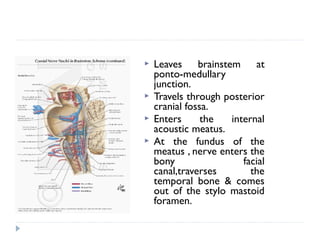
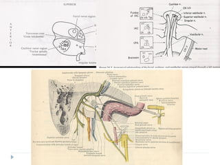
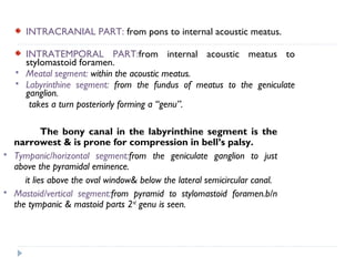
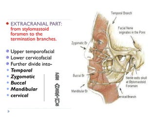
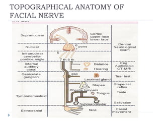
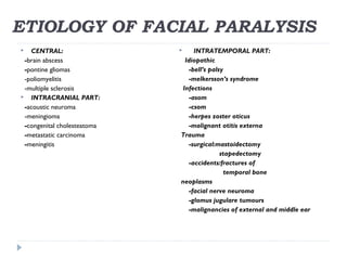
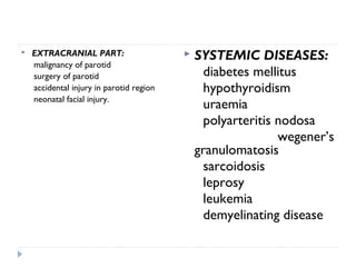
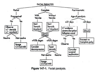
The facial nerve is the 7th cranial nerve that has both motor and sensory functions. It has a complex anatomical course through the skull and face. Facial paralysis can result from lesions anywhere along this course. Bell's palsy is the most common cause of acute facial paralysis, believed to be due to a viral infection causing inflammation where the nerve exits the skull. Other potential causes include trauma, tumors, infections, and systemic diseases. Treatment depends on the underlying cause but often includes corticosteroids for Bell's palsy and surgery for decompression or repair of severed nerve segments.
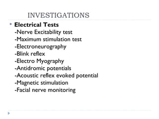
![ NERVE EXCITABILITY TEST [NET]
-When the difference between 2 sides exceed 3.5 MA the test is positive
for degeneration.
-Degeneration of fibres cannot be detected earlier than 48 to 72 hours of
its commencement
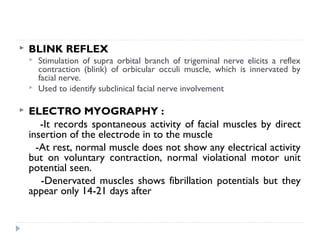
MAXIMUM STIMULATION TEST [MST]
-The movements on the paralysed side are subjectively expressed as a
percentage (0%, 25%, 50%, 75% & 100%) of the movement on the healthy
side.
ELECTRO NEUROGRAPHY [ENOG]
Evoked electromyography
Nerve is stimulated and the compound action potentials from facial muscles are
recorded and measured objectively & compared with normal side.
The average difference in healthy is only 3%
> 30% considered as abnormal](https://image.slidesharecdn.com/facialnerveparalysis-dr-160412065700/85/Facial-nerve-paralysis-dr-davis-11-04-16-34-320.jpg)


![Cells and Organs of immune system [Autosaved].pptx](https://cdn.slidesharecdn.com/ss_thumbnails/cellsandorgansofimmunesystemautosaved-260123152717-ea0cb261-thumbnail.jpg?width=640&height=640&fit=bounds)


