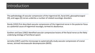
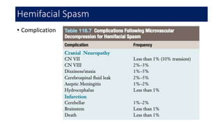
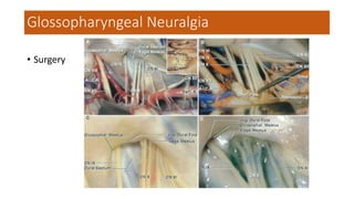
Microvascular Decompression
Glen Sandi Saapang
Supervisor:
Prof. Dr. dr. Tjokorda Gde Bagus Mahadewa, Sp. BS (K) Spinal
Introduction
The pathobiology of vascular compression of the trigeminal (V), facial (VII), glossopharyngeal
(IX), and vagus (X) nerves underlies a number of related neurologic disorders.
Dandy (1932) first described vascular compression of the trigeminal nerve in the posterior fossa
as a potential cause of trigeminal neuralgia, termed tic douloureux.
Gardner and Sava (1962) identified vascular compressive lesions of the facial nerve as the likely
underlying etiology of hemifacial spasm
Jannetta (1967) used the microscope to systematically study vascular compression of cranial
nerves, termed microvascular decompression (MVD).
Trigeminal Neuralgia
• Most common cranial nerve vascular compression syndrome.
• Female : Male = 2:1
• Peak onset 50-70yo
• Most common affected branches : V2 and/ V3
• Unilateral pain (1% patients bilateral)
Clinical presentasion:
• Proxysmal, intense, intermittent pain
• Described as an intense stabbing or electrical shock-like sensation,
lasting seconds to minutes
• Triggered by light cutaneous stimuli
• Episodic free of pain between pain attacks
• Occasionally patients suffer from both trigeminal neuralgia and
hemifacial spam, known as tic convulsif.
Trigeminal Neuralgia
• Differential diagnosis
• Multiple sclerosis: can causes paroxysmal attacks of Trigeminal facial pain
• Isolated orbital pain syndromes (low onset, hours to days)
• Cluster headache (insidious onset, runny nose, ptosis or watery eye)
• Postherpetic neuralgia (constant pain with vesicles)
• Dental disease (site spesific)
• TMJ dysfunction
• Temporal arteritis (tenderness over STA)
• Posttraumatic neuralgia
Trigeminal Neuralgia
Imaging: High-resolution MRI of posterior fossa
• T1
• sequence: whole brain axial and sagittal (volumetric when possible)
• purpose: anatomical, brain screen
• T2
• sequence: axial, limited to posterior fossa (medulla to the upper pons) with thin slices (e.g. 3D
CISS and FIESTA)
• purpose: assess the N. trigeminal from their origin at the mid pons anteriorly, through
the prepontine cistern until the Meckel’s cave (trigeminal ganglion)
• the most common cause of trigeminal neuralgia is an enlarged looping artery (most commonly the superior
cerebellar artery) or vein pressing on the trigeminal nerve at the CPA , which is best depicted by this sequence
• FLAIR
• sequence: whole brain axial
• purpose: white matter signal abnormality such as in multiple sclerosis
• T1 C+ (Gd)
Trigeminal Neuralgia
Medical management
• First-line medications: (blockade of voltage-sensitive sodium channels,
quelling hyperexcited neurons)
• Carbamazepine, started dose 2x100mg, increased by 100mg/day until pain control is
achieved or toxicity develops. Typical maintenance dose 300-800mg/day
• Oxcarbazepine, started dose 2x150mg. Increased by 300mg every 3 days until pain
relief. Maintenance dose 2 x 300—600mg
• Second-line medications:
• Baclofen, 10-80mg/day (GABAB receptors agonist, depresses excitatory neuroal
activity)
• Lamotrigine, initial dose 25mg/day, titrated up to 200-400mg/day
• Other: phenytoin, clonazepam, gabapentin, pregabalin, topiramate, leviracetam,
valproate.
Trigeminal Neuralgia
Surgical indications: Fail medical management
Trigeminal Neuralgia
Positioning
supralateral cerebellar approach  Blue arrow for TN
infralateral cerebellar or infrafloccular approach  Green Arrow for HSF and GPN
Neurovascular conflict:
variation
Postoperative care
• Observe any neurologic sign of posterior fossa pathology, due to
either cerebellar swelling or hematoma
• Oral intake
• Initiate DVT prophylaxis on the 1st postop day
• Surgical dressing is removed on 2nd postop day
• Headache postop controlled with narcotic analgetic
• Discharge by second or third postop day
Trigeminal Neuralgia
Operative results
• Operative findings
Trigeminal Neuralgia
• Outcomes
Factor associated with outcome:
• Quality and nature of pain before
surgery
• Classic episodic, paroxysmal preop
pain  excellent outcome
Trigeminal Neuralgia
• Surgical Complication
Trigeminal Neuralgia
Hemifacial Spasm
Clinical features
• Most frequently on female
• Mean age of onset of 45 yo
• Repetitive, painless paroxysmal twitching of facial muscles (starts with
the orbicularis oculi and periorbital muscles and then slowly progresses
to middle and lower facial musculature).
• Significantly worsens during period of stress or anxiety  psychogenic
or emotional problems
EMF findings
• 5-20 rhythmic burst discharge/
second, can lead to 150-250
bursts / second
Hemifacial Spasm
Diferential diagnosis
• Blepharospasm (bilateral)
• Synkinetic movements after bell’s
palsy (during nerve regeneration)
• Facial myokymia (worm-like
movements, associated intrinsic
brainstem pathology)
Hemifacial Spasm
Medical management
• Botulinum toxin injection (release acetylcholine from presynaptic
nerve terminal).
• Carbamazepine, gabapentin, clonazepam, and baclofen
Hemifacial Spasm
Surgical indications
• Good health
• Younger than 75yo
Surgical options
• MVD (best choice)
• Neurotomy
• Distal nerve avulsion
• Facial nerve sectioning (with or without anastomosis to hypoglossal or
accessory nerve)
Hemifacial Spasm
Perioperative considerations
• Lateral or park bench position with modification rotating head 10-20
degress toward to floor (improve exposure of the lower cranial
nerves)
• Intraoperative monitoring of N.VII (EMG) dan N.VIII (Brain stem
auditory evoked respons) is recomended.
Hemifacial Spasm
Operative findings and results
Hemifacial Spasm
• Complication
Hemifacial Spasm
Glossopharyngeal Neuralgia
• Less common than TN, incidence 0,2-0,7/100.000/people/year
• female more frequently (2:1), more common in the left side
• Symptom onset most common observed in fifth decade
• Most idiopathic GN (severe demyelination and degeneration of N. IX
and X)
• Secondary GN occure due to compression by vascular, tumor CPA,
calcified stylohyoid, MS, pagets disease, etc
Diagnosis:
• Confirmed with topical cocaine (10%) in trigger zone  relief from
painful 1-2 hours after stimulation
Imaging:
• High-resolution MRI of the posterior fossa
Medical management: carbamazepien, etc.
Glossopharyngeal Neuralgia
• Surgery
Glossopharyngeal Neuralgia
THANK YOU

Microvascular Decompression.pptx

  • 1.
    Microvascular Decompression Glen SandiSaapang Supervisor: Prof. Dr. dr. Tjokorda Gde Bagus Mahadewa, Sp. BS (K) Spinal
  • 2.
    Introduction The pathobiology ofvascular compression of the trigeminal (V), facial (VII), glossopharyngeal (IX), and vagus (X) nerves underlies a number of related neurologic disorders. Dandy (1932) first described vascular compression of the trigeminal nerve in the posterior fossa as a potential cause of trigeminal neuralgia, termed tic douloureux. Gardner and Sava (1962) identified vascular compressive lesions of the facial nerve as the likely underlying etiology of hemifacial spasm Jannetta (1967) used the microscope to systematically study vascular compression of cranial nerves, termed microvascular decompression (MVD).
  • 3.
    Trigeminal Neuralgia • Mostcommon cranial nerve vascular compression syndrome. • Female : Male = 2:1 • Peak onset 50-70yo • Most common affected branches : V2 and/ V3 • Unilateral pain (1% patients bilateral)
  • 5.
    Clinical presentasion: • Proxysmal,intense, intermittent pain • Described as an intense stabbing or electrical shock-like sensation, lasting seconds to minutes • Triggered by light cutaneous stimuli • Episodic free of pain between pain attacks • Occasionally patients suffer from both trigeminal neuralgia and hemifacial spam, known as tic convulsif. Trigeminal Neuralgia
  • 6.
    • Differential diagnosis •Multiple sclerosis: can causes paroxysmal attacks of Trigeminal facial pain • Isolated orbital pain syndromes (low onset, hours to days) • Cluster headache (insidious onset, runny nose, ptosis or watery eye) • Postherpetic neuralgia (constant pain with vesicles) • Dental disease (site spesific) • TMJ dysfunction • Temporal arteritis (tenderness over STA) • Posttraumatic neuralgia Trigeminal Neuralgia
  • 7.
    Imaging: High-resolution MRIof posterior fossa • T1 • sequence: whole brain axial and sagittal (volumetric when possible) • purpose: anatomical, brain screen • T2 • sequence: axial, limited to posterior fossa (medulla to the upper pons) with thin slices (e.g. 3D CISS and FIESTA) • purpose: assess the N. trigeminal from their origin at the mid pons anteriorly, through the prepontine cistern until the Meckel’s cave (trigeminal ganglion) • the most common cause of trigeminal neuralgia is an enlarged looping artery (most commonly the superior cerebellar artery) or vein pressing on the trigeminal nerve at the CPA , which is best depicted by this sequence • FLAIR • sequence: whole brain axial • purpose: white matter signal abnormality such as in multiple sclerosis • T1 C+ (Gd) Trigeminal Neuralgia
  • 9.
    Medical management • First-linemedications: (blockade of voltage-sensitive sodium channels, quelling hyperexcited neurons) • Carbamazepine, started dose 2x100mg, increased by 100mg/day until pain control is achieved or toxicity develops. Typical maintenance dose 300-800mg/day • Oxcarbazepine, started dose 2x150mg. Increased by 300mg every 3 days until pain relief. Maintenance dose 2 x 300—600mg • Second-line medications: • Baclofen, 10-80mg/day (GABAB receptors agonist, depresses excitatory neuroal activity) • Lamotrigine, initial dose 25mg/day, titrated up to 200-400mg/day • Other: phenytoin, clonazepam, gabapentin, pregabalin, topiramate, leviracetam, valproate. Trigeminal Neuralgia
  • 10.
    Surgical indications: Failmedical management Trigeminal Neuralgia
  • 11.
  • 15.
    supralateral cerebellar approach Blue arrow for TN infralateral cerebellar or infrafloccular approach  Green Arrow for HSF and GPN
  • 17.
  • 18.
    Postoperative care • Observeany neurologic sign of posterior fossa pathology, due to either cerebellar swelling or hematoma • Oral intake • Initiate DVT prophylaxis on the 1st postop day • Surgical dressing is removed on 2nd postop day • Headache postop controlled with narcotic analgetic • Discharge by second or third postop day Trigeminal Neuralgia
  • 19.
    Operative results • Operativefindings Trigeminal Neuralgia
  • 20.
    • Outcomes Factor associatedwith outcome: • Quality and nature of pain before surgery • Classic episodic, paroxysmal preop pain  excellent outcome Trigeminal Neuralgia
  • 21.
  • 22.
    Hemifacial Spasm Clinical features •Most frequently on female • Mean age of onset of 45 yo • Repetitive, painless paroxysmal twitching of facial muscles (starts with the orbicularis oculi and periorbital muscles and then slowly progresses to middle and lower facial musculature). • Significantly worsens during period of stress or anxiety  psychogenic or emotional problems
  • 23.
    EMF findings • 5-20rhythmic burst discharge/ second, can lead to 150-250 bursts / second Hemifacial Spasm
  • 25.
    Diferential diagnosis • Blepharospasm(bilateral) • Synkinetic movements after bell’s palsy (during nerve regeneration) • Facial myokymia (worm-like movements, associated intrinsic brainstem pathology) Hemifacial Spasm
  • 26.
    Medical management • Botulinumtoxin injection (release acetylcholine from presynaptic nerve terminal). • Carbamazepine, gabapentin, clonazepam, and baclofen Hemifacial Spasm
  • 27.
    Surgical indications • Goodhealth • Younger than 75yo Surgical options • MVD (best choice) • Neurotomy • Distal nerve avulsion • Facial nerve sectioning (with or without anastomosis to hypoglossal or accessory nerve) Hemifacial Spasm
  • 28.
    Perioperative considerations • Lateralor park bench position with modification rotating head 10-20 degress toward to floor (improve exposure of the lower cranial nerves) • Intraoperative monitoring of N.VII (EMG) dan N.VIII (Brain stem auditory evoked respons) is recomended. Hemifacial Spasm
  • 32.
    Operative findings andresults Hemifacial Spasm
  • 33.
  • 34.
    Glossopharyngeal Neuralgia • Lesscommon than TN, incidence 0,2-0,7/100.000/people/year • female more frequently (2:1), more common in the left side • Symptom onset most common observed in fifth decade • Most idiopathic GN (severe demyelination and degeneration of N. IX and X) • Secondary GN occure due to compression by vascular, tumor CPA, calcified stylohyoid, MS, pagets disease, etc
  • 35.
    Diagnosis: • Confirmed withtopical cocaine (10%) in trigger zone  relief from painful 1-2 hours after stimulation Imaging: • High-resolution MRI of the posterior fossa Medical management: carbamazepien, etc. Glossopharyngeal Neuralgia
  • 36.
  • 37.
  • 39.
  • 40.