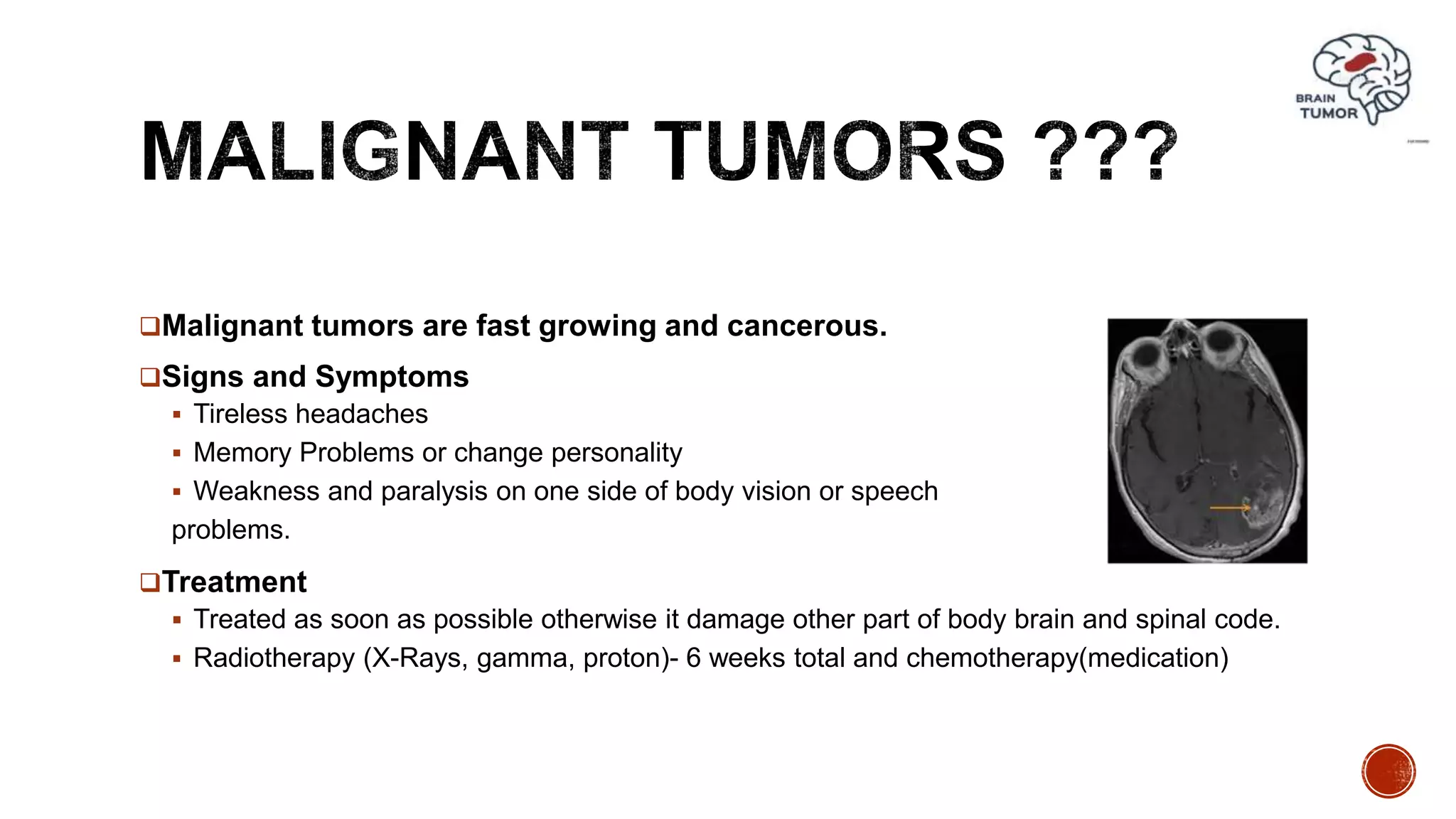
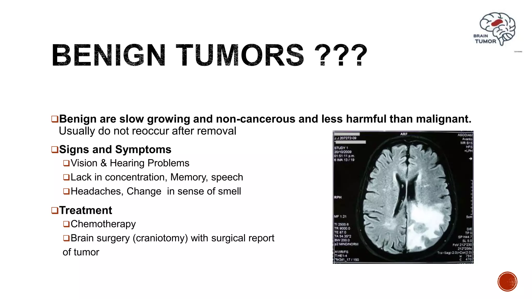
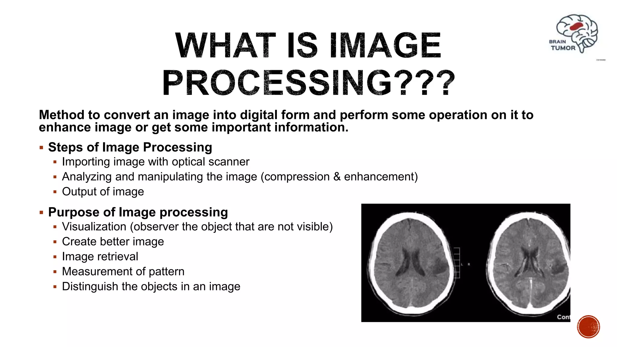
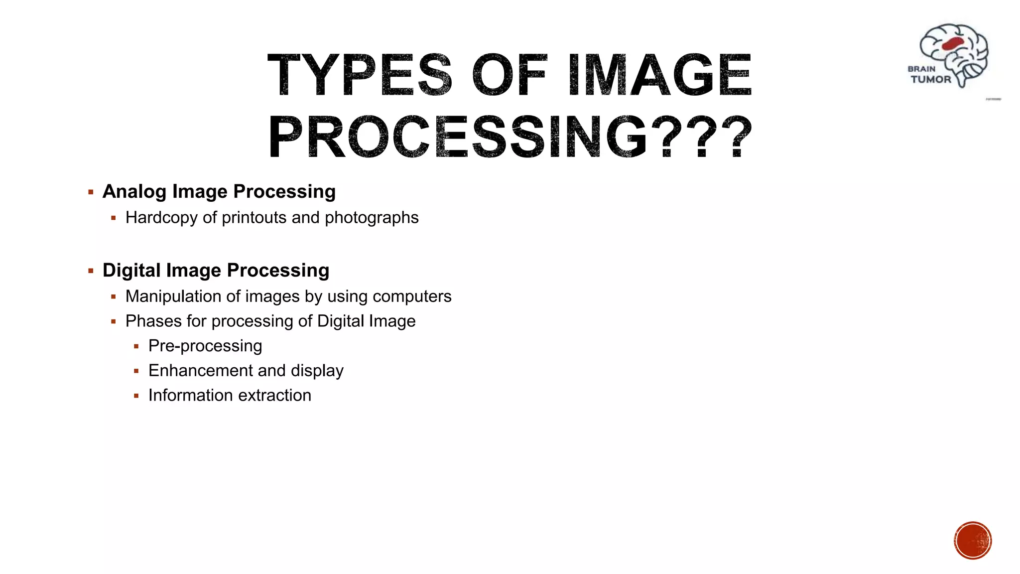
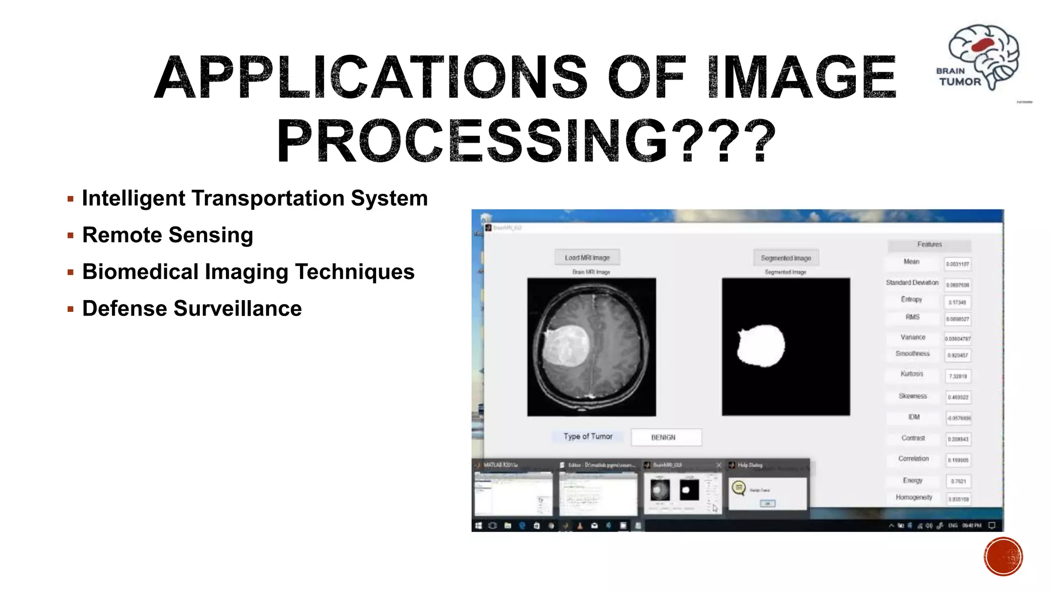
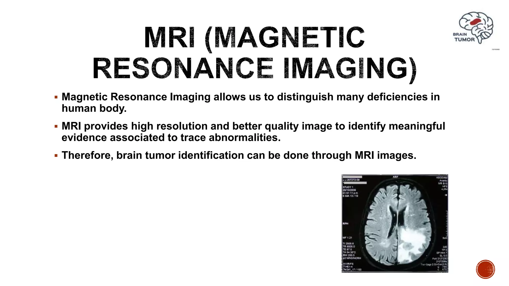
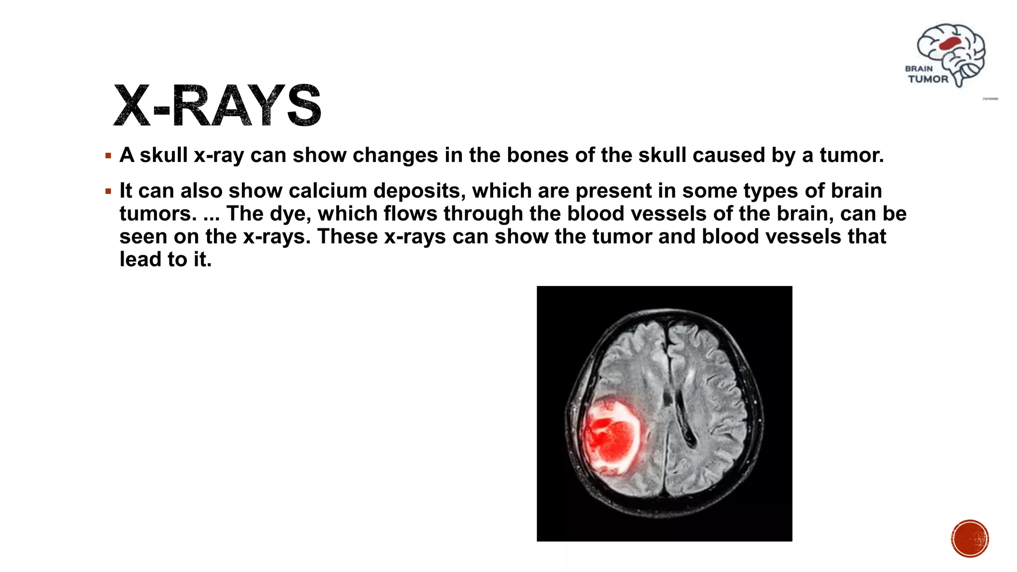
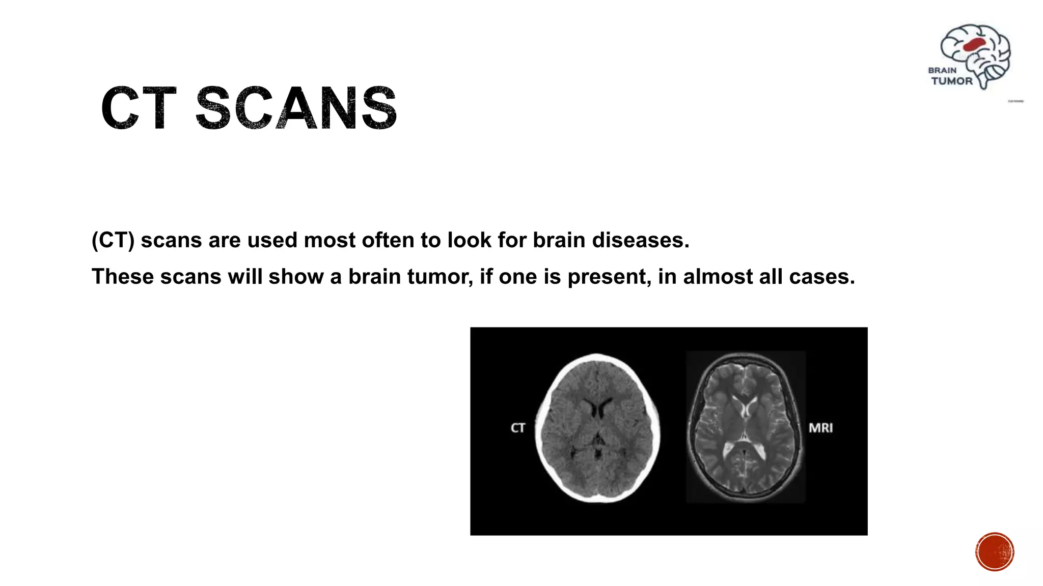
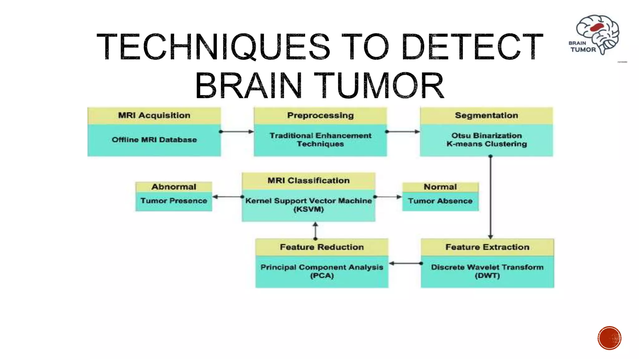
By: Muhammad Ayaz Farid Shah
Muhammad Khaleel Afzal
Muhammad Tayyab Husain
Muhmmad Mazhar
“Abnormal growth of tissues in the brain which affect proper brain functions is
considered as a brain tumor.”
 Brain tumor is an abandoned growth that is affecting no of peoples all over the
world.
 It is not only limited with the old age people but also detected in the early age.
 Most analysis and research tell us that numbers of death is cause because user
is not known or unaware for detection of Tumor.
 Early detection of the brain tumor is possible with the advancement of machine
learning (ML) and image processing (IP) .
Two types of brain tumors have been identified.
 Malignant tumors
 Benign tumors
Malignant tumors are fast growing and cancerous.
Signs and Symptoms
 Tireless headaches
 Memory Problems or change personality
 Weakness and paralysis on one side of body vision or speech
problems.
Treatment
 Treated as soon as possible otherwise it damage other part of body brain and spinal code.
 Radiotherapy (X-Rays, gamma, proton)- 6 weeks total and chemotherapy(medication)
Benign are slow growing and non-cancerous and less harmful than malignant.
Usually do not reoccur after removal
Signs and Symptoms
Vision & Hearing Problems
Lack in concentration, Memory, speech
Headaches, Change in sense of smell
Treatment
Chemotherapy
Brain surgery (craniotomy) with surgical report
of tumor
Method to convert an image into digital form and perform some operation on it to
enhance image or get some important information.
 Steps of Image Processing
 Importing image with optical scanner
 Analyzing and manipulating the image (compression & enhancement)
 Output of image
 Purpose of Image processing
 Visualization (observer the object that are not visible)
 Create better image
 Image retrieval
 Measurement of pattern
 Distinguish the objects in an image
 Analog Image Processing
 Hardcopy of printouts and photographs
 Digital Image Processing
 Manipulation of images by using computers
 Phases for processing of Digital Image
 Pre-processing
 Enhancement and display
 Information extraction
 Intelligent Transportation System
 Remote Sensing
 Biomedical Imaging Techniques
 Defense Surveillance
These technologies allow us to detect even the smallest defects in the human body.
 MRI
 X-rays
 CT Scan
 SPECT (Single-photon emission computed tomography)
 Ultrasound
 Magnetic Resonance Imaging allows us to distinguish many deficiencies in
human body.
 MRI provides high resolution and better quality image to identify meaningful
evidence associated to trace abnormalities.
 Therefore, brain tumor identification can be done through MRI images.
 A skull x-ray can show changes in the bones of the skull caused by a tumor.
 It can also show calcium deposits, which are present in some types of brain
tumors. ... The dye, which flows through the blood vessels of the brain, can be
seen on the x-rays. These x-rays can show the tumor and blood vessels that
lead to it.
(CT) scans are used most often to look for brain diseases.
These scans will show a brain tumor, if one is present, in almost all cases.
 Pre-processing
 Segmentation
 Feature Extraction
 Classification
Pre-processing and Enhancement of an image
 First step of image processing it is used to enhance the chances of detecting the suspicious
region.
 Finer details of the images are enhanced and noise is removed from the image.
Image Segmentation
 Breaking down an image into small parts
 It consists of separating tumor tissues from normal brain tissues.
 Segmentation is performed to make analysis easier
 Boundary or Beginning Approach
 Edge approach
 Region approach
Feature Extraction
 Extraction as size
 Extraction as shape
 Composition
 Location Image
Imageprocessinginbraintumordetection 190316110830

Imageprocessinginbraintumordetection 190316110830

  • 1.
    By: Muhammad AyazFarid Shah Muhammad Khaleel Afzal Muhammad Tayyab Husain Muhmmad Mazhar
  • 2.
    “Abnormal growth oftissues in the brain which affect proper brain functions is considered as a brain tumor.”
  • 3.
     Brain tumoris an abandoned growth that is affecting no of peoples all over the world.  It is not only limited with the old age people but also detected in the early age.  Most analysis and research tell us that numbers of death is cause because user is not known or unaware for detection of Tumor.  Early detection of the brain tumor is possible with the advancement of machine learning (ML) and image processing (IP) .
  • 4.
    Two types ofbrain tumors have been identified.  Malignant tumors  Benign tumors
  • 5.
    Malignant tumors arefast growing and cancerous. Signs and Symptoms  Tireless headaches  Memory Problems or change personality  Weakness and paralysis on one side of body vision or speech problems. Treatment  Treated as soon as possible otherwise it damage other part of body brain and spinal code.  Radiotherapy (X-Rays, gamma, proton)- 6 weeks total and chemotherapy(medication)
  • 6.
    Benign are slowgrowing and non-cancerous and less harmful than malignant. Usually do not reoccur after removal Signs and Symptoms Vision & Hearing Problems Lack in concentration, Memory, speech Headaches, Change in sense of smell Treatment Chemotherapy Brain surgery (craniotomy) with surgical report of tumor
  • 7.
    Method to convertan image into digital form and perform some operation on it to enhance image or get some important information.  Steps of Image Processing  Importing image with optical scanner  Analyzing and manipulating the image (compression & enhancement)  Output of image  Purpose of Image processing  Visualization (observer the object that are not visible)  Create better image  Image retrieval  Measurement of pattern  Distinguish the objects in an image
  • 8.
     Analog ImageProcessing  Hardcopy of printouts and photographs  Digital Image Processing  Manipulation of images by using computers  Phases for processing of Digital Image  Pre-processing  Enhancement and display  Information extraction
  • 9.
     Intelligent TransportationSystem  Remote Sensing  Biomedical Imaging Techniques  Defense Surveillance
  • 10.
    These technologies allowus to detect even the smallest defects in the human body.  MRI  X-rays  CT Scan  SPECT (Single-photon emission computed tomography)  Ultrasound
  • 11.
     Magnetic ResonanceImaging allows us to distinguish many deficiencies in human body.  MRI provides high resolution and better quality image to identify meaningful evidence associated to trace abnormalities.  Therefore, brain tumor identification can be done through MRI images.
  • 12.
     A skullx-ray can show changes in the bones of the skull caused by a tumor.  It can also show calcium deposits, which are present in some types of brain tumors. ... The dye, which flows through the blood vessels of the brain, can be seen on the x-rays. These x-rays can show the tumor and blood vessels that lead to it.
  • 13.
    (CT) scans areused most often to look for brain diseases. These scans will show a brain tumor, if one is present, in almost all cases.
  • 14.
     Pre-processing  Segmentation Feature Extraction  Classification
  • 15.
    Pre-processing and Enhancementof an image  First step of image processing it is used to enhance the chances of detecting the suspicious region.  Finer details of the images are enhanced and noise is removed from the image.
  • 16.
    Image Segmentation  Breakingdown an image into small parts  It consists of separating tumor tissues from normal brain tissues.  Segmentation is performed to make analysis easier  Boundary or Beginning Approach  Edge approach  Region approach
  • 17.
    Feature Extraction  Extractionas size  Extraction as shape  Composition  Location Image