The document describes a new surgical technique called biplane double-supported screw fixation (BDSF) for treating femoral neck fractures in patients with osteoporosis. BDSF involves placing two screws in different coronal planes to provide stronger fixation than conventional methods. It establishes two supporting points - the femoral calcar and proximal diaphysis cortex - to better distribute loads. Early results found BDSF achieved bone union in 97.6% of patients and had a lower failure rate compared to conventional fixation. The technique provides improved stability and is particularly suitable for unstable fractures in osteoporotic bone.
Hoffa's Fracture: Diagnosis, management & New Classification System by BAGARI...Vaibhav Bagaria
Hoffa's Fracture - coronal split fracture of distal femur, its diagnosis, management strategy, a new classification and tips and tricks of management. First described Hoffa, a new classification system by Bagaria et al helps plan the surgery for these tricky fracture. The most crucial step is not to miss these fractures in ER.
Can read freely here
https://sethiortho.blogspot.com/
Challenges and Solutions in
Management of Distal Humerus Fractures
Epidemiology
Anatomy
Classification
Controversies and Recent studies
Approach
Implants selection
Plate configuration
Ulnar nerve transposition
Role of total elbow arthroplasty in DHF
Role of hemiarthroplasty in DHF
Metaphyseal comminution –
Anatomic complexity of the distal humerus
Positioning of the plates
TBW –
Skin closure
Osteoporotic nature of the bone –
Less BMD/Thin metaphysis
Screw Pullout strength is low
DHF account for 2% of all adult fractures
The common pattern of fracture
Intraarticular and involves both columns
Bimodal distribution
Peak incidence in young male and in older female patients
Young male – High-velocity injury
Older female - Osteoporosis
The distal humerus is flattened and expanded bony structure
It is composed of lateral and medial columns with the trochlea situated between these columns.
The location of the trochlea is central rather than medial
Formed by Medial SCR + M/Epicondyle
The distal end has 450 angulation with humeral shaft
M/ Epicondyle gives attachment for MCL & Common Flexor Origin
The MCL originates from the undersurface of the medial epicondyle where it is vulnerable to excessive dissection
Ulnar nerve
Formed by Lateral SCR and L/Epicondyle and Capitulum
Distal end has 200 with humeral shaft
L/ epicondyle gives attachment for LCL & common extensor origin
Its posterior surface is non articular and can be used as a site for a plate fixation
The lateral column curves anteriorly
Placement of a straight plate on the posterolateral surface of the humerus risks straightening of distal humerus.
The medial column including the medial epicondyle is in line with the humeral shaft.
It forms the center of the triangle
It has 30 - 80 – external rotation & 250 anterior divergent with the shaft
It forms a 40 - 80 degree valgus direction
X-ray -
Anterior-posterior view
lateral View
Traction View – This can help to define articular fragments and aid in pre-operative classification of the fracture.
NCCT – Elbow
Articular surfaces
Position of the fracture fragments
useful for identifying impacted fracture fragments that make reduction challenging
Olecranon Osteotomy Approach – 52-57%
Triceps sparing VS Olecranon osteotomy approach
The lateral column was often the first to fail as a result of excessive varus forces acting on the elbow during normal activities of daily living. Small anterior-posterior diameter
Smaller diameter of the humerus, permitting only one or two short screws for fixation.
Interruption of blood supply to the lateral column
blood supply to the lateral column is also derived from posterior segmental vessels. Sagittal plane plating has less risk of injuring these structures, which may improve the chances of union
Deformity: It’s the position of a limb/Joint, from which it cannot be brought back to its normal anatomical position.
Described as abnormalities of :
Length
Angulation
Rotation
Translation
Combination
Safe surgical dislocation for femoral head fractures.dr mohamed ashraf,dr rah...drashraf369
femoral head fractures are very complex fractures that need immediate and prompt surgical intervention.conventional surgical appproaches to hip may lead to short and long term complications.dr mohamed ashraf ,dr rahul thampi et al are presenting their experience with gantz safe surgical dislocation approach to surgical management of femoral head fractures
Ethanol (CH3CH2OH), or beverage alcohol, is a two-carbon alcohol
that is rapidly distributed in the body and brain. Ethanol alters many
neurochemical systems and has rewarding and addictive properties. It
is the oldest recreational drug and likely contributes to more morbidity,
mortality, and public health costs than all illicit drugs combined. The
5th edition of the Diagnostic and Statistical Manual of Mental Disorders
(DSM-5) integrates alcohol abuse and alcohol dependence into a single
disorder called alcohol use disorder (AUD), with mild, moderate,
and severe subclassifications (American Psychiatric Association, 2013).
In the DSM-5, all types of substance abuse and dependence have been
combined into a single substance use disorder (SUD) on a continuum
from mild to severe. A diagnosis of AUD requires that at least two of
the 11 DSM-5 behaviors be present within a 12-month period (mild
AUD: 2–3 criteria; moderate AUD: 4–5 criteria; severe AUD: 6–11 criteria).
The four main behavioral effects of AUD are impaired control over
drinking, negative social consequences, risky use, and altered physiological
effects (tolerance, withdrawal). This chapter presents an overview
of the prevalence and harmful consequences of AUD in the U.S.,
the systemic nature of the disease, neurocircuitry and stages of AUD,
comorbidities, fetal alcohol spectrum disorders, genetic risk factors, and
pharmacotherapies for AUD.
Ozempic: Preoperative Management of Patients on GLP-1 Receptor Agonists Saeid Safari
Preoperative Management of Patients on GLP-1 Receptor Agonists like Ozempic and Semiglutide
ASA GUIDELINE
NYSORA Guideline
2 Case Reports of Gastric Ultrasound
Pulmonary Thromboembolism - etilogy, types, medical- Surgical and nursing man...VarunMahajani
Disruption of blood supply to lung alveoli due to blockage of one or more pulmonary blood vessels is called as Pulmonary thromboembolism. In this presentation we will discuss its causes, types and its management in depth.
These simplified slides by Dr. Sidra Arshad present an overview of the non-respiratory functions of the respiratory tract.
Learning objectives:
1. Enlist the non-respiratory functions of the respiratory tract
2. Briefly explain how these functions are carried out
3. Discuss the significance of dead space
4. Differentiate between minute ventilation and alveolar ventilation
5. Describe the cough and sneeze reflexes
Study Resources:
1. Chapter 39, Guyton and Hall Textbook of Medical Physiology, 14th edition
2. Chapter 34, Ganong’s Review of Medical Physiology, 26th edition
3. Chapter 17, Human Physiology by Lauralee Sherwood, 9th edition
4. Non-respiratory functions of the lungs https://academic.oup.com/bjaed/article/13/3/98/278874
Lung Cancer: Artificial Intelligence, Synergetics, Complex System Analysis, S...Oleg Kshivets
RESULTS: Overall life span (LS) was 2252.1±1742.5 days and cumulative 5-year survival (5YS) reached 73.2%, 10 years – 64.8%, 20 years – 42.5%. 513 LCP lived more than 5 years (LS=3124.6±1525.6 days), 148 LCP – more than 10 years (LS=5054.4±1504.1 days).199 LCP died because of LC (LS=562.7±374.5 days). 5YS of LCP after bi/lobectomies was significantly superior in comparison with LCP after pneumonectomies (78.1% vs.63.7%, P=0.00001 by log-rank test). AT significantly improved 5YS (66.3% vs. 34.8%) (P=0.00000 by log-rank test) only for LCP with N1-2. Cox modeling displayed that 5YS of LCP significantly depended on: phase transition (PT) early-invasive LC in terms of synergetics, PT N0—N12, cell ratio factors (ratio between cancer cells- CC and blood cells subpopulations), G1-3, histology, glucose, AT, blood cell circuit, prothrombin index, heparin tolerance, recalcification time (P=0.000-0.038). Neural networks, genetic algorithm selection and bootstrap simulation revealed relationships between 5YS and PT early-invasive LC (rank=1), PT N0—N12 (rank=2), thrombocytes/CC (3), erythrocytes/CC (4), eosinophils/CC (5), healthy cells/CC (6), lymphocytes/CC (7), segmented neutrophils/CC (8), stick neutrophils/CC (9), monocytes/CC (10); leucocytes/CC (11). Correct prediction of 5YS was 100% by neural networks computing (area under ROC curve=1.0; error=0.0).
CONCLUSIONS: 5YS of LCP after radical procedures significantly depended on: 1) PT early-invasive cancer; 2) PT N0--N12; 3) cell ratio factors; 4) blood cell circuit; 5) biochemical factors; 6) hemostasis system; 7) AT; 8) LC characteristics; 9) LC cell dynamics; 10) surgery type: lobectomy/pneumonectomy; 11) anthropometric data. Optimal diagnosis and treatment strategies for LC are: 1) screening and early detection of LC; 2) availability of experienced thoracic surgeons because of complexity of radical procedures; 3) aggressive en block surgery and adequate lymph node dissection for completeness; 4) precise prediction; 5) adjuvant chemoimmunoradiotherapy for LCP with unfavorable prognosis.
MANAGEMENT OF ATRIOVENTRICULAR CONDUCTION BLOCK.pdfJim Jacob Roy
Cardiac conduction defects can occur due to various causes.
Atrioventricular conduction blocks ( AV blocks ) are classified into 3 types.
This document describes the acute management of AV block.
These lecture slides, by Dr Sidra Arshad, offer a quick overview of physiological basis of a normal electrocardiogram.
Learning objectives:
1. Define an electrocardiogram (ECG) and electrocardiography
2. Describe how dipoles generated by the heart produce the waveforms of the ECG
3. Describe the components of a normal electrocardiogram of a typical bipolar leads (limb II)
4. Differentiate between intervals and segments
5. Enlist some common indications for obtaining an ECG
Study Resources:
1. Chapter 11, Guyton and Hall Textbook of Medical Physiology, 14th edition
2. Chapter 9, Human Physiology - From Cells to Systems, Lauralee Sherwood, 9th edition
3. Chapter 29, Ganong’s Review of Medical Physiology, 26th edition
4. Electrocardiogram, StatPearls - https://www.ncbi.nlm.nih.gov/books/NBK549803/
5. ECG in Medical Practice by ABM Abdullah, 4th edition
6. ECG Basics, http://www.nataliescasebook.com/tag/e-c-g-basics
Title: Sense of Smell
Presenter: Dr. Faiza, Assistant Professor of Physiology
Qualifications:
MBBS (Best Graduate, AIMC Lahore)
FCPS Physiology
ICMT, CHPE, DHPE (STMU)
MPH (GC University, Faisalabad)
MBA (Virtual University of Pakistan)
Learning Objectives:
Describe the primary categories of smells and the concept of odor blindness.
Explain the structure and location of the olfactory membrane and mucosa, including the types and roles of cells involved in olfaction.
Describe the pathway and mechanisms of olfactory signal transmission from the olfactory receptors to the brain.
Illustrate the biochemical cascade triggered by odorant binding to olfactory receptors, including the role of G-proteins and second messengers in generating an action potential.
Identify different types of olfactory disorders such as anosmia, hyposmia, hyperosmia, and dysosmia, including their potential causes.
Key Topics:
Olfactory Genes:
3% of the human genome accounts for olfactory genes.
400 genes for odorant receptors.
Olfactory Membrane:
Located in the superior part of the nasal cavity.
Medially: Folds downward along the superior septum.
Laterally: Folds over the superior turbinate and upper surface of the middle turbinate.
Total surface area: 5-10 square centimeters.
Olfactory Mucosa:
Olfactory Cells: Bipolar nerve cells derived from the CNS (100 million), with 4-25 olfactory cilia per cell.
Sustentacular Cells: Produce mucus and maintain ionic and molecular environment.
Basal Cells: Replace worn-out olfactory cells with an average lifespan of 1-2 months.
Bowman’s Gland: Secretes mucus.
Stimulation of Olfactory Cells:
Odorant dissolves in mucus and attaches to receptors on olfactory cilia.
Involves a cascade effect through G-proteins and second messengers, leading to depolarization and action potential generation in the olfactory nerve.
Quality of a Good Odorant:
Small (3-20 Carbon atoms), volatile, water-soluble, and lipid-soluble.
Facilitated by odorant-binding proteins in mucus.
Membrane Potential and Action Potential:
Resting membrane potential: -55mV.
Action potential frequency in the olfactory nerve increases with odorant strength.
Adaptation Towards the Sense of Smell:
Rapid adaptation within the first second, with further slow adaptation.
Psychological adaptation greater than receptor adaptation, involving feedback inhibition from the central nervous system.
Primary Sensations of Smell:
Camphoraceous, Musky, Floral, Pepperminty, Ethereal, Pungent, Putrid.
Odor Detection Threshold:
Examples: Hydrogen sulfide (0.0005 ppm), Methyl-mercaptan (0.002 ppm).
Some toxic substances are odorless at lethal concentrations.
Characteristics of Smell:
Odor blindness for single substances due to lack of appropriate receptor protein.
Behavioral and emotional influences of smell.
Transmission of Olfactory Signals:
From olfactory cells to glomeruli in the olfactory bulb, involving lateral inhibition.
Primitive, less old, and new olfactory systems with different path
TEST BANK for Operations Management, 14th Edition by William J. Stevenson, Ve...kevinkariuki227
TEST BANK for Operations Management, 14th Edition by William J. Stevenson, Verified Chapters 1 - 19, Complete Newest Version.pdf
TEST BANK for Operations Management, 14th Edition by William J. Stevenson, Verified Chapters 1 - 19, Complete Newest Version.pdf
New Directions in Targeted Therapeutic Approaches for Older Adults With Mantl...i3 Health
i3 Health is pleased to make the speaker slides from this activity available for use as a non-accredited self-study or teaching resource.
This slide deck presented by Dr. Kami Maddocks, Professor-Clinical in the Division of Hematology and
Associate Division Director for Ambulatory Operations
The Ohio State University Comprehensive Cancer Center, will provide insight into new directions in targeted therapeutic approaches for older adults with mantle cell lymphoma.
STATEMENT OF NEED
Mantle cell lymphoma (MCL) is a rare, aggressive B-cell non-Hodgkin lymphoma (NHL) accounting for 5% to 7% of all lymphomas. Its prognosis ranges from indolent disease that does not require treatment for years to very aggressive disease, which is associated with poor survival (Silkenstedt et al, 2021). Typically, MCL is diagnosed at advanced stage and in older patients who cannot tolerate intensive therapy (NCCN, 2022). Although recent advances have slightly increased remission rates, recurrence and relapse remain very common, leading to a median overall survival between 3 and 6 years (LLS, 2021). Though there are several effective options, progress is still needed towards establishing an accepted frontline approach for MCL (Castellino et al, 2022). Treatment selection and management of MCL are complicated by the heterogeneity of prognosis, advanced age and comorbidities of patients, and lack of an established standard approach for treatment, making it vital that clinicians be familiar with the latest research and advances in this area. In this activity chaired by Michael Wang, MD, Professor in the Department of Lymphoma & Myeloma at MD Anderson Cancer Center, expert faculty will discuss prognostic factors informing treatment, the promising results of recent trials in new therapeutic approaches, and the implications of treatment resistance in therapeutic selection for MCL.
Target Audience
Hematology/oncology fellows, attending faculty, and other health care professionals involved in the treatment of patients with mantle cell lymphoma (MCL).
Learning Objectives
1.) Identify clinical and biological prognostic factors that can guide treatment decision making for older adults with MCL
2.) Evaluate emerging data on targeted therapeutic approaches for treatment-naive and relapsed/refractory MCL and their applicability to older adults
3.) Assess mechanisms of resistance to targeted therapies for MCL and their implications for treatment selection
HOT NEW PRODUCT! BIG SALES FAST SHIPPING NOW FROM CHINA!! EU KU DB BK substit...GL Anaacs
Contact us if you are interested:
Email / Skype : kefaya1771@gmail.com
Threema: PXHY5PDH
New BATCH Ku !!! MUCH IN DEMAND FAST SALE EVERY BATCH HAPPY GOOD EFFECT BIG BATCH !
Contact me on Threema or skype to start big business!!
Hot-sale products:
NEW HOT EUTYLONE WHITE CRYSTAL!!
5cl-adba precursor (semi finished )
5cl-adba raw materials
ADBB precursor (semi finished )
ADBB raw materials
APVP powder
5fadb/4f-adb
Jwh018 / Jwh210
Eutylone crystal
Protonitazene (hydrochloride) CAS: 119276-01-6
Flubrotizolam CAS: 57801-95-3
Metonitazene CAS: 14680-51-4
Payment terms: Western Union,MoneyGram,Bitcoin or USDT.
Deliver Time: Usually 7-15days
Shipping method: FedEx, TNT, DHL,UPS etc.Our deliveries are 100% safe, fast, reliable and discreet.
Samples will be sent for your evaluation!If you are interested in, please contact me, let's talk details.
We specializes in exporting high quality Research chemical, medical intermediate, Pharmaceutical chemicals and so on. Products are exported to USA, Canada, France, Korea, Japan,Russia, Southeast Asia and other countries.
Surat @ℂall @Girls ꧁❤8527049040❤꧂@ℂall @Girls Service Vip Top Model Safe
F technique for fracture neck femur
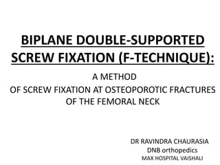
1. BIPLANE DOUBLE-SUPPORTED
SCREW FIXATION (F-TECHNIQUE):
A METHOD
OF SCREW FIXATION AT OSTEOPOROTIC FRACTURES
OF THE FEMORAL NECK
DR RAVINDRA CHAURASIA
DNB orthopedics
MAX HOSPITAL VAISHALI
2. INTRODUCTION
• The femur neck fracture is one of the most common
traumatic injury in the elderly.
• Osteosynthesis is associated with poor results in 21–46%.
• Osteosynthesis failure can be due to insufficient
reduction, unstable fixation or poor-quality osteoporotic
bone.
3. CONVENTIONAL FIXATION
• Multiple cannulated cancellous screw have a
compression effect at the fracture site. It also avoids
re displacement and rotation.
• Three cancellous screws, placed parallel to each other
and parallel to the femoral neck axis, (complication 20
to 48%) (Asnis 1994, Lu-Yao 1994, Tidermark 2003, Blomfeldt 2005, Rogmark 2006,
Gjertsen 2010.
4. NUMBER OF SCREWS
• Cannulated cancellous screws are thick in diameter.
• Diameter of the neck of the femur and the width of the
lateral cortex is relatively smaller in Indian patients.
• Therefore it is not always possible to fix femoral neck
fractures with three cannulated cancellous screws.
• Springer et al suggested that the fourth screw adds little
in additional fixation.
5. • Peripheral placement of screws in the femoral neck
(rather than central placement) to improve the strength of
fixation.
• Mizrahi et al specifically recommended an inverted
triangle configuration as it was stronger construct.
• The apex-proximal configuration was found to slip in the
cancellous bone because insufficient cortical purchase.
6. The starting position is above the
level of the lesser trochanter, and
the apex-proximal configuration
of the screws can be seen.
(B) A subtrochanteric femur
fracture resulted after a fall from
the standing position
Subtrochanteric femur fractures after screw fixation have
been documented in all age groups.
7. • A triangular screw configuration with its apex distal
would withstand greater forces than an apex-proximal
configuration.
• A larger stress riser is created when two screws are
placed distally (because of the
larger cortical defect), and
fracture will occur at lower
loads in this area.
8. FAILURE OF CONVENTIONAL FIXATION
• Lack of stability of the construction regarding
varus stress
• Lack of sliding phenomenon.
• Inability to move the entry point of the screws
distally into the solid diaphyseal cortex
9. • This may be attributed to several factors e.g. the entry
points below the level of the lesser trochanter, screws
placed too close together, and violation of the lateral cortex
with multiple guide-wire .
• This leads to the development of a stress riser along the
lateral aspect of the proximal femur.
10. BIPLANE DOUBLE-SUPPORTED SCREW
FIXATION
• BDSF implements two calcar buttressed
screws, oriented in different coronal
inclinations and intended to provide sufficient
stability during various physical activities.
11. • It provides strongest possible posterior cortical support
for the fixation construct.
• Biomechanically, the most effective component is the
distal screw placed at steeper angle and supported on a
large area.
12. The distal screw (red) is placed in the dorsal oblique plane, whereas the middle (blue) and
proximal screw (grey) are oriented in the anterior oblique plane. The distal and the middle
screws are calcar-buttressed with coronal inclinations of 150°–165° and 130°–140°,
respectively.
13. • It establish two supporting points. The solid cortex of
the calcar acts as a medial supporting point for the
screws. This supporting point works under pressure.
• The entry points of the distal and the middle screws
in the solid cortex of the proximal diaphysis, acts as a
lateral supporting point for the two screws.
14. BDSF ADVANTAGES
1) Provide antero-posterior bending stability of the neck.
2) Due to the biplane placement, enough space for a third
screw is provided.
3) due to the increase in the distance between the two
supporting points, the weight borne by the bone is
reduced.
4) The entry points of the screws are positioned wide apart
from each other so the tensile forces spread over a
greater surface of the lateral cortex and thus the risk of a
subtrochanter fracture decreases significantly.
5) The screw, placed at a highly increased angle, works in a
direction close to the direction of the loading force.
15. INDICATION
• Patient in which primary arthroplasty is contraindicated
Fractures of the Garden types from I to IV.
• Implants: 7.3-mm self-tapping cannulated screws.
16. REDUCTION
• Closed reduction (mild traction, slight abduction and
internal rotation of the limb).
• criteria for acceptable reposition: no varus, and valgus
alignment of 0°–15° on AP view, maximum
displacement of 2 mm, up to 20° ventral and 10° dorsal
angular displacement on lateral view.
• Open reduction, through Watson–Jones approach
17. TECHINIQUE
• A straight lateral incision is performed, starting at the
level of the lower border of the greater trochanter,
with a distal length of 6–10 cm. Following a direct
lateral transmuscular approach, a stripping of the
periosteum of the lateral diaphysis over a distance of
6–7 cm is performed.
18. PLACEMENT AND POSITIONING OF GUIDING WIRES
• First: 5–7 cm distally from the lower border of the
greater trochanter, inclined at an angle of 150°–165°
and directed posteriorly, so that after it touches onto
the “calcar” tangentially on AP view, the wire goes
into the dorsal third of femoral head.
19. • The second guiding wire is placed is at 2–4 cm
proximally from the distal wire, inclined at an angle of
130°–140°, directed anteriorly so that it goes in frontal
one-third of the femoral head (on lateral view) and into
the distal one-third of the femoral head (on AP view).
20. • Third : 1.5–2.0 cm proximally from the middle wire
and parallel to it. It goes into the front one-third of the
femoral head (on lateral view) and into the proximal
one-third of the femoral head (on AP view).
21. INSERTION OF SCREWS
• Measurement of the screw lengths and drilling with a 5-
mm cannulated reamer.
• The middle and proximal screws are placed first because
they are perpendicular to the fracture surface.
• Before placing the middle and distal screws, overdrill their
holes in the lateral cortex using a 7-mm cannulated
reamer.
• Then foot traction is released, impaction of the fracture
with an additional tightening up of the screws.
• Finally, the distal screw is placed.
• All three screws are inserted subchondrally (less than 5
mm), and no screw is placed in the central zone of the
femoral neck on lateral view.
22. AFTER-TREATMENT
• FWB Mobilization can be done after surgery without
limitations in the range of motion.
• In younger patients(< 55 years) PWB (30 kg) during the
first 8 weeks. (enough frictional stability at the fracture
site can not be achieved by intraoperative impaction
because of their dense bone).
23. Comparision b/w conventional and BDSF
The load acting at point A is pressure in a distal direction and denoted as A = FL/a
The load acting at point B is pressure in a proximal direction and denoted as B = A - F.
Static models of the implant. a A beam on elastic foundation; b A simple beam with an
overhanging end.
F load, L = length of beam; a distance between points A and B
24. CONVENTIONAL METHODS
• The screws are often located in the soft cancellous bone
near the axis of the femoral neck, without any cortical
support.
• Due to the lack of two solid supporting points, the
implant works like a beam on an elastic foundation.
• The elastic foundation is realized by the cancellous bone.
25.
26. RESULTS
• 42 males and 165 females, aged 75.7 ± 10.3 (range 49–
99)
• The average follow-up period was 29.6 months,
• Five complications were developed within less than 12
months.
• Radiographical results
• The bone union was seen in 97.6%
• One case with pseudoarthrosis and 1 nonunion with
AVN (Garden IV fracture).
• Fixation failure occurred in 5 patients.
27.
28. CONCLUSION
• Femoral neck fracture stability can be substantially
increased applying BDSF due to better cortical screw
support and screw orientation.
• The more unstable fracture, the better BDSF stability
is in comparison to CFIX.
29. REFERENCE
• Filipov O. Biplane double-supported screw fixation (F-technique): a
method of screw fixation at osteoporotic fractures of the femoral
neck. Eur J Orthop Surg Traumatol 2011;21:539–43.
• Thiele OC, Eckhardt C, Linke B, Schneider E, Lill CA. Factors affecting
the stability of screws in human cortical osteoporotic bone. A
cadaver study. J Bone Joint Surg (Br) 2007;89-B(5):701–5.
• Lindequist S, Waldemark T, Eriksson SA, Samnegard E. Screw
positions in femoral neck fractures. Comparison of two different
screw positions in cadavers. Acta Orthop Scand 1993;64:67–70
• von Bahr V, Syk B, Walheim G. Osteosynthesis of femoral neck
fracture using screws. Acta Chir Scand 1974;140:277–82.
• Gurusamy K, Parker MJ, Rowlands TK. The complications of
displaced intracapsular fractures of the hip: the effect of screw
positioning and angulation on fracture healing. J Bone Joint Surg
(Br) 2005;87(5):632–4.