Download to read offline
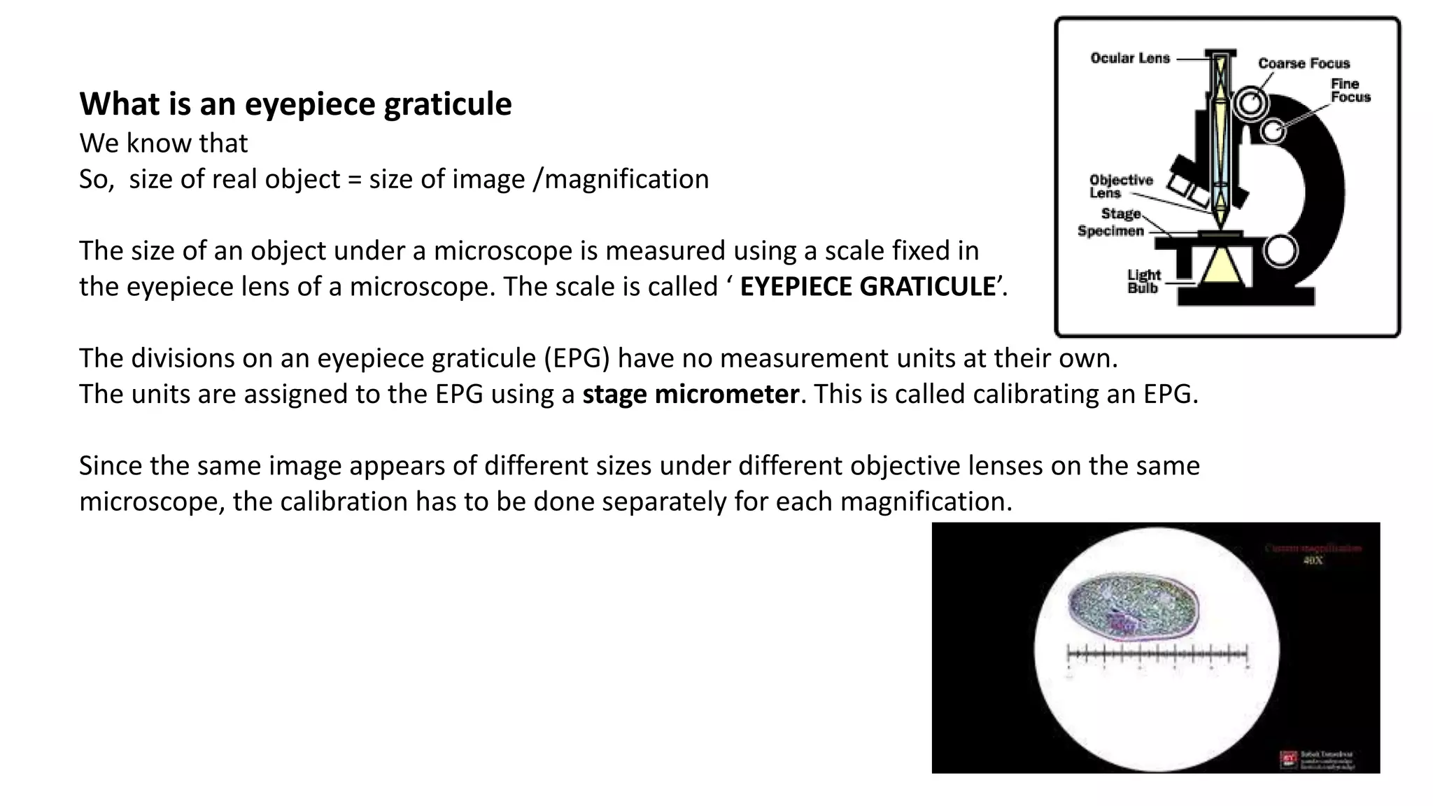

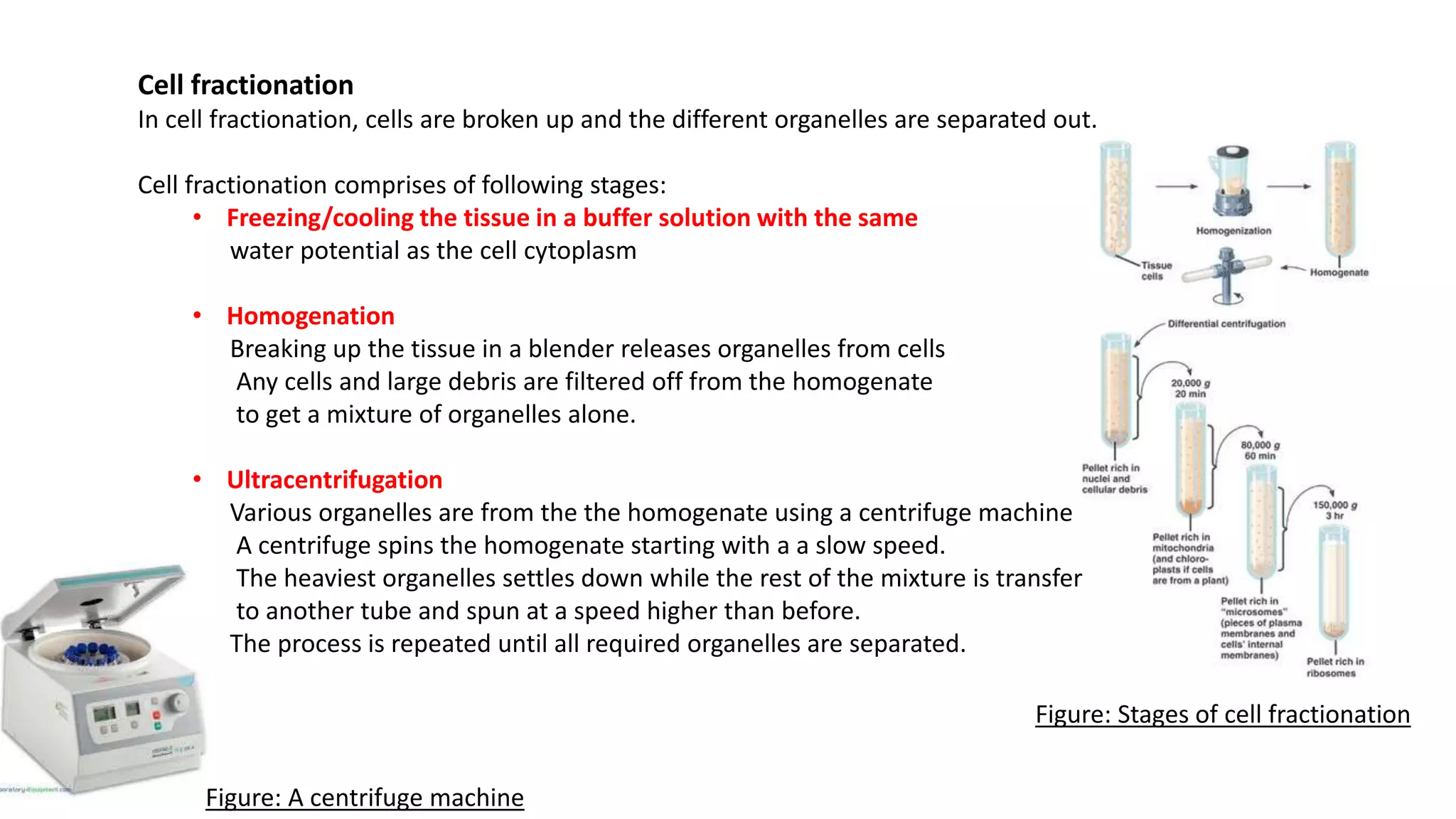
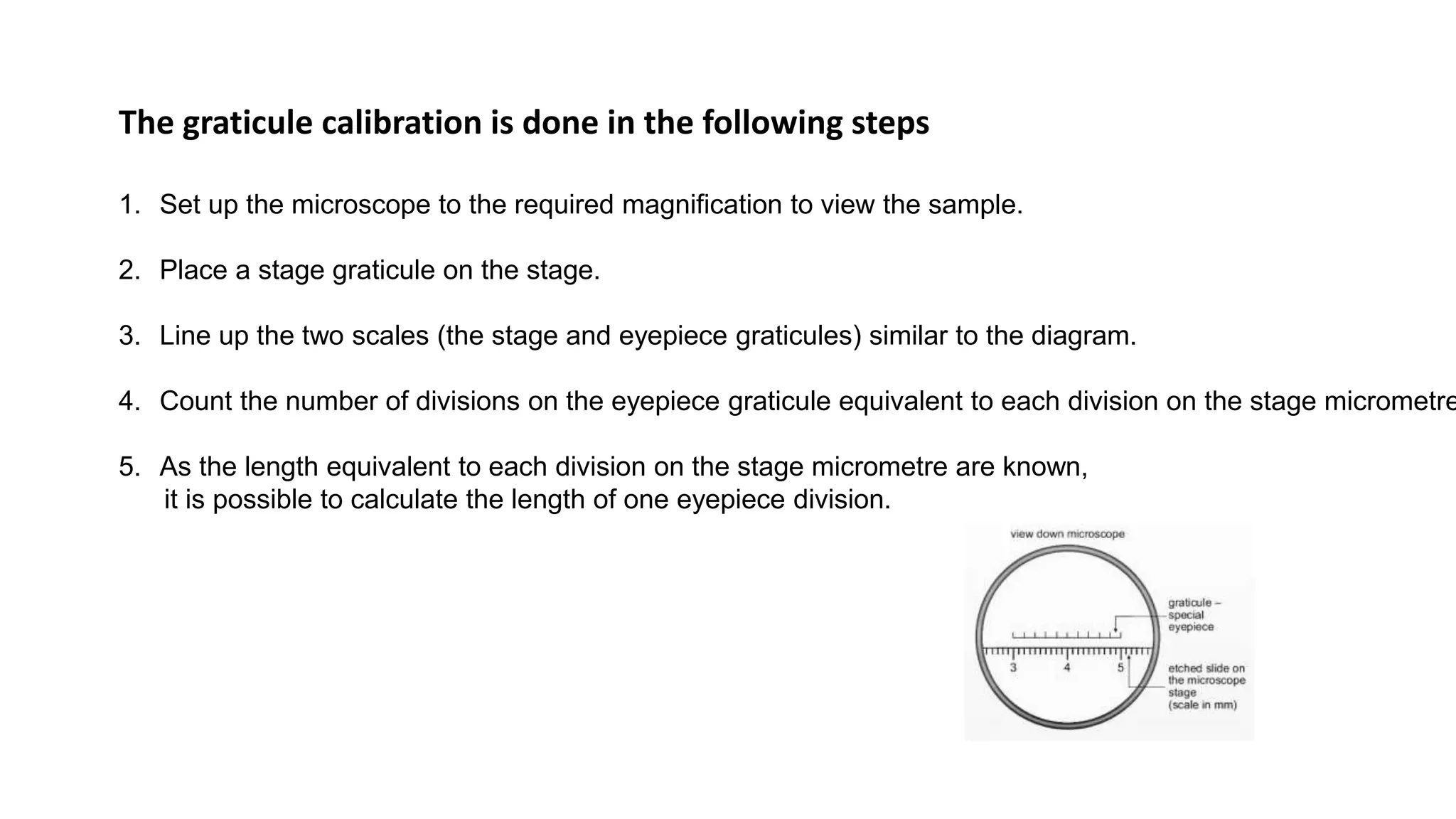
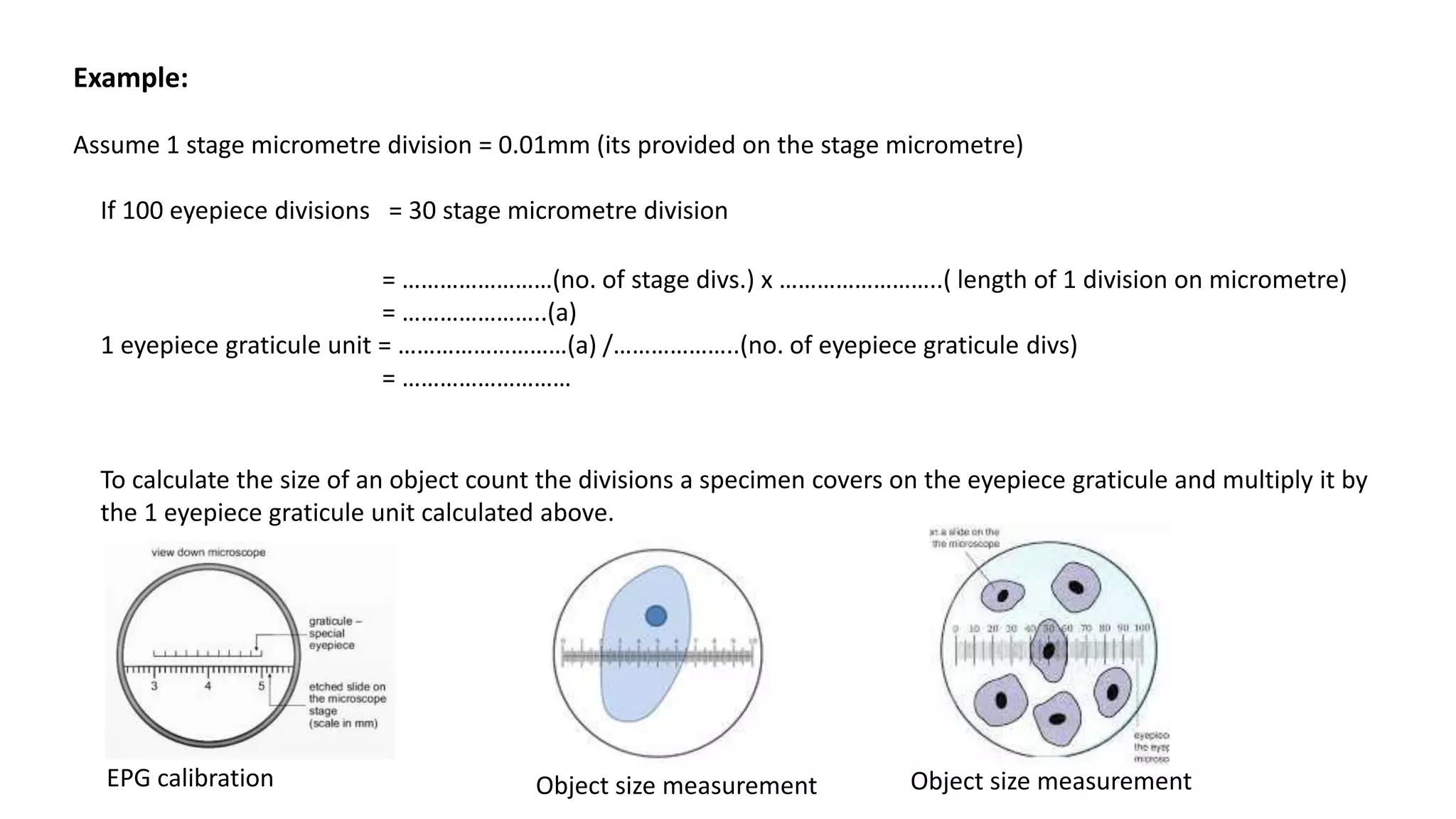


The document discusses two topics: 1. Measuring the size of objects under a microscope using an eyepiece graticule. An eyepiece graticule must be calibrated for each magnification by comparing it to a stage micrometer. 2. The process of cell fractionation which separates cell components by breaking open cells and centrifuging to separate organelles based on density.


