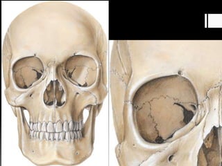
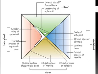
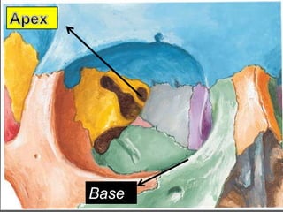
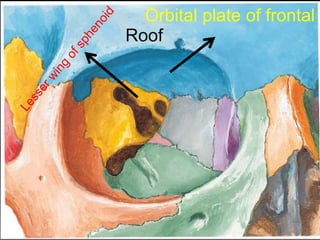
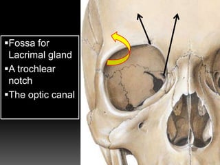
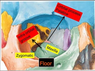
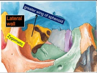
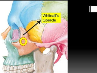
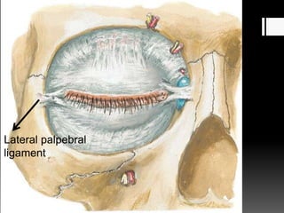
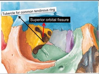
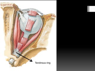
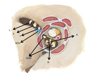
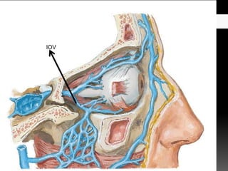
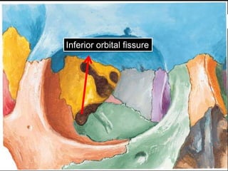
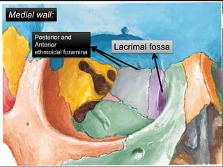
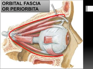
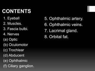
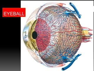
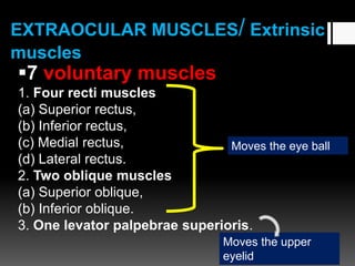
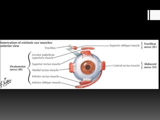
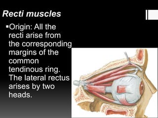
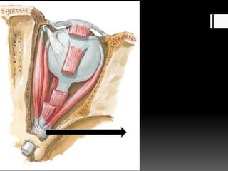
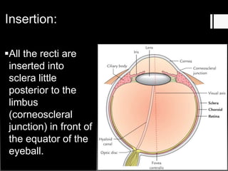
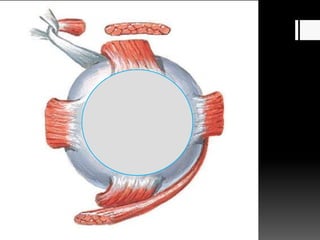
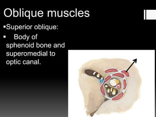
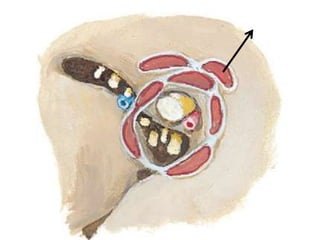
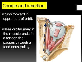
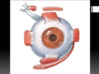
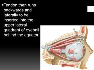
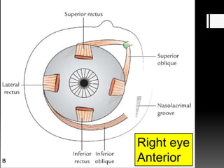
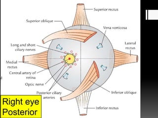
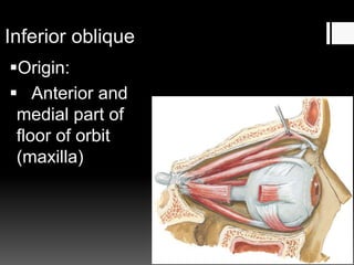
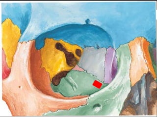
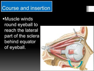
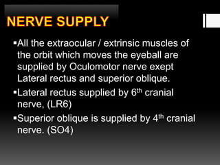
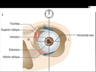
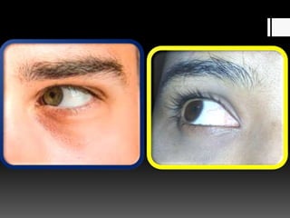
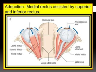
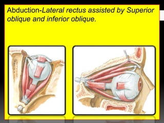
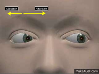
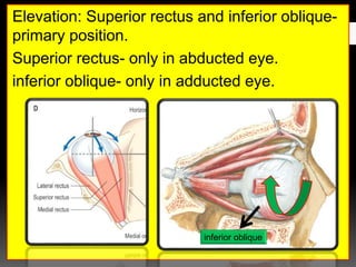
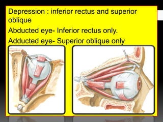
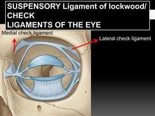
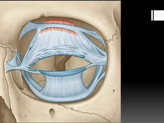
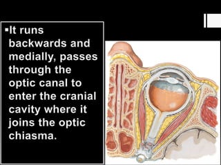
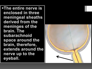
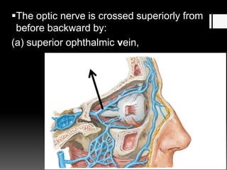
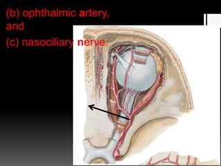
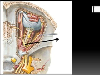
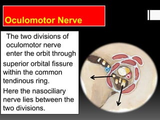
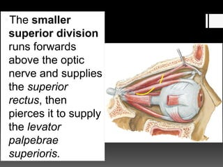
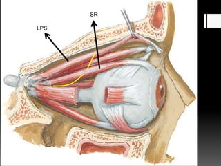
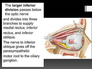
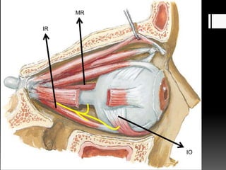
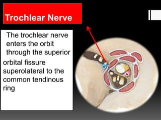
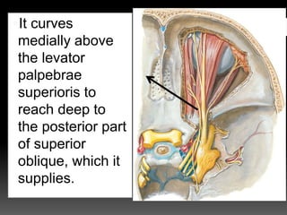
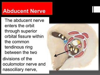
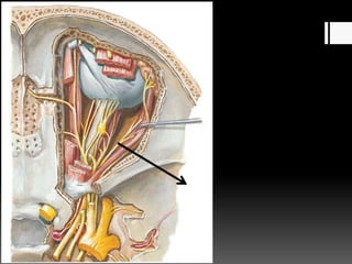
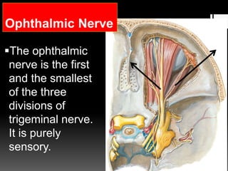
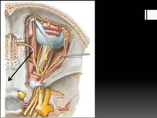
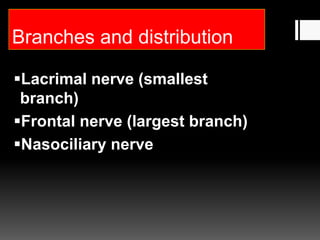
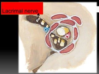
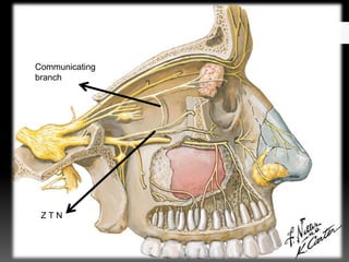
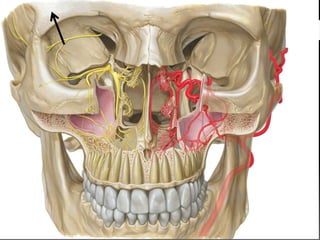
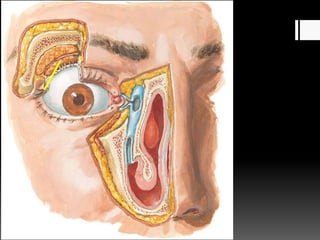
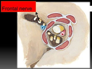
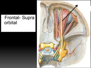
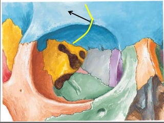
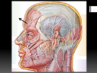
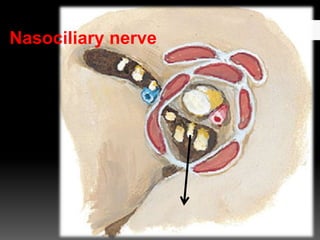
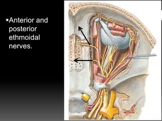
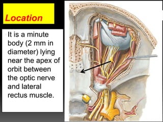
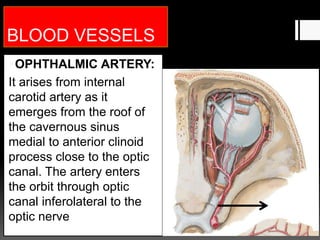
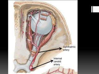
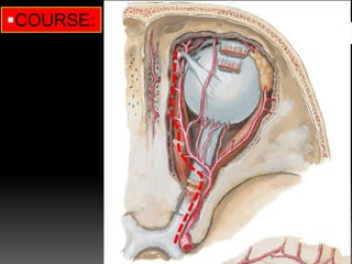
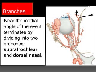
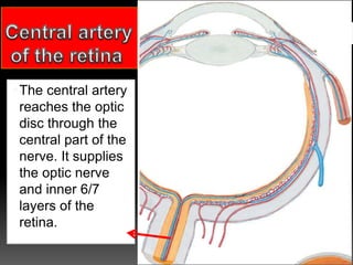
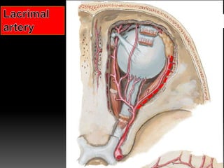
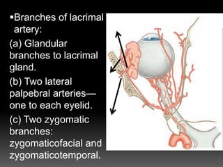
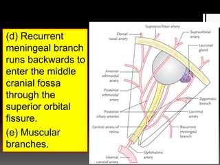
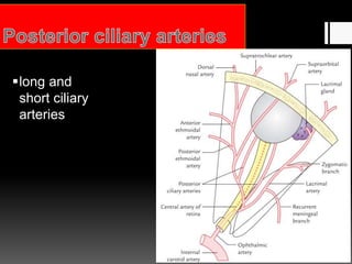
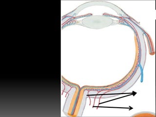
Base
Boundaries of the orbit
Roof
Floor
Lateral wall
Medial wall
Roof
Orbital plate of frontal
Fossa for
Lacrimal gland
A trochlear
notch
The optic canal
Floor
Zygomatic
Whitnall’s
tubercle
Lateral palpebral
ligament
Superior orbital fissure
Tendinous ring
L
F
T
SOV
N
OcM
A
IOV
Inferior orbital fissure
Medial wall:
Lacrimal fossa
Posterior and
Anterior
ethmoidal foramina
ORBITAL FASCIA
OR PERIORBITA
CONTENTS
1. Eyeball
2. Muscles.
3. Fascia bulbi.
4. Nerves
(a) Optic
(b) Oculomotor
(c) Trochlear
(d) Abducent
(e) Ophthalmic
(f) Ciliary ganglion.
5. Ophthalmic artery.
6. Ophthalmic veins.
7. Lacrimal gland.
8. Orbital fat.
EYEBALL
Cornea
Sclera
EXTRAOCULAR MUSCLES/ Extrinsic
muscles
7 voluntary muscles
1. Four recti muscles
(a) Superior rectus,
(b) Inferior rectus,
(c) Medial rectus,
(d) Lateral rectus.
2. Two oblique muscles
(a) Superior oblique,
(b) Inferior oblique.
3. One levator palpebrae superioris.
Moves the eye ball
Moves the upper
eyelid
There are three involuntary/smooth
muscles
superior tarsal or Muller's muscle,
Inferior tarsal and orbitalis.
Recti muscles
Origin: All the
recti arise from
the corresponding
margins of the
common
tendinous ring.
The lateral rectus
arises by two
heads.
Insertion:
All the recti are
inserted into
sclera little
posterior to the
limbus
(corneoscleral
junction) in front of
the equator of the
eyeball.
Oblique muscles
Superior oblique:
 Body of
sphenoid bone and
superomedial to
optic canal.
Course and insertion
Runs forward in
upper part of orbit.
Near orbital margin
the muscle ends in
a tendon the
passes through a
tendinous pulley.
Tendon then runs
backwards and
laterally to be
inserted into the
upper lateral
quadrant of eyeball
behind the equator.
Right eye
Anterior
Right eye
Posterior
Inferior oblique
Origin:
 Anterior and
medial part of
floor of orbit
(maxilla)
Course and insertion
Muscle winds
round eyeball to
reach the lateral
part of the sclera
behind equator
of eyeball.
All the extraocular / extrinsic muscles of
the orbit which moves the eyeball are
supplied by Oculomotor nerve exept
Lateral rectus and superior oblique.
Lateral rectus supplied by 6th cranial
nerve, (LR6)
Superior oblique is supplied by 4th cranial
nerve. (SO4)
AXES OF MUSCLES
vss
vss
vss
Visual axis
25 ˚
In abducted eye, sup recti and inferior recti coincides with
optical axis.
In adducted eye optical axis coincides with the
axis of oblique muscles.
51 ̊
Axis of movement
Elevation and
depression:
around the
transverse axis
passing through
the equator.
Adduction and
abduction:
Around the
vertical axis
passing through
the equator.
Rotation (torsion): around the anteroposterior
axis extending from anterior pole to posterior
pole of the eyeball.
When 12 o’clock position of the cornea rotates
medially, it is called intorsion and when it
rotates laterally, it called extorsion.
Eye movements produced by muscles
No movement is done by a single muscle.
While some muscles act as prime movers,
others act as synergists
Adduction- Medial rectus assisted by superior
and inferior rectus.
Abduction-Lateral rectus assisted by Superior
oblique and inferior oblique.
Abduction Adduction
Elevation: Superior rectus and inferior oblique-
primary position.
Superior rectus- only in abducted eye.
inferior oblique- only in adducted eye.
inferior oblique
Depression : inferior rectus and superior
oblique
Abducted eye- Inferior rectus only.
Adducted eye- Superior oblique only
Abduction
Adduction
Intorsion- superior rectus, superior oblique.
Extorsion- Inferior rectus, inferior oblique.
Associated Movements of the
Eyeball
Conjugate movements: when both the eyes
move in the same direction with visual axes
being parallel.
superduction
subduction
Disconjugate movements: when
the axes of both eyes converge or
diverge.
Levator
palpebrae
superioris muscle
Origin: from
undersurface of the
lesser wing of
sphenoid at the
apex of the orbit,
above the common
tendinous ring by a
narrow tendon.
INSERTION:
 the muscle broadens as it passes forwards from
its origin and divides into three lamellae:
Upper lamella consisting of skeletal muscle
penetrates the orbital septum passes through the
fibres of orbicularis oculi to be inserted into the
skin of upper eyelid.
Intermediate lamella consisting of smooth muscle
(superior tarsal muscle) is inserted on to the
upper border of the superior tarsal plate.
Lower lamella consisting of
connective tissue is inserted on to
the superior fornix of the conjunctiva.
The fascia
bulbi (Tenon’s
capsule) is a
loose
membranous
sheath that
envelops the
eyeball and
extends from
optic nerve to
the
sclerocorneal
junction.
 It is separated from the sclera by the
episcleral space.
The Tenon’s capsule forms a socket for
the eyeball to facilitate free ocular
movements.
Lateral check ligament
Medial check ligament
SUSPENSORY Ligament of lockwood/
CHECK
LIGAMENTS OF THE EYE
Clinical aspect
• If suspensory ligament of the
eye remains intact when the
floor of the orbit is fractured or
the maxilla is removed
surgically, the eyeball does not
sag.
NERVES OF THE ORBIT
Somatic and autonomic motor and
somatic sensory nerves are found in
the orbit.
The oculomotor, trochlear and
abducent nerves supply the extraocular
muscles.
The sensory nerves within the orbit are
the optic, ophthalmic and maxillary
nerves
Parasympathetic fibres from the
oculomotor nerve supply sphincter
pupillae and the ciliary muscle
(ciliaris) via the ciliary ganglion.
facial nerve - lacrimal gland and
choroid via the pterygopalatine
ganglion.
Sympathetic fibres - dilator pupillae.
Infraorbital and Zygomatic Nerves
OPTIC NERVE
This nerve of sight is
4 cm long. It is made
up of about 1 million
myelinated nerve
fibres. The optic nerve
emerges from the
eyeball, 3 or 4 mm
nasal to the posterior
pole of the eyeball.
Relations
The central artery and vein of the
retina pierce the optic nerve
inferomedially about 1.25 cm behind
the eyeball.
The optic nerve is crossed superiorly from
before backward by:
(a) superior ophthalmic vein,
(b) ophthalmic artery,
and
(c) nasociliary nerve.
Oculomotor Nerve
The two divisions of
oculomotor nerve
enter the orbit through
superior orbital fissure
within the common
tendinous ring.
Here the nasociliary
nerve lies between the
two divisions.
The smaller
superior division
runs forwards
above the optic
nerve and supplies
the superior
rectus, then
pierces it to supply
the levator
palpebrae
superioris.
LPS
SR
The larger inferior
division passes below
the optic nerve
and divides into three
branches to supply
medial rectus, inferior
rectus, and inferior
oblique.
The nerve to inferior
oblique gives off the
parasympathetic
motor root to the ciliary
ganglion.
IR
IO
MR
Trochlear Nerve
The trochlear nerve
enters the orbit
through the superior
orbital fissure
superolateral to the
common tendinous
ring
It curves
medially above
the levator
palpebrae
superioris to
reach deep to
the posterior part
of superior
oblique, which it
supplies.
The abducent nerve
enters the orbit
through superior
orbital fissure within
the common
tendinous ring
between the two
divisions of the
oculomotor nerve and
nasociliary nerve,
Abducent Nerve
The ophthalmic
nerve is the first
and the smallest
of the three
divisions of
trigeminal nerve.
It is purely
sensory.
Ophthalmic Nerve
Branches and distribution
Lacrimal nerve (smallest
branch)
Frontal nerve (largest branch)
Nasociliary nerve
Lacrimal nerve
Communicating
branch
Z T N
Frontal nerve
Frontal- Supra
orbital
Supra trochlear
Nasociliary nerve
Anterior and
posterior
ethmoidal
nerves.
Branches of ant
ethmoidal nerve
Infratrochlear nerve
It is a peripheral
parasympathetic
ganglion,
connected with
the nasociliary
nerve.
Ciliary Ganglion
Although topographically it is
connected to the nasociliary nerve
from ophthalmic division of the
trigeminal, but functionally it is
connected to the oculomotor nerve.
Location
It is a minute
body (2 mm in
diameter) lying
near the apex of
orbit between
the optic nerve
and lateral
rectus muscle.
Roots
Three roots enter its posterior end.
Motor (parasympathetic) root:
Preganglionic- Oculomotor
Post ganglionic- The postganglionic
parasympathetic fibres arise from the cells
of the ganglion and pass through short ciliary
nerves to supply the ciliary muscle and
sphincter pupillae.
Accessory
oculomotor
(Edinger-
Westphal) nucleus
OMN
CG
SCN
Sensory root:
It is derived from nasociliary
nerve. It consists of sensory fibres
(for pain, touch, and temperature)
from eyeball, which pass through
the ciliary ganglion without relay.
Sympathetic root:
Dilator
Pupillae &
Blood vessels
Branches
 8-10 Short ciliary
nerves
Clinical aspects
The ciliary ganglion is blocked to
produce dilatation of pupil before
cataract extraction.
BLOOD VESSELS
OPHTHALMIC ARTERY:
It arises from internal
carotid artery as it
emerges from the roof of
the cavernous sinus
medial to anterior clinoid
process close to the optic
canal. The artery enters
the orbit through optic
canal inferolateral to the
optic nerve
Ophthalmic
artery
Internal
carotid
artery
COURSE:
Branches
Near the medial
angle of the eye it
terminates by
dividing into two
branches:
supratrochlear
and dorsal nasal.
The central artery
reaches the optic
disc through the
central part of the
nerve. It supplies
the optic nerve
and inner 6/7
layers of the
retina.
Clinical aspects
The central artery of retina is an
example of a typical end artery. Its
damage produces sudden total
blindness on the side of the lesion.
Branches of lacrimal
artery:
(a) Glandular
branches to lacrimal
gland.
(b) Two lateral
palpebral arteries—
one to each eyelid.
(c) Two zygomatic
branches:
zygomaticofacial and
zygomaticotemporal.
(d) Recurrent
meningeal branch
runs backwards to
enter the middle
cranial fossa
through the
superior orbital
fissure.
(e) Muscular
branches.
long and
short ciliary
arteries
Supraorbital
artery
Posterior
ethmoidal
artery
Anterior
ethmoidal
artery
Dorsal
(external)
nasal artery
Supratrochle
ar artery
Medial
palpebral
branches
Superior
ophthalmic vein:
Inferior
ophthalmic vein: Cavernous sinus
Clinical aspects
Because the ophthalmic veins drain into
cavernous sinus and communicate with
the extracranial veins, they act as routes
through which infection can spread from
outside to inside the cranial cavity.
Strabismus/squint: Unilateral
paralysis of an individual muscle
due to involvement of the nerves
produces strabismus or squint
(deviation of eye to the opposite
side) and may result in diplopia
(double vision).
Paralysis of levator palpebrae superioris:
Paralysis of levator palpebrae superioris due to
involvement of the oculomotor nerve leads to
complete ptosis (drooping of upper eyelid).
To be continued

Orbit