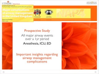
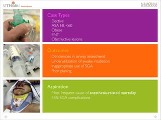
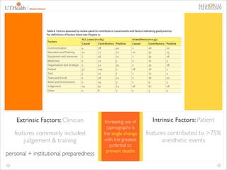
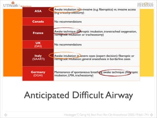
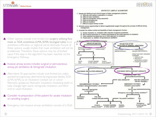
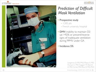
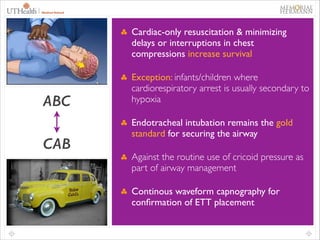
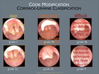
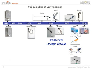
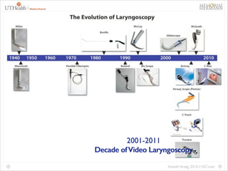
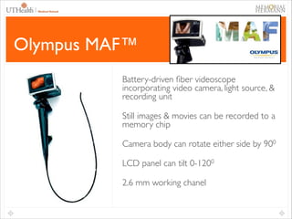
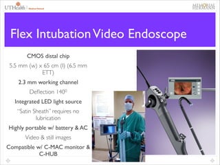
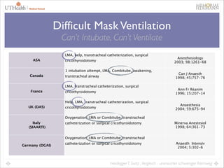
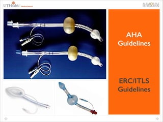
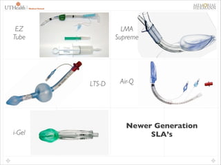
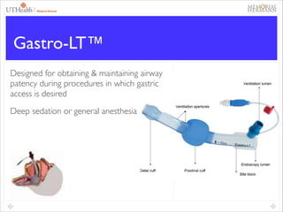
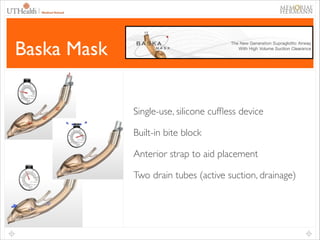
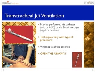
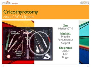
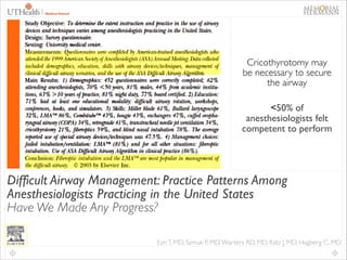
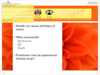
This document provides an overview of difficult airway management concepts. It begins by outlining the scope of the problem, noting that 1-3% of general anesthetics involve an unanticipated difficult airway. Key factors that contribute to difficult airways are discussed, including patient characteristics and clinician experience. A variety of airway devices and techniques to secure the airway are presented, including video laryngoscopy, fiberoptic intubation, supraglottic airways, and surgical options like cricothyrotomy. The document emphasizes the importance of thorough pre-operative airway assessment and having a well-thought out plan for managing difficult airway scenarios.
The document discusses the posterior fossa, including its boundaries, contents, blood supply, clinical presentation of lesions, and considerations for anesthesia. The posterior fossa is bounded anteriorly by the clivus and petrous bone, posteriorly by the occipital bone, and laterally by the temporal bone. It contains the cerebellar hemispheres, brainstem, and cranial nerves III-XII. Lesions can cause a variety of signs and symptoms depending on location, including ataxia, nystagmus, limb weakness, and cranial nerve deficits. Anesthesia for posterior fossa surgery requires careful monitoring and positioning to maintain stability while allowing surgical access.
Double lumen tubes were developed in the 1950s-60s to enable lung isolation during thoracic surgery. The Carlens and Bryce-Smith tubes were some of the earliest designs, featuring curves and cuffs to isolate the left or right mainstem bronchus. Modern tubes like the Robertshaw are widely used and come in varying sizes from 26-41 French. Placement requires careful advancement and confirmation via auscultation, cuff inflation, and bronchoscopy to avoid malposition and injury. Double lumen tubes allow selective ventilation and treatment of each lung but require replacement with a single tube after surgery.
Guillain Barre’ syndrome(GBS) and Anesthesia considerationTenzin yoezer
Patients with GBS need special care when coming to the surgery. They have a high risk of aspiration, airway compromise, autonomic instability, altered response to NMBs. It is the duty of the anesthesia providers to recognize those problems and minimize the complications.
Respiratory Physiology & Respiratory Function During AnesthesiaDang Thanh Tuan
This document summarizes respiratory physiology and function during anesthesia. It discusses factors related to respiratory function including gravity-determined distribution of perfusion and ventilation. It also covers non-gravitational determinants of pulmonary vascular resistance and blood flow distribution. Finally, it examines oxygen and carbon dioxide transport through the lungs.
One-lung ventilation (OLV) is used for thoracic surgeries to isolate one lung from the other. It requires skill to place lung isolation equipment like double-lumen endotracheal tubes (DLT) and prevent hypoxemia. DLTs have two lumens allowing independent ventilation of each lung. Placement is checked by auscultation and bronchoscopy to ensure proper position before surgery. Complications can include airway damage if the tube is malpositioned or overinflated. Careful technique and monitoring are needed for safe OLV.
Caudal anesthesia involves injecting local anesthetic into the caudal canal of the sacrum to provide pain relief below the umbilicus. It can be used alone or with general anesthesia for surgeries involving the perineum, anus, rectum, or lower extremities. The technique involves identifying the sacral hiatus and inserting a needle, with ultrasound or fluoroscopy guidance available. Potential complications include dural puncture, nerve injury, and local anesthetic toxicity. The level of pain relief varies significantly among patients.
This document discusses respiratory function and its importance to anesthesia. It covers topics like cellular respiration, aerobic vs anaerobic respiration, muscles of respiration, mechanisms of ventilation, lung volumes, compliance, and factors that affect respiration. The speaker is Dr. Tipu and the event is being coordinated by Dr. Shivali Pandey.
The document discusses the management of difficult airways. It defines difficult mask ventilation and difficult laryngoscopy/intubation. It describes various tests that can be used to assess a difficult airway, such as the Mallampati test, thyromental distance, sternomental distance, and neck mobility tests. Radiographic predictors of a difficult airway are also discussed, along with causes of difficult intubation related to patient anatomy and various medical conditions.
One Lung Ventilation (OLV) is a technique used during thoracic surgery to isolate one lung allowing it to function independently. There are several methods to achieve OLV including using a double lumen endotracheal tube, Univent tube, or endobronchial blockers. OLV provides benefits like protecting the healthy lung during surgery on the other lung but also causes physiological changes and risks like hypoxemia. Care must be taken to properly position the lung isolation device and monitor for complications during OLV.
The document discusses airway assessment for anesthesia. It defines the upper and lower airways and provides details on relevant anatomy. Key points of airway assessment are identified including patient history, external examination focusing on dentition, head and neck mobility. Specific tests like Mallampati score, thyromental distance and range of motion are described. The document emphasizes the importance of thorough airway assessment prior to procedures to anticipate difficult intubation. Advanced assessment methods involving imaging and fiberoptics are also mentioned.
This document discusses the Laryngeal Mask Airway (LMA), including its uses, sizes, advantages, and disadvantages compared to other airway devices. It also covers complications associated with the LMA like laryngospasm and negative pressure pulmonary edema. Laryngospasm is described as a protective reflex that can be triggered by light anesthesia or airway irritation. The management of laryngospasm involves securing the airway and administering muscle relaxants if needed. Negative pressure pulmonary edema is discussed as a rare complication of laryngospasm where high negative intrathoracic pressures cause fluid accumulation in the lungs.
Pre-oxygenation is: safe, simple, cheap, effective, well-tolerated. This article provides a compelling argument in favour of pre-oxygenation prior to all general anaesthesia.
This document provides an overview of the anatomy of the epidural space. It discusses the boundaries, contents, size, and structures that must be penetrated to access the epidural space. Key points include that the epidural space lies between the spinal meninges and vertebral canal, contains connective tissue, fat, blood vessels and spinal nerves. It varies in size from 1-6mm depending on the spinal region. To access it requires penetrating the skin, ligaments and ligamentum flavum in the midline.
1. Difficult intubation can result in significant morbidity and mortality, so proper prediction allows time for equipment selection and experienced personnel.
2. Several physical exam findings can predict difficult airways, including reduced neck mobility, short chin-to-hyoid or thyromental distances, large tongue size, and reduced mouth opening.
3. Scoring systems like Mallampati, Wilson, and Cormack-Lehane grading can synthesize exam findings to predict difficult mask ventilation, intubation, or laryngoscopic views. Physical indicators are combined in some group indices to improve predictive power.
The document discusses the difficult airway, including its definition, causes, assessment, and management. It defines difficult ventilation and difficult intubation. Causes can be related to the anesthesiologist, equipment, or patient factors like congenital syndromes or acquired conditions. Assessment involves history, physical exam including airway indices like Mallampati score, and radiologic evaluation. Management includes preparing a difficult airway cart and having alternate plans for securing the airway.
Obesity presents unique challenges for anaesthesia. Obese patients have decreased lung volumes which increases the risk of hypoxemia during induction and intubation. Preoxygenation in a slightly head-up position can help reduce this risk. Intubation may be difficult due to obesity related anatomical changes. Regional anaesthesia can also be challenging due to obscured landmarks and extensive adipose tissue. Postoperatively, obese patients are at higher risk of respiratory failure, DVT, and wound infections requiring close monitoring. Careful consideration of dosing, positioning, and postoperative monitoring is needed to safely manage anaesthesia for obese patients.
This document discusses the circle system used in anesthesia. It describes the components of the circle system including the absorber, canisters, unidirectional valves, fresh gas inlet, adjustable pressure limiting valve, and reservoir bag. It explains how the circle system works and how it can be configured as a closed, semi-closed, or semi-open system depending on the fresh gas flow. It also discusses the advantages and disadvantages of the circle system and components like the absorber, how it neutralizes carbon dioxide, and factors that influence compound A and carbon monoxide formation.
The questions asked in the Anaesthesiology viva examination are presented in this presentation which will be useful for the post-graduates appearing for the M.D-Anaesthesia examination.
This document discusses anaesthesia considerations for EHPVO (extrahepatic portal venous obstruction) and meso-Rex shunt surgery. EHPVO is a non-cirrhotic cause of portal hypertension most common in children, while IPH (idiopathic portal hypertension) typically affects adults. Key differences are noted. Meso-Rex shunt restores hepatic blood flow more physiologically than non-physiological shunts. Anaesthesia must consider issues like malnutrition, anemia, ascites, and potential for bleeding or thrombosis. Careful monitoring is needed due to fluid shifts and potential liver or cardiac dysfunction.
The document provides guidelines from the American Society of Regional Anesthesia (ASRA) on placing and removing epidural catheters in patients taking anticoagulant and antiplatelet drugs. It lists recommended minimum times to wait after the last dose of various medications before catheter placement and removal, as well as when to restart anticoagulation therapy after removal. The medications are grouped into heparins, low molecular weight heparins, factor Xa and direct thrombin inhibitors, antiplatelet agents, fibrinolytics, and glycoprotein IIb/IIIa inhibitors. For each group, it provides the drug names and recommended waiting times.
This document discusses the anatomy, physiology, and functions of the liver as they relate to anesthesia. It begins with an overview of hepatic anatomy including gross and microscopic structure, blood supply, and drainage. It then covers hepatic blood flow regulation by intrinsic and extrinsic factors and how anesthesia can affect blood flow. The major sections discuss hepatic functions such as metabolism, synthesis, and detoxification. In particular, it notes the liver's roles in glucose regulation, protein and lipid metabolism, coagulation factor production, and bilirubin metabolism.
This document discusses various supraglottic airway devices that can be used for airway management during anesthesia and difficult airway situations. It begins by outlining the advantages of supraglottic devices over endotracheal intubation. It then provides detailed classifications and descriptions of numerous supraglottic devices, including LMAs, i-gels, laryngeal tubes, and more. Placement techniques, sizing considerations, and diagnostic tests for ensuring proper placement are also reviewed. The document serves as a comprehensive guide to the use of supraglottic airway devices for airway management.
The document discusses the case of a 26-year-old female patient who is 36 weeks pregnant with mitral stenosis. She presents with palpitations, breathlessness, and fatigue. Her history and examination are consistent with mild mitral stenosis of rheumatic origin, as confirmed by echocardiogram findings of a mitral valve area of 2.0 cm2 and transvalvular pressure of 8 mm Hg. The discussion centers on the pathophysiology, diagnosis, and management of mitral stenosis, including the plan for regional anesthesia for her elective caesarean section.
Intro to Hypoxic pulmonary vasoconstriction Arun Shetty
Hypoxic pulmonary vasoconstriction, a seldom heard phenomenon but very effective physiologic property which helps lungs utilise ventilation to the maximum
The majority of pediatric airway emergencies occur in children under 1 year old and are primarily caused by upper airway obstruction from infectious diseases like viral croup. The pediatric airway has unique anatomical features like a higher larynx and narrower subglottic airway that make it more prone to obstruction. Initial management focuses on airway stabilization through suction, positioning, oxygen therapy, and supportive care. Further treatment depends on the specific condition but may include nebulization, intubation, tracheostomy, or endoscopic evaluation and intervention. Outcomes are generally good with resolution of acute issues and management of any underlying structural abnormalities.
Cardiac output can be measured through various invasive and non-invasive methods. The pulmonary artery catheter using thermodilution is still considered the gold standard but is invasive. Minimally invasive methods include lithium dilution, pulse contour analysis devices, esophageal Doppler, and transesophageal echocardiography. Non-invasive methods include partial gas rebreathing, thoracic bioimpedance, and Doppler ultrasound. The ideal monitor is accurate, continuous, non-invasive and provides reliable measurements during different physiological states.
This document discusses recognition and management of difficult airways. It defines difficult airway as difficulty with mask ventilation or tracheal intubation. It discusses incidence, risk factors, assessment techniques like Mallampati classification and thyromental distance, and management strategies including awake intubation, use of airway adjuncts like LMA, and the ASA difficult airway algorithm which provides a structured approach. The key points are assessing risk factors, having proper equipment and backup plans, and pursuing oxygenation throughout management of a difficult airway.
Pediatric laryngeal and subglottic stenosis can be graded using the Cotton system. Congenital subglottic stenosis is classified as membranous or cartilaginous based on histopathology. Endoscopic management is effective for early stenosis but open surgery is needed for more advanced cases. Mitomycin C has shown promise in reducing restenosis after laryngotracheal reconstruction surgery. Careful assessment and antimicrobial coverage are important for decannulation and preventing complications.
Overlapping MAF is a modification of the original MAF technique used in the management of high anal fistulas. This simple modification showed to improve the success rate in 10% more than the original technique.
1) Airway management in trauma patients presents unique challenges beyond simply placing an endotracheal tube, with outcomes dependent on the provider's ability to anticipate difficulties and have a safe plan.
2) There is conflicting evidence on whether early definitive airway management in the prehospital setting saves lives, with some studies finding benefits and others finding increased mortality.
3) Anatomic and physiologic factors must both be considered when planning airway management for trauma patients, and advanced techniques do not necessarily mean better outcomes.
This document discusses airway management in the ICU for an obese patient with pneumonia who is deteriorating. It covers assessing the airway, sedation options like midazolam and fentanyl, difficult airway tools like bougies and LMAs, and special considerations for obese, rheumatoid arthritis, head/neck cancer, and trauma patients. The key is to oxygenate, assess ability to ventilate and intubate, call for help, and position the patient while preparing airway adjuncts and securing the airway.
This document proposes a new algorithm for extubating patients called "VSS+4S+2S". It notes that while considerable research has focused on intubation, relatively little attention has been given to developing standardized guidelines and protocols for safe extubation. The proposed algorithm aims to improve extubation practices, minimize failures, and provide a practical framework for teaching residents. It consists of evaluating Vital Signs Stability, then Stent, Strength, Spontaneous breathing, and Suctioning the patient while still under anesthesia. Once these criteria are met, anesthesia is discontinued and the return of Swallowing and ability to Secure the airway are assessed before full extubation. The goal is to establish standardized steps to guide ext
Airway and Anesthetic Management of the Traumatized Patient.pptxHadi Munib
The document discusses airway and anesthetic management of traumatized patients with maxillofacial injuries. It emphasizes the importance of securing the airway as the top priority during initial assessment and treatment. The initial airway assessment involves evaluating airway patency, ventilation, oxygenation, and vital signs. Specific factors that can complicate airway management in maxillofacial trauma patients are also reviewed, including mandibular fractures, dental injuries, hemorrhage, and soft tissue swelling. A thorough history including allergies, medications, and injury details is also important to guide appropriate anesthetic strategies.
Abandoning difficult airway algorithms to improve patient safety | Richard Le...scanFOAM
The document discusses abandoning difficult airway algorithms and improving patient safety. Some key points:
1) Predicting difficult intubation is difficult and algorithms often fail to accurately predict difficulty. Most predicted difficult cases turn out to be easy.
2) Video laryngoscopy and new airway devices have reduced risks that were previously associated with direct laryngoscopy. Physiologically difficult airways in critically ill patients remain high risk.
3) First pass success is important for reducing risks. Checklists, checklists, checklists along with optimizing oxygenation, positioning and teamwork can help achieve this. Understanding priorities is more important than memorizing algorithms.
4) Adverse events often result from hypo
This document discusses anaesthesia considerations for cleft lip and palate surgery. It begins by describing the types of orofacial clefts and the importance of treating them. It then discusses pre-operative assessment, focusing on identifying other congenital anomalies or syndromes, assessing for difficult airways, and considering nutrition, chronic airway issues, and premedication. Intra-operative considerations include induction, potential difficult mask ventilation or laryngoscopy, and appropriate tube selection. Managing difficult airways is an important part of the anaesthetic plan.
1) The document describes a case of awake fibreoptic intubation in a parturient with an expanding soft palate hematoma. Nasotracheal intubation was chosen and successfully performed with the aid of a videolaryngoscope and fiberoptic scope.
2) Videolaryngoscopes provide improved glottic views compared to direct laryngoscopy but performance varies depending on the type of difficult airway and clinical setting. Certain videolaryngoscopes may be better suited for specific airway challenges such as trauma, cervical spine immobilization, or difficult environmental conditions.
3) A study comparing videolaryngoscope performance to direct laryngoscopy in simulated difficult int
Airway assessment is important for identifying patients at risk of a difficult airway. Several tests can be used including Mallampati scoring, mouth opening, neck mobility, and thyromental distance. A difficult airway is when facemask ventilation or intubation is not possible using conventional methods. It is important to prepare for difficult airway scenarios by having proper equipment and involving senior help. Identifying difficult airway risks pre-operatively allows time for planning alternative strategies to ensure patient safety.
Endolymphatic Hydrops Surgery talk NOTSA 2022 (FINAL) .pptxKristyRae1
Endolymphatic hydrops, or excess fluid buildup in the inner ear, can cause Meniere's disease which features episodes of vertigo, hearing loss, and tinnitus. Surgical interventions aim to reduce these symptoms by addressing the endolymphatic sac, but their efficacy compared to placebo is unclear due to the natural fluctuations of the disease. A landmark 1981 sham study found that a simple mastoidectomy was as effective as endolymphatic sac surgery, casting doubt on surgeries' benefits. However, advocates argue surgeries still help when medical options fail, though more research is needed to prove their efficacy while avoiding harm.
- Balloon sinuplasty is a minimally invasive technique for treating sinusitis using balloon catheters to dilate sinus ostia rather than conventional endoscopic sinus surgery.
- Studies show balloon sinuplasty improves symptoms in selected patients with chronic sinusitis and is safe, with minimal adverse effects. However, longer term data is still needed to define its optimal role and indications.
- While initial data is promising for symptom relief and preservation of sinus anatomy compared to traditional FESS, balloon sinuplasty may not eliminate the need for conventional sinus surgery in all patients.
This document discusses strategies for managing difficult airways in emergency situations. It begins by outlining goals of predicting difficult airways, having appropriate plans, and confidence in "can't intubate, can't ventilate" situations. It then discusses factors that can help identify difficult airways through past medical history, physical exam findings like thyromental distance, and classifications like Mallampati. The document emphasizes having alternative plans for airway management if intubation fails, such as BVM ventilation, supraglottic airways, or surgical techniques. It stresses the importance of paramedics feeling prepared with multiple airway options when facing emergent difficult airway scenarios.
Methods: Retrospectively, the file records of the patients who underwent sleeve gastrectomy were examined. Demographic features, Body Mass Index (BMI), the mouth opening, Mallampati score, thyromental distance, sternomental distance, neck circumference measurements and videolaryngoscopic examination results were recorded Results: In a total of 140 consecutive patients (58 male, 82 female) were included in the study. The mean age of the study participants was 35.40 ± 9.78 and the mean BMI of the patients was 44.33 ± 7.52 kg/m2
. The mean mouth opening of the patients was 4.82 ± 0.54 cm
and the mean neck circumference was 43.52 ± 4.66 cm. The mean thyromental distance was 8.02 ± 1.00 cm and the mean sternomental distance was16.58 ± 1.53 cm. Difficult intubation was determined in 8 (5.7%) patients. In logistic regression analysis, age (p : 0.446), gender (p : 0.371), BMI (p : 0.947), snoring (p : 0.567), sleep apnea (p : 0.218), mouth opening (p : 0.687), thyromental distance (p :0.557), sternomental (p : 0.596) and neck circumference (p : 0.838) were not the independent predictors of difficult intubation. However, Mallampati score (p : 0.001) and preoperative direct laryngoscopy findings (p : 0.037) performed in outpatient clinic were the significant
predictors of difficult intubation. Interestingly, all patients with grade 4 laryngoscopy findings had difficult intubation.
This document summarizes a technique for performing local/regional anesthesia for thyroid surgery. It begins by describing the patient who will undergo a parathyroidectomy due to hypercalcemia from hyperparathyroidism. It then provides background on the history and development of performing thyroid surgery under local/regional anesthesia. It proceeds to describe the relevant anatomy of the cervical plexus and its branches. It concludes by outlining the technique for performing a superficial cervical plexus block, including patient position, landmarks, local anesthetic used, and injection points along the posterior border of the sternocleidomastoid muscle at the level of the external jugular vein. The summary is provided in 3 sentences or less as requested.
TEST BANK For Basic and Clinical Pharmacology, 14th Edition by Bertram G. Kat...rightmanforbloodline
TEST BANK For Basic and Clinical Pharmacology, 14th Edition by Bertram G. Katzung, Verified Chapters 1 - 66, Complete Newest Version.
Integrating Ayurveda into Parkinson’s Management: A Holistic ApproachAyurveda ForAll
Explore the benefits of combining Ayurveda with conventional Parkinson's treatments. Learn how a holistic approach can manage symptoms, enhance well-being, and balance body energies. Discover the steps to safely integrate Ayurvedic practices into your Parkinson’s care plan, including expert guidance on diet, herbal remedies, and lifestyle modifications.
- Video recording of this lecture in English language: https://youtu.be/kqbnxVAZs-0
- Video recording of this lecture in Arabic language: https://youtu.be/SINlygW1Mpc
- Link to download the book free: https://nephrotube.blogspot.com/p/nephrotube-nephrology-books.html
- Link to NephroTube website: www.NephroTube.com
- Link to NephroTube social media accounts: https://nephrotube.blogspot.com/p/join-nephrotube-on-social-media.html
Does Over-Masturbation Contribute to Chronic Prostatitis.pptxwalterHu5
In some case, your chronic prostatitis may be related to over-masturbation. Generally, natural medicine Diuretic and Anti-inflammatory Pill can help mee get a cure.
Promoting Wellbeing - Applied Social Psychology - Psychology SuperNotesPsychoTech Services
A proprietary approach developed by bringing together the best of learning theories from Psychology, design principles from the world of visualization, and pedagogical methods from over a decade of training experience, that enables you to: Learn better, faster!
Basavarajeeyam is a Sreshta Sangraha grantha (Compiled book ), written by Neelkanta kotturu Basavaraja Virachita. It contains 25 Prakaranas, First 24 Chapters related to Rogas& 25th to Rasadravyas.
Here is the updated list of Top Best Ayurvedic medicine for Gas and Indigestion and those are Gas-O-Go Syp for Dyspepsia | Lavizyme Syrup for Acidity | Yumzyme Hepatoprotective Capsules etc
1. 1
Difficult
Airway Management
Current Concepts
CARIN A. HAGBERG, MD
JOSEPH C. GABEL PROFESSOR & CHAIR ∣ DEPT. OF ANESTHESIOLOGY
THE UNIVERSITY OF TEXAS MEDICAL SCHOOL AT HOUSTON
MEDICAL DIRECTOR ∣ PERIOPERATIVE SERVICES
MEMORIAL HERMANN HOSPITAL, HOUSTON, TX
5. 5
Scope of the Problem
Local: 25k GA’s performed
-
250-75 possible unanticipated DA/DIs
per yr
National: 46k ASA members
-
46k DIs per yr
-
Doesn’t consider other clinical
settings/nonmember care providers
International: HUGE problem
In patients undergoing
GA, 1-3% incidence of
unanticipated DA
6. 6
Prospective Study
All major airway events
over a 1yr period
Anesthesia, ICU, ED
Important insights regarding
airway management
complications
7. 7
Case Types
Elective
ASA I-II, <60
Obese
ENT
Obstructive lesions
Outcomes
Deficiencies in airway assessment
Underutilization of awake intubation
Inappropriate use of SGA
Poor planing
Aspiration
Most frequent cause of anesthesia-related mortality
56% SGA complications
8. 8
Extrinsic Factors: Clinician
features commonly included
judgement & training
personal + institutional preparedness
Increasing use of
capnography is
the single change
with the greatest
potential to
prevent deaths
Intrinsic Factors: Patient
features contributed to >75%
anesthetic events
10. 10
Prediction of Difficult Tracheal Intubation
Time for a Paradigm Change
Langeron O, MD, PhD, Cuvillon P, MD, Ibanez-Esteve C, MD,
Lenfant F, MD, PhD, Riou B, MD, PhD, LeManach Y, MD, PhD
Anesthesiology 2012; 117:1123-33
11. 11
Gray Zone
Important to assess risk of DI
beyond a dichotomous approach
Patients should be identified as low,
intermediate, & high risk
Implement an airway management
strategy accordingly
18. 18
Other options include (not limited to): surgery utilizing face
mask or SGA anesthesia (LMA, ILMA, laryngeal tube), local
anesthesia infiltration or regional nerve blockade. Pursuit of
these options usually implies that mask ventilation will not be
problematic. Therefore, these options may be of limited
value if this step in the algorithm has been reached via the
Emergency Pathway.
Invasive airway access includes surgical or percutaneous
airway, jet ventilation, & retrograde intubation.
Alternative DI approaches include (not limited to): videoassisted laryngoscopy, alternative laryngoscope blades, SGA
(LMA, ILMA) as an intubation conduit (w/ or w/out
fiberoptic guidance), fiberoptic intubation, intubating stylet or
tube changer, light wand, retrograde intubation, and blind
oral or nasal intubation.
Consider re-preparation of the patient for awake intubation
or canceling surgery.
Emergency non-invasive airway ventilation consists of a SGA.
Anesthesiology 2013 118:251-70.
19. 19
Prediction of Difficult
Mask Ventilation
‣ Prospective study
- 1,502 pts
- French university hospital
‣ DMV: inability to maintain O2
sat >92% or prevent/reverse
signs of inadequate ventilation
during PPMV under GA
‣ Incidence 5%
Langeron O, MD, PhD, Masso E, MD,
Huraux C, MD, Guggiari M, Bianchi
A, MD, Coriat, MD, Riou B, MD, PhD
Anesthesiology 2009; 92:1229-36
20. 20
Difficult Mask Ventilation
Pre-Operative Risk Factors
M: mask seal
O: BMI >26 kg/m2
A: Age >55 yrs
N: Lack of teeth
S: History of snoring
>2 risk factors
markedly increases risk
Langeron O, MD et al. Anesthesiology 2000; 92:1229-36
21. 21
Prediction & Outcomes:
Impossible Mask Ventilation
Review of 50,000 Anesthetics
53,04 BMV attempts
(2004-08)
77 Impossible BMV (0.15%)
Inability to exchange air during BMV
attempts, despite multiple providers, airway
adjustments, or NMB
Independent Predictors
M: mask seal
O: mouth opening (III or IV)
A: adult male
N: neck radiation
S: history of snoring
>3 risk factors markedly
increase risk for IMV
Sachin K, MD, MBA et al. Anesthesiology 2009; 110
24. 24
Cardiac-only resuscitation & minimizing
delays or interruptions in chest
compressions increase survival
ABC
!
CAB
Exception: infants/children where
cardiorespiratory arrest is usually secondary to
hypoxia
Endotracheal intubation remains the gold
standard for securing the airway
Against the routine use of cricoid pressure as
part of airway management
Continous waveform capnography for
confirmation of ETT placement
25. 25
Recommendations for Continuous
Capnography
‣ All patients undergoing advanced
life support
‣ Undergoing or recovering from
moderate or deep sedation
‣ In all anesthetized patients,
regardless of the airway device
used
‣ All patients whose trachea is
intubated, regardless of patient
location
http://www.aagbi.org/sites/default/files May, 2011
32. 32
Original Research
Telemedicine & Telepresence for Prehospital & Remote Hospital Tracheal
Intubation Using a GlideScope™ Videolaryngoscope: A Model for TeleIntubation
Sakles JC, MD, FACEP, Mosler J, MD, Hadeed G, MPH, Hudson
M, MD, Valenzuela T, MD, Latifi R, MD, FACS
Telemedicine & e-Health, April 2011
33. !
!
!
Dr. Thomas M. Hemmerling
from McGill University
Health Centre has created
the world’s first intubation
robot, called the Kepler
Intubation System (KIS), a
robotic arm with a video
laryngoscope that’s
controlled via a joystick.
!
!
Intubation bot lets doctors safely shove tubes
down unconscious human throats
By Michael Gorman, Apr 16th 2011
http://www.engadget.com/2011/04/16/intubation-botlets-doctors-safely-shove-tubes-down-unconscious/
36. 36
Hybrid Scopes
Sensa Scope®
-
Rigid S-shaped endoscope
-
Stererable tip
-
Built-in camera & LED light source
-
Connects to a video monitor to all full screen image
-
Miniaturized CMOS chip allows for high image quality
Video Rifl Scope™
-
Rigid video styler
-
Articulating tip 1350
-
Powered solely by lithium CR-123 batteries
-
LCD screen that rotates 1800
-
Miniaturized CMOS chip allows for high image quality
37. 37
Olympus MAF™
Battery-driven fiber videoscope
incorporating video camera, light source, &
recording unit
Still images & movies can be recorded to a
memory chip
Camera body can rotate either side by 900
LCD panel can tilt 0-1200
2.6 mm working chanel
38. 38
®
AMBU
aSCOPE™
aSCOPE™
Sterile & single-use flexible fiberoptic
scope
-
5.3 mm (>6.0 ETT)
63 cm length
New camera technology
Lightweight, ergonomic handle
Reusable screen, Ambu aScope™ monitor
Always available, no cleaning & repairs, no
cross contamination
39. 39
Flex Intubation Video Endoscope
CMOS distal chip
5.5 mm (w) x 65 cm (l) (6.5 mm
ETT)
2.3 mm working channel
Deflection 1400
Integrated LED light source
“Satin Sheath” requires no
lubrication
Highly portable w/ battery & AC
Video & still images
Compatible w/ C-MAC monitor &
C-HUB
44. 44
Gastro-LT™
Designed for obtaining & maintaining airway
patency during procedures in which gastric
access is desired
Deep sedation or general anesthesia
48. 48
Considerations
Using SGA as Conduit for Intubation
‣
Type of device
- Simple SGA vs Intubating SGA
‣
Difficult airway scenario
- Predicted vs Unpredicted
- Elective vs Emergent
‣
Technique
- Awake vs Asleep
- Blind +/- Bougie
- FOB +/- Aintree exchange catheter
‣
Exchange or leave in place
‣
Equipment cost & availability
49. 49
Aintree Airway Exchange Catheter
‣
Polyethylene, 1cm markings
‣
19 Fr, 56 cm, straight distal tip
‣
Hollow, allows FOB passage (4mm
scope; distal 3mm free)
‣
3 distal ports & luer-lock connector
for jet ventilation
‣
Used for exchange of SGAs
‣
Limitations of LMA
- Length, narrowness, aperture bars
50. POCKET Bougie™
‣
14 Fr (4.7 mm) solid intubation guide
‣
Balanced rigidity, flexibility, & memory
w/ no metal core
‣
Double-sided depth markings
‣
Tactiglide technology
‣
Designed to fit into a pocket
51. 51
Difficult Airway Society
Pediatric Difficult Airway Guidelines
‣
Target audience is non-specialists
- Wish to learn or maintain pediatric
airway skills
- Rehearse unexpected difficult
airway scenarios
- Teach good practice
‣
Developed 3 separate algorithms,
1-8 yo
- DMV after routine induction
- Unanticipated DTI as above
- CICV after paralysis
‣
Grade I evidence minimal
52. 52
Failure to manage the airway continues to be
among the leading anesthesia-related causes of
adverse outcomes in obstetrics
53. “Often we speak of
the safety of modern
anesthesiology; it is
safe because of the
committment to learn
from previous errors,
to discover new
techniques &
equipment, and to
perform at the
highest possible level
each and every day”
54. History of Airway Techniques
Gum Elastic Bougie
1949
Miller Blade
1941
FOB Intubation
1972
Retrograde Intubation
1960
Macintosh Blade
1943
First SGA
1981
TTJV
1971
Lighted Stylet
1958
Bullard
1989
Bonfils
1983
Cricothyrotomy
comeback
1976
54
UpsherScope
Glidescope
1996
2003
ASA DA
DCI Video
Sensascope
1993
2002
2007
WuScope
1994
ASA DA
2003
Shikani
1996
McGrath
2005
55. 55
Retrograde Intubation
Techniques include
classic, silk, guide wire
(≥ 70 cm), and FOB
‣ Techniques: classic, silk, guide wire, &
Safe, effective and fast
when technique is familiar
FOB
Useful whenever anatomic
limitations obscure glottic
opening (pathology, CSI,
upper airway trauma)
‣ Safe, effective, & fast when
technique is familiar
CAN VENTILATE situations
‣ Useful whenever anatomic
limitations obscure glottic opening
(pathology, CSI, upper airway
trauma)
‣ CAN VENTILATE situations
56. flexible)
Technique varies with type
of procedure
Transtracheal Vigilance is of the essence
Jet Ventilation
‣ May be performed via catheter
Enkor AEC) or via bronchoscope
(cric oxygen flow modulator
(rigid or flexible)
OPEN THE AIRWAY !!!!
‣ Techniques vary with type of
procedure
‣ Vigilance is of the essence
May be performed via a
catheter (cric or AEC) or via
a bronchoscope (rigid
‣ OPEN THE AIRWAY!!! or
flexible)
Technique varies with type
of procedure
Vigilance is of the essence
56
58. Cricothyrotomy may
be necessary to secure
the airway
!
<50% of
anesthesiologists felt
competent to perform
Difficult Airway Management: Practice Patterns Among
Anesthesiologists Practicing in the United States
Have We Made Any Progress?
Ezri T, MD, Szmuk P, MD, Warters RD, MD, Katz J, MD, Hagberg C, MD
59. 59
‣
Needle cric rescue technique of
choice
!
‣
Often unsuccessful
- Barotrauma
- BD
- Death
!
‣
Practitioner must be experienced.
Institute early!!
60. 60
Trauma
‣ Bag-mask ventilation during RSI
‣ Cricoid pressure
Curved blunt
dilator
Tracheal
hook
‣ Manual in-line immobilization
‣ ASA Difficult Airway Guidelines
Trousseau
tracheal dilator
Final CVCI option in
airway algorithms
Methods include need
percutaneous, and sur
Perform in inferior port
Universal cricothyrotom
catheter set
Studies are lacking
‣ Role of anesthesiologist
Movement of the neck d
Ease of cric with MILS
Neurological deterioratio
61. 61
Summary
‣ Algorithms only serve as guidelines
‣ Be cognizant of predictors of the
DA
‣ Equipment must be available
‣ Acquire & maintain advanced
airway management skills
‣ Do what works best for you
‣ You CAN make a difference!!
62. 62
Aphorisms
Practice is the best of all instructors.
The better you are, the luckier you become.
We live a life of choice, not chance.
ASA NEWSLETTER Abouleish EI. Moments With The Pen. <www.momentswiththepen.com>
64. 64
BVM Ventilation Prior to Intubation
Difficult to achieve adequate
preoxygenation
High risk of arterial desaturation
Pre-existing conditions
-
Obesity
Lung injury
Altered LOC
Combativeness
65. 65
Cricoid Pressure
Removed as a Level I recommendation
May worsen laryngoscopic view
Impair bag-valve mask
(BVM/ventilation)
Not reduce incidence of aspiration
Recommendation: Apply throughout
induction and intubation attempts if
necessary, alter/remove to ease
intubation or SGA insertion.
66. 66
Cervical Spine Manual In-Line Stabilization
(MILS)
Inferior view/longer time or
failure to secure airway
Recommended by ATLS
guidelines
No outcome data
demonstrating inferior
Benefits should be balanced
against potential for hypoxic
damage
67. 67
Video Laryngoscopy
Does VL reduce cervical
motion compared to DL in
patients w/ known or
suspected CSI?
!
Is there improved intubation
success rate in the trauma
patient?
68. 68
Airway Management Controversies
Trauma Care
!
‣
Common problems
-
!
!
Hemodynamic instability
Time pressure
Lack of patient cooperation
Risk of aspiration
Need for cervical spine protection
Facial injuries
Limited options (can’t wake up/
cancel case)