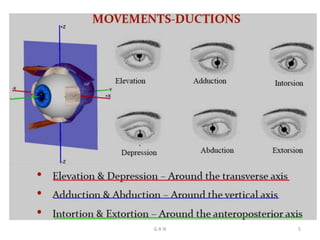
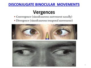
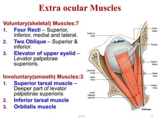
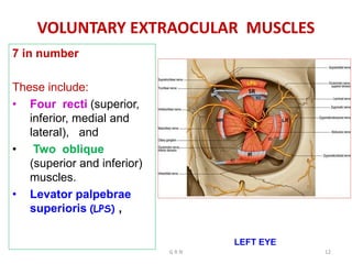
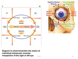
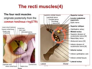
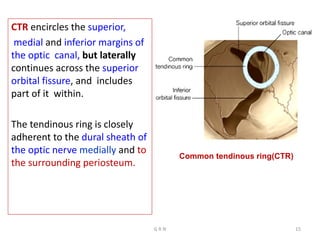
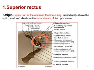
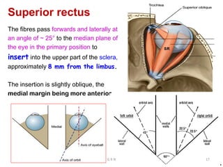
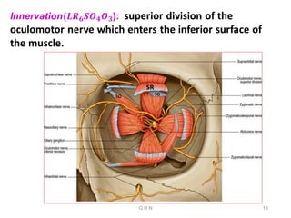
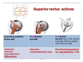
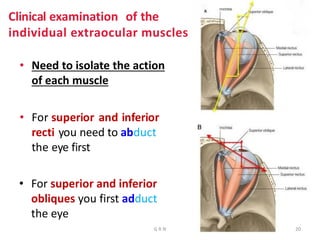
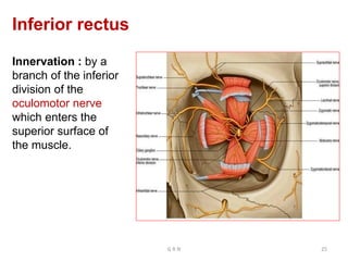
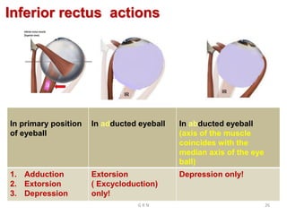
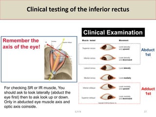
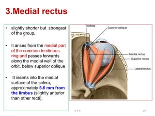
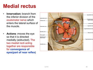
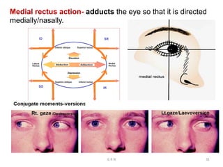
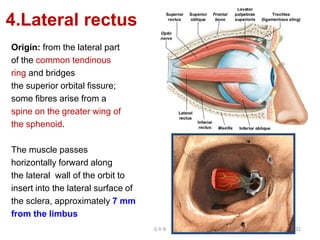
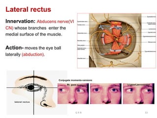
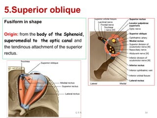
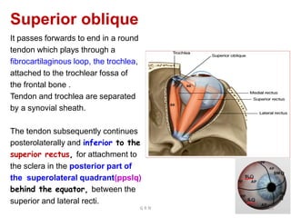
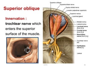
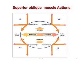
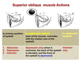
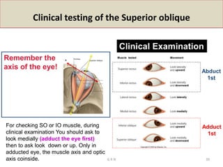
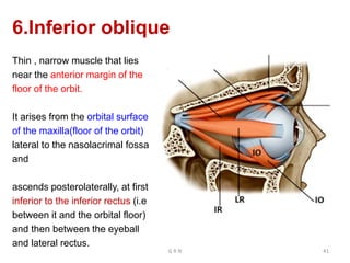
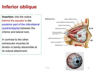
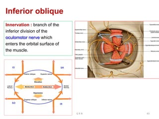
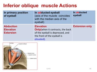
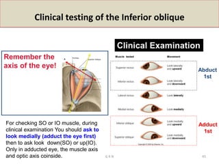
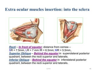
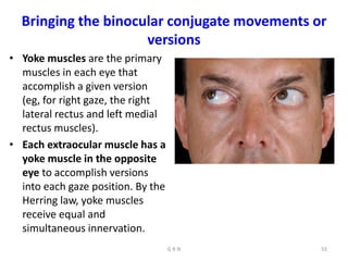
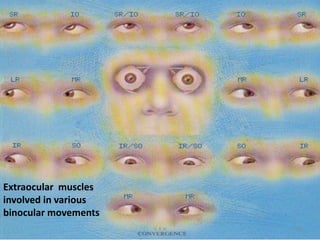
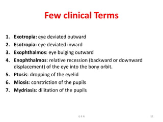
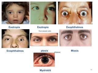
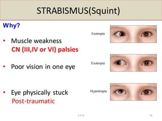
This document provides information about the extraocular muscles. It describes the seven voluntary extraocular muscles - the four rectus muscles (superior, inferior, medial, lateral), and two oblique muscles (superior, inferior). It also describes the levator palpebrae superioris muscle. For each muscle, it details their origin, course, innervation, and actions. Clinical testing techniques for each muscle are also outlined. The summary focuses on identifying the seven voluntary extraocular muscles and briefly mentioning their attachments and functions.
This document describes the muscles and structures in the back of the neck. It discusses the superficial and deep muscles in the back of the neck, including the trapezius, levator scapulae, splenius capitis, and suboccipital muscles. It then focuses on the suboccipital triangle, bounded superiorly by the rectus capitis posterior major and minor, superolaterally by the obliquus capitis superior, and inferiorly by the obliquus capitis inferior. The suboccipital triangle contains the suboccipital nerve, vertebral artery, and venous plexus and is the site of cisternal puncture to access the cisterna magna through the
The cavernous sinus is a pair of venous channels located on each side of the body of the sphenoid bone in the middle cranial fossa. It is approximately 2 cm long and 1 cm wide. Several important structures pass through or are located within the cavernous sinus, including the internal carotid artery, cranial nerves III, IV, V1, and VI, and the inferior hypophyseal arteries. The cavernous sinus drains into various venous channels including the superior and inferior petrosal sinuses, which connect to the transverse sinus and internal jugular vein. Cavernous sinus thrombosis is a potential complication caused by sepsis that can spread from the face or paranasal sinuses,
This document discusses the anatomy of the face, including boundaries, skin layers, fascia, muscles, nerves, arteries, veins, and lymphatic drainage. It describes key facial muscles like the orbicularis oculi and orbicularis oris. The motor innervation of the face is outlined, with the facial nerve supplying muscles. The arterial blood supply is dominated by the facial artery. Applied anatomy concepts like Bell's palsy and trigeminal neuralgia are also briefly mentioned.
The document describes the anatomy of the bony orbit and the extraocular muscles within. It discusses the seven bones that make up the bony orbit, including the frontal, zygomatic, maxillary, ethmoid, sphenoid, lacrimal and palatine bones. It describes the roof, floor, medial wall and lateral wall of the orbit. It then discusses the extraocular muscles, including the four rectus muscles, two oblique muscles and levator palpebrae superioris. It details the origin, insertion, nerve supply and actions of each muscle. Finally, it discusses some clinical implications like strabismus and Horner's syndrome.
The parotid gland is the largest salivary gland. It is wedge-shaped and located below the ear, between the ramus of the mandible and sternocleidomastoid muscle. The parotid gland has three surfaces - lateral, anteromedial, and posteromedial. It is divided into superficial and deep lobes by the facial nerve branching through it. The gland has relations superiorly to the skin, fascia and branches of the great auricular nerve. Inferiorly it relates to the masseter, medial pterygoid and ramus. The facial nerve and its branches pass through the substance of the gland. Lymph drains from the parotid via preauricular
The lacrimal apparatus consists of the lacrimal gland, lacrimal drainage system, and associated structures that produce and drain tears from the eye.
The lacrimal gland develops from surface ectoderm and is located above and lateral to the eye. Tears produced by the gland drain through puncta and canaliculi into the lacrimal sac, then through the nasolacrimal duct into the nose.
The lacrimal sac is lodged in the lacrimal fossa of the medial orbital wall. It connects to the nasolacrimal duct, which courses posteriorly and laterally through bone to drain into the nasal cavity. Coordinated blinking and pressure differences aid
The scalp has five layers - skin, deep connective tissue, aponeurosis, loose connective tissue, and periosteum. It is innervated by branches of the trigeminal nerve and cervical nerves and supplied by branches of the external carotid and ophthalmic arteries. The occipitofrontalis muscle allows movement and raising of the eyebrows. Lymph drains from the scalp to occipital, cervical, mastoid, parotid, preauricular, and submandibular lymph nodes.
The internal surface of the cranial base has three large depressions called cranial fossae: the anterior, middle, and posterior cranial fossae. The anterior fossa is the highest and lodges parts of the frontal lobes. The middle fossa is butterfly-shaped and contains the sella turcica. The posterior fossa is the largest and deepest, lodging the cerebellum, pons, and medulla oblongata. Various foramina and sinuses penetrate the cranial fossae to allow passage of nerves, vessels and CSF. Dural folds such as the falx cerebri and tentorium cerebelli further subdivide the cranial cavity.
The scalp receives its blood supply from branches of the external carotid artery and the ophthalmic artery. The main arteries supplying the scalp are the superficial temporal artery, the posterior auricular artery, and the occipital artery. Venous drainage of the scalp occurs through the external jugular vein and anterior jugular veins. Sensory innervation of the scalp is provided by branches of the trigeminal nerve and cervical nerves depending on the location on the scalp.
The cavernous sinus is located in the middle cranial fossa on either side of the body of the sphenoid bone and sella turcica. It contains the internal carotid artery and cranial nerves III, IV, V1, and VI. The cavernous sinus drains into the angular vein, pterygoid venous plexus, and intercavernous sinuses which connect the two cavernous sinuses. Clinical implications include spread of infection or thrombosis from the face into the cavernous sinus via connections to facial veins.
introduction to skull, parts of skull, bones involved forming skull, different views of skull, norma basalis, anterio cranial middle cranial and posterior cranial fossa, clinical aspects of cranial fossa, foramens present in the cranial fossa
The fourth ventricle is located in the posterior cranial fossa between the pons and cerebellum. It has an triangular outline in sagittal section and rhomboidal shape in horizontal section. It contains five recesses and has superior, inferior, and lateral angles. Its boundaries include the inferior cerebellar peduncle laterally and superior cerebellar peduncle superiorly. It has a roof formed by the convergence of superior cerebellar peduncles and floor formed by the posterior surfaces of the pons and medulla, featuring a median sulcus and medial eminence.
The document discusses two muscles of the eye - the levator palpebrae superioris and the orbicularis oculi. The levator palpebrae superioris originates from the lesser wing of the sphenoid bone and elevates the upper eyelid. It has a collection of smooth muscle fibers that help maintain eyelid elevation. The orbicularis oculi has two parts - the palpebral part that closes the eyelids gently and the orbital part that closes the eyelids forcefully. Both parts originate around the orbit and insert into the eyelids and are innervated by the facial nerve.
The lacrimal apparatus is responsible for producing and draining tears. It consists of the lacrimal gland, conjunctival sac, lacrimal puncta, canaliculi, lacrimal sac, and nasolacrimal duct. The lacrimal gland produces tears which flush the conjunctival sac and nourish the cornea. Tears drain through the puncta and canaliculi into the lacrimal sac and then the nasolacrimal duct, which empties into the nose. Obstruction of the nasolacrimal duct can cause inflammation of the lacrimal sac due to retained tears.
The document discusses the venous drainage of the head and neck. It begins by defining veins and their role in transporting deoxygenated blood. It then describes the different types of veins and the structure of vein walls. The document discusses the development of the venous system during embryogenesis. It provides details on specific veins that drain the head, face, neck and brain, such as the facial vein, supraorbital vein, maxillary vein, and internal and external jugular veins. It notes that facial veins have no valves and connect to the cavernous sinus, so infections can spread from facial veins to intracranial sinuses.
The meninges are the three membranes - the dura mater, arachnoid mater, and pia mater - that cover and protect the brain and spinal cord. The dura mater is the outermost and toughest layer. It forms folds such as the falx cerebri and tentorium cerebelli. Between the dura mater and arachnoid mater is the subdural space, while between the arachnoid mater and pia mater is the subarachnoid space, which contains cerebrospinal fluid. The pia mater is the innermost layer and closely adheres to the brain surface. The choroid plexus produces cerebrospinal fluid which circulates and
The three main structures of the eyelid are the skin, orbicularis oculi muscle, and tarsal plate. The orbicularis oculi muscle is responsible for eyelid closure and blinking. Below the muscle sits the tarsal plate, a dense fibrous structure that provides structure to the eyelid. In the upper eyelid, the levator palpebrae superioris and Müller muscle act as retractors to open the eyelid. The document describes the anatomy and structures of the eyelid in detail.
The cervical plexus is a network of nerves formed by the ventral rami of cervical spinal nerves C1-C4. It provides sensory and motor innervation to the neck and shoulder regions. The cervical plexus gives rise to cutaneous branches that innervate the skin of the neck and shoulders, including the lesser occipital, great auricular, transverse cervical, and supraclavicular nerves. It also forms motor branches like the ansa cervicalis that innervate neck muscles. The phrenic nerve, which originates from C3-C5 and innervates the diaphragm, is another important branch of the cervical plexus.
The document describes the anatomy and physiology of the lacrimal apparatus. It discusses the following key points:
- The lacrimal apparatus includes the lacrimal gland and associated drainage system for tear production and transport.
- The main structures are the lacrimal gland, puncta, canaliculi, lacrimal sac, and nasolacrimal duct. Tears drain from the puncta through these structures and into the nose.
- The lacrimal gland secretes tears in response to sensory nerve stimulation. Contraction of surrounding muscles aids in drainage and prevents backflow through the puncta.
- Obstruction at any point can cause excess tearing (epiphora) by
The document discusses the anatomy of the anterior triangle of the neck. It begins by outlining the boundaries and contents of the anterior triangle. It then describes how the triangle is divided into four smaller triangles - the submental, submandibular, carotid, and muscular triangles - by the digastric and omohyoid muscles. Each smaller triangle's boundaries, floor, contents, and structures are defined in detail. Key structures discussed include the thyroid gland, carotid sheath, carotid sinus, and carotid body. Blood supply and lymphatic drainage of the thyroid gland are also summarized.
The document discusses the extraocular muscles of the eye. It describes the six extraocular muscles - the four rectus muscles (medial, lateral, superior, inferior) and two oblique muscles (superior, inferior) that control eye movement. It details the origin, insertion, nerve supply and action of each individual muscle. Collectively, these muscles enable movements like elevation, depression, adduction, abduction, intorsion and extorsion of the eyeball. The levator palpebrae superioris muscle is also described, which is responsible for elevating the upper eyelid. Various clinical tests and conditions involving these muscles like strabismus, ptosis and nerve palsies are mentioned.
Contents of vertebral canal and its applied aspects mbbsmgmcri1234
The document summarizes the contents of the vertebral canal, including the spinal cord, meninges (dura mater, arachnoid mater, pia mater), blood supply, and applied anatomy of epidural anesthesia and lumbar puncture. Key structures mentioned include the spinal cord terminating at L1-L3, the cauda equina formed by nerve roots, the subarachnoid space containing cerebrospinal fluid, and the valveless vertebral venous system that allows blood flow in either direction.
Bony orbits are Quadrangular truncated pyramids with Anterior cranial fossa above and the maxillary sinuses below.
in this presentation we study the detailed anatomy of the arbit, the bones, relations of each wall, the contents, the apertures, orbital fissures and structures passing, fascia, septa and the surgical spaces of the orbit
This document discusses the muscles of the eye. It describes two types of muscles: intraocular muscles that change the shape of the lens and size of the pupil, and extraocular muscles that control eye movement. The six extraocular muscles are identified along with their nerve supply, origin, insertion and actions. Diagrams illustrate the individual and coordinated movements of the eyes. Finally, some common disorders involving the eye muscles are listed.
The ophthalmic artery originates from the internal carotid artery as it leaves the cavernous sinus. It passes through the optic canal and orbits the eye, dividing into branches that supply the eye and surrounding structures. The central artery of the retina arises in the optic canal and supplies the retina. The lacrimal artery runs to the lacrimal gland. Other branches include the supraorbital, anterior and posterior ethmoidal, meningeal, muscular, and dorsal nasal and supratrochlear arteries which supply the surrounding areas. The ciliary arteries pierce the sclera to supply the choroid, iris, and ciliary body. The ophthalmic artery and its branches provide the main blood supply to
The neck is divided into anterior and posterior triangles by the sternocleidomastoid muscle.
The anterior triangle contains the sternocleidomastoid muscle, which attaches to the sternum, clavicle, and mastoid process. Contracting one side tilts the head to that side and rotates it opposite, while contracting both flexes the neck and extends the head.
The posterior triangle is bounded anteriorly by the sternocleidomastoid, posteriorly by the trapezius muscle, and contains nerves like the brachial plexus and arteries like the subclavian. It is an important area for identifying structures during procedures like carotid endarterectomy.
Lateral ventricle of Brain. By Dr.N.Mugunthan.M.Smgmcri1234
Lateral ventricle of brain. Lecture by Dr.N.Mugunthan.
Associate Professor,
Mahatma Gandhi Medical College & Research Institute,
Sri Balaji Vidyapeeth, Pondicherry.
Anatomy of extraocular muscles and ocular motilityvanya kodali
The document summarizes key anatomical and physiological details of the extraocular muscles and eye movements:
1. It describes the bony orbit anatomy, six extraocular muscles and their actions, innervation and blood supply. The four rectus muscles control horizontal and vertical eye movements, while the two oblique muscles enable torsional movements.
2. The document outlines uniocular and binocular eye movements including versions, vergences, and diagnostic positions of gaze. Hering's and Sherrington's laws govern coordinated eye movements between the eyes.
3. Supranuclear control systems like saccadic, smooth pursuit, vergence and vestibulo-ocular pathways mediate voluntary and reflexive eye movements
The document discusses the extraocular muscles that control eye movement. It describes the six extraocular muscles - the four rectus muscles (medial, lateral, superior, inferior) and two oblique muscles (superior, inferior). It discusses their origins, innervation, blood supply, axes of rotation, actions, and roles as agonists, synergists and antagonists. It also covers concepts like position of rest, muscle planes, vergence movements, anomalies like tropias and phorias, and tests to evaluate eye movement.
you can get information about the extraocular muscles which are responsible for the movement of the eyes in different direction, near and distance.
you will know how many extraocular muscles and how they work....
you will get information about the different position of gazes....
The document describes the extra-ocular muscles that control eye movement and pupil size. It discusses both the intrinsic muscles that control the lens and pupil, and the extrinsic muscles involved in eye movement. The six extrinsic muscles include the superior, inferior, medial, and lateral rectus muscles, and the superior and inferior oblique muscles. Each muscle is described in terms of its origin, insertion, nerve supply, and primary action. The document also covers ocular movements including versions, ductions, and vergences. Laws of ocular motility like Hering's law of equal innervation are also summarized.
ANATOMY AND PHYSIOLOGY OF EXTRAOCULAR MUSCLES.pptxUttaraBhat
This document summarizes the muscles of the orbit. It describes the voluntary muscles - the four recti muscles and two oblique muscles, as well as the involuntary levator palpebrae superioris muscle. For each muscle, it provides details on origin, insertion, nerve supply, and actions based on classical concepts. It also discusses the fascial structures surrounding the muscles, including the fascial sheaths, intermuscular septa, and expansions. Finally, it covers the basic physiology and kinematics of eye movements including versions, ductions, vergences, and laws governing ocular motility such as Hering's law and Sherrington's law.
The document discusses the anatomy of the orbit and eye. It identifies the bones surrounding the orbit, extraocular muscles and their nerve supply. It describes the flow of blood into and out of the orbit and traces the autonomic innervation of the eyeball and lacrimal gland. Clinical tests of extraocular muscle function are contrasted with the individual muscle actions. Effects of cranial nerve damage are also predicted.
The document discusses the anatomy and actions of the extraocular muscles, including their origins, nerve and blood supply, and primary, secondary, and tertiary actions. It also covers several fundamental laws governing ocular motility, such as Listing's Law, Hering's Law of Equal Innervation, and Sherrington's Law of Reciprocal Innervation. The actions of the extraocular muscles and these governing laws have important clinical significance in understanding and treating conditions like strabismus and ocular motility disorders.
This document discusses the extraocular muscles. It begins with an overview of the extraocular muscles and their nerve supply. It then provides more detailed information on the individual muscles, including their origin, insertion, blood supply, innervation and actions. It discusses concepts like Listing's law, Hering's law of equal innervation and Sherrington's law of reciprocal innervation. Clinical significance and applications to conditions like strabismus are also mentioned.
The extraocular muscles include 6 rectus muscles and 2 oblique muscles that control movement of the eyeball. The rectus muscles are the superior, inferior, medial, and lateral rectus and act to move the eye in specific directions. The oblique muscles are the superior and inferior oblique and produce torsional movements as their primary action with secondary actions of moving the eye. All the muscles are supplied by specific cranial nerves with some variations in their blood supply.
The document summarizes the motor apparatus of the eye including the extraocular muscles, their actions, innervation and coordination for binocular vision. It describes the positioning and movement of the eyes, convergence and accommodation reflexes, and grades of binocular single vision including fusion and stereopsis. The extraocular muscles work in synkinesis to move the eyes conjugately or disjunctively according to Hering's and Sherrington's laws of innervation.
The document discusses the anatomy and functions of the extraocular muscles. It defines key terminology used to describe the muscles and their actions, including agonist, antagonist, synergist, and yoke muscles. It describes the primary, secondary and tertiary actions of each of the six extraocular muscles and their roles in monocular and binocular eye movements including ductions, versions, and vergences. Cardinal positions of gaze and diagnostic positions of gaze are also outlined.
This lecture includes anatomy, Physiology of Extra Ocular Muscle, kindly share it with colleagues and like it. I will share more lectures related to eye anatomy and optometry.
Thank You.
The document summarizes the anatomy and physiology of the extraocular muscles (EOMs). It describes the 7 EOMs, their origins, insertions, actions, and nerve supply. The EOMs allow for precise eye movements through arrangements of fibers and innervation that provide both rapid and fatigue-resistant function. Their coordinated actions follow laws of ocular motility to produce conjugate and vergent eye movements in the cardinal gazes.
1. There are 14 extraocular muscles that control eye movement, including 6 muscles in each eyeball and 1 muscle in each eyelid.
2. The extraocular muscles originate from structures like the annulus of Zinn and insert into areas on the eyeball to facilitate movements.
3. Cranial nerves like the oculomotor, trochlear, and abducens nerves provide motor innervation to the extraocular muscles.
This document discusses eye movements and the anatomy of the extraocular muscles. It describes the different types of eye movements including smooth pursuit, saccades, fixation, and reflex eye movements. It also outlines the actions and innervation of the individual extraocular muscles - the four rectus muscles and two oblique muscles. The document provides details on motor units, fiber types, and recruitment patterns in extraocular muscles.
The document describes the anatomy of the orbit and its contents. It discusses the bones that form the boundaries of the orbit, including the roof, floor, lateral wall, and medial wall. It then describes the structures contained within the orbit, including the extraocular muscles, nerves, blood vessels, lacrimal gland, and orbital fat. It provides details on the origins, insertions, innervation and actions of each extraocular muscle. It also discusses the clinical implications of injuries or disorders that may affect structures in the orbit.
The document discusses the extraocular muscles and their nerve supply, actions, and clinical presentations of nerve palsies. It describes the origins, insertions, innervations and primary actions of the 4 recti muscles and 2 oblique muscles. Figure 1 shows ptosis and limitations of eye movements in 3rd nerve palsy. Figures 2 and 3 demonstrate the clinical findings in 4th nerve and 6th nerve palsies, respectively. Ophthalmoplegia is defined as paralysis of the extraocular muscles resulting in double vision.
The document discusses paralytic strabismus, including:
1) Hering's law of equal innervation and Sherington's law of reciprocal innervation which are important in diagnosing paralytic strabismus.
2) The sequelae of ocular muscle palsy including overactions and underactions of muscles.
3) Methods for investigating incomitant strabismus including cover tests, motility examination, and Hess screen plots to identify the affected muscle.
orbit anatomy along with its borders and contents. extra ocular muscles and their attachment , and their actions, along with their innervation. strabismus and squint. damage of occulomotor, trochlear and abducent nerve sign and symptoms of the patient.
The extraocular muscles are the six muscles that control movement of the eye and one muscle that controls eyelid elevation. The actions of the six muscles responsible for eye movement depend on the position of the eye at the time of muscle contraction.
The development of teeth (odontogenesis) is a complex process where teeth form from embryonic cells and grow and erupt into the oral cavity. It begins around the 6th week of gestation with the formation of the primary epithelial band in the upper and lower jaws, from which the dental lamina develops and projects into the underlying mesenchyme to form tooth buds. These buds develop through the bud, cap, and bell stages to form the crown and root structures. Various cell types differentiate and interact to form enamel, dentin, cementum, and the periodontium. Root formation occurs after crown formation is complete, guided by Hertwig's epithelial root sheath which determines the root shape. Teeth continue developing and
.Explain the extent, relations, blood supply, nerve supply, lymphatic drainage of the trachea.
3. Explain the applied anatomy of the trachea.
4. Define esophagus, explain the beginning, course, relations, constrictions, termination, nerve supply,
blood supply, and lymphatic drainage of esophagus
5. Explain the applied anatomy of the esophagus
6. Define thoracic duct, explain the origin, course, relations, termination, tributaries, and areas of
drainage of the thoracic duct
7. Explain the applied anatomy of the thoracic duct
1. The ventricular system consists of a series of cavities within the brain that produce and circulate cerebrospinal fluid. It includes the lateral ventricles, third ventricle, and fourth ventricle.
2. The lateral ventricles are located within the cerebral hemispheres. Each has 5 parts - the anterior horn, body, posterior horn, inferior horn, and are lined with ependyma and choroid plexus.
3. The third ventricle is located between the thalami in the diencephalon. It has walls formed by the thalamus, hypothalamus, and structures like the lamina terminalis. Its roof contains the choroid plex
the fibers present in the cerebellar peduncles
the applied anatomy of the cerebellum
the microscopic structure of the cerebellum, mossy, and climbing fibers
The document provides information about the anatomy and functions of the cerebellum. It can be summarized as follows:
1. The cerebellum is located in the posterior cranial fossa and is separated into two hemispheres and a median vermis. It has three lobes and three functional subdivisions that control balance, coordination of movements, and muscle tone.
2. The cerebellum connects to the brainstem via three cerebellar peduncles - superior, middle, and inferior - which carry afferent and efferent fibers. Deep within the cerebellum are four nuclei - dentate, globose, emboliformis, and fastigius.
3. The cerebell
This document provides an overview of the radiological anatomy of the upper limb. It includes images and descriptions of bones and joints in the shoulder, elbow, and wrist regions. The learning objectives are to identify bones and their parts on radiographs of the upper limb and explain the related applied anatomy. Images show normal anatomy as well as fractures and dislocations of the clavicle, shoulder, elbow, and wrist. Descriptions provide details on anatomical structures and landmarks visible in the radiographs.
This document provides an overview of radiological anatomy of the lower limb through images and descriptions of key bones and joints. It includes radiographs and explanations of the pelvis, hip, femur, knee, ankle, foot, and specific injuries or conditions like fractures of the femur or fifth metatarsal. The goal is to teach identification of bones and their features on radiographs and related clinical anatomy. Key structures highlighted include the sacrum, femoral head, patella, talus, and calcaneus as well as joints like the hip, knee, ankle, and Lisfranc joint.
The document describes the boundaries and contents of the bony orbit. It details the bones that form the roof, floor, medial wall and lateral wall of the orbit, including the frontal bone, sphenoid bone, lacrimal bone, maxilla, palatine bone and zygomatic bone. The orbit contains the eyeball, extraocular muscles, cranial nerves III, IV, VI, the ophthalmic and maxillary branches of cranial nerve V, the ciliary ganglion, ophthalmic vessels and lacrimal apparatus.
This slide is special for master students (MIBS & MIFB) in UUM. Also useful for readers who are interested in the topic of contemporary Islamic banking.
How to Build a Module in Odoo 17 Using the Scaffold MethodCeline George
Odoo provides an option for creating a module by using a single line command. By using this command the user can make a whole structure of a module. It is very easy for a beginner to make a module. There is no need to make each file manually. This slide will show how to create a module using the scaffold method.
How to Setup Warehouse & Location in Odoo 17 InventoryCeline George
In this slide, we'll explore how to set up warehouses and locations in Odoo 17 Inventory. This will help us manage our stock effectively, track inventory levels, and streamline warehouse operations.
How to Manage Your Lost Opportunities in Odoo 17 CRMCeline George
Odoo 17 CRM allows us to track why we lose sales opportunities with "Lost Reasons." This helps analyze our sales process and identify areas for improvement. Here's how to configure lost reasons in Odoo 17 CRM
Executive Directors Chat Leveraging AI for Diversity, Equity, and InclusionTechSoup
Let’s explore the intersection of technology and equity in the final session of our DEI series. Discover how AI tools, like ChatGPT, can be used to support and enhance your nonprofit's DEI initiatives. Participants will gain insights into practical AI applications and get tips for leveraging technology to advance their DEI goals.
Walmart Business+ and Spark Good for Nonprofits.pdfTechSoup
"Learn about all the ways Walmart supports nonprofit organizations.
You will hear from Liz Willett, the Head of Nonprofits, and hear about what Walmart is doing to help nonprofits, including Walmart Business and Spark Good. Walmart Business+ is a new offer for nonprofits that offers discounts and also streamlines nonprofits order and expense tracking, saving time and money.
The webinar may also give some examples on how nonprofits can best leverage Walmart Business+.
The event will cover the following::
Walmart Business + (https://business.walmart.com/plus) is a new shopping experience for nonprofits, schools, and local business customers that connects an exclusive online shopping experience to stores. Benefits include free delivery and shipping, a 'Spend Analytics” feature, special discounts, deals and tax-exempt shopping.
Special TechSoup offer for a free 180 days membership, and up to $150 in discounts on eligible orders.
Spark Good (walmart.com/sparkgood) is a charitable platform that enables nonprofits to receive donations directly from customers and associates.
Answers about how you can do more with Walmart!"
বাংলাদেশের অর্থনৈতিক সমীক্ষা ২০২৪ [Bangladesh Economic Review 2024 Bangla.pdf] কম্পিউটার , ট্যাব ও স্মার্ট ফোন ভার্সন সহ সম্পূর্ণ বাংলা ই-বুক বা pdf বই " সুচিপত্র ...বুকমার্ক মেনু 🔖 ও হাইপার লিংক মেনু 📝👆 যুক্ত ..
আমাদের সবার জন্য খুব খুব গুরুত্বপূর্ণ একটি বই ..বিসিএস, ব্যাংক, ইউনিভার্সিটি ভর্তি ও যে কোন প্রতিযোগিতা মূলক পরীক্ষার জন্য এর খুব ইম্পরট্যান্ট একটি বিষয় ...তাছাড়া বাংলাদেশের সাম্প্রতিক যে কোন ডাটা বা তথ্য এই বইতে পাবেন ...
তাই একজন নাগরিক হিসাবে এই তথ্য গুলো আপনার জানা প্রয়োজন ...।
বিসিএস ও ব্যাংক এর লিখিত পরীক্ষা ...+এছাড়া মাধ্যমিক ও উচ্চমাধ্যমিকের স্টুডেন্টদের জন্য অনেক কাজে আসবে ...
This presentation was provided by Steph Pollock of The American Psychological Association’s Journals Program, and Damita Snow, of The American Society of Civil Engineers (ASCE), for the initial session of NISO's 2024 Training Series "DEIA in the Scholarly Landscape." Session One: 'Setting Expectations: a DEIA Primer,' was held June 6, 2024.
Pollock and Snow "DEIA in the Scholarly Landscape, Session One: Setting Expec...
Extraocular muscles
1. Extraocular muscles
All the information, including the images and pics
collected from various sources is strictly for
teaching and helping the students in learning.
G R N 1
Dr. G. RAVINDRANATH MBBS; MS
PROFESSOR OF ANATOMY,
NRI INSTITUTE OF MEDICAL SCIENCES
2. Learning objectives
1. Describe the attachments, nerve supply, actions of
muscles of the orbit
2. Explain the applied anatomy of muscles of the orbit
G R N 2
3. Monocular eye movements- Ductions
• Monocular eye movements are called ductions
• Movement of the eyeball nasally/medially is adduction;
temporal/lateral movement is- abduction.
• Elevation of the eye is termed as sursumduction (supraduction)&
depression of the eye- deorsumduction (infraduction)
• Incycloduction(intorsion) is nasal rotation of the vertical meridian;
excycloduction (extorsion) - temporal rotation of the vertical
meridian.
G R N 3
6. Binocular eye movements(BOM)
• Binocular eye movements are either conjugate
(versions) or disconjugate (vergences).
• conjugate movements (Versions)- are movements
of both eyes in the same direction(E.g. right gaze in
which both eyes move to the right- dextroversion and
levoversion is movement of both eyes to the left).
- Similarly sursumversion(supraversion) and
deorsumversion(infraversion) are elevation and
depression of both eyes, respectively.
G R N 6
7. Conjugate binocular movements -
(versions)
1. Dextroversion / right gaze
2. Laevoversion / left gaze
3. Sursumversion / elevation / up gaze
4. Deorsumversion / depression / down gaze
5. Dextroelevation / gaze up and right
6. Dextrodepression / gaze down and right
7. Laevoelevation / gaze up and left
8. Laevodepression / gaze down and left
9. Dextrocycloversion – top of the eye rotates to the right
10. Laevocycloversion – top of the eye rotates to the left
G R N 7
11. Voluntary(skeletal) Muscles:7
1. Four Recti – Superior,
inferior, medial and lateral.
2. Two Oblique – Superior &
inferior.
3. Elevator of upper eyelid –
Levator palpebrae
superioris.
Involuntary(smooth) Muscles:3
1. Superior tarsal muscle –
Deeper part of levator
palpebrae superioris
2. Inferior tarsal muscle
3. Orbitalis muscle
Extra ocular Muscles
G R N 11
12. VOLUNTARY EXTRAOCULAR MUSCLES
7 in number
These include:
• Four recti (superior,
inferior, medial and
lateral), and
• Two oblique
(superior and inferior)
muscles.
• Levator palpebrae
superioris (LPS) ,
LEFT EYE
G R N 12
13. Diagram to show/remember the action of
individual extraocular muscles
irrespective of the right or left eye G R N 13
14. The four recti muscles
originate posteriorly from the
common tendinous ring(CTR)
The recti muscles(4)
G R N 14
15. CTR encircles the superior,
medial and inferior margins of
the optic canal, but laterally
continues across the superior
orbital fissure, and includes
part of it within.
The tendinous ring is closely
adherent to the dural sheath of
the optic nerve medially and to
the surrounding periosteum.
Common tendinous ring(CTR)
G R N 15
16. 1.Superior rectus
Origin- upper part of the common tendinous ring, immediately above the
optic canal and also from the dural sheath of the optic nerve.
G R N 16
17. Superior rectus
The fibres pass forwards and laterally at
an angle of ~ 25° to the median plane of
the eye in the primary position to
insert into the upper part of the sclera,
approximately 8 mm from the limbus.
The insertion is slightly oblique, the
medial margin being more anterior.
G R N 17
20. • Need to isolate the action
of each muscle
• For superior and inferior
recti you need to abduct
the eye first
• For superior and inferior
obliques you first adduct
the eye
Clinical examination of the
individual extraocular muscles
G R N 20
21. Clinical testing of the superior rectus
Clinical Examination
Adduct
1st
Abduct
1st
Remember the
axis of the eye!
For checking SR or IR muscle, during
clinical examination You ask the patient
or individual to look laterally (abduct
the eye first) then ask to look up or
down. Only in abducted eye, the
muscle axis and optic/eye axis coincide. G R N 21
24. 2.Inferior rectus
Arises from the common tendinous
ring, below the optic canal.
It runs along the orbital floor in a similar
direction to superior rectus (i.e., forwards
and laterally) and
inserts obliquely into the sclera ~ 6.5
mm from the limbus
G R N 24
25. Inferior rectus
Innervation : by a
branch of the inferior
division of the
oculomotor nerve
which enters the
superior surface of
the muscle.
G R N 25
26. Inferior rectus actions
In primary position
of eyeball
In adducted eyeball In abducted eyeball
(axis of the muscle
coincides with the
median axis of the eye
ball)
1. Adduction
2. Extorsion
3. Depression
Extorsion
( Excycloduction)
only!
Depression only!
G R N 26
27. Clinical testing of the inferior rectus
Clinical Examination
Adduct
1st
Abduct
1st
Remember the
axis of the eye!
For checking SR or IR muscle, You
should ask to look laterally (abduct the
eye first) then to ask look up or down.
Only in abducted eye muscle axis and
optic axis coinside.
G R N 27
29. 3.Medial rectus
• slightly shorter but strongest
of the group.
• It arises from the medial part
of the common tendinous
ring and passes forwards
along the medial wall of the
orbit, below superior oblique
.
• It inserts into the medial
surface of the sclera,
approximately 5.5 mm from
the limbus (slightly anterior
than other recti).
G R N 29
30. Medial rectus
• Innervation: branch from
the inferior division of the
oculomotor nerve which
enters the lateral surface of
the muscle.
• Actions :moves the eye
so that it is directed
medially (adducted). The
two medial recti acting
together are responsible
for convergence of
eyes(part of near reflex)
G R N 30
31. Medial rectus action- adducts the eye so that it is directed
medially/nasally.
G R N 31
32. 4.Lateral rectus
Origin: from the lateral part
of the common tendinous
ring and bridges
the superior orbital fissure;
some fibres arise from a
spine on the greater wing of
the sphenoid.
The muscle passes
horizontally forward along
the lateral wall of the orbit to
insert into the lateral surface of
the sclera, approximately 7 mm
from the limbus
G R N 32
33. Lateral rectus
Innervation: Abducens nerve(VI
CN) whose branches enter the
medial surface of the muscle.
Action- moves the eye ball
laterally (abduction).
G R N 33
34. 5.Superior oblique
Fusiform in shape
Origin: from the body of the Sphenoid,
superomedial to the optic canal and
the tendinous attachment of the superior
rectus.
G R N 34
35. Superior oblique
It passes forwards to end in a round
tendon which plays through a
fibrocartilaginous loop, the trochlea,
attached to the trochlear fossa of
the frontal bone .
Tendon and trochlea are separated
by a synovial sheath.
The tendon subsequently continues
posterolaterally and inferior to the
superior rectus, for attachment to
the sclera in the posterior part of
the superolateral quadrant(ppslq)
behind the equator, between the
superior and lateral recti.
G R N 35
38. Superior oblique muscle Actions
In primary position
of eyeball
In adducted eyeball
(axis of the muscle coincides
with the median axis of the
eyeball)
In abducted
eyeball
1. Abduction
2. Depression
3. Intorsion
Depression only (when it
contracts, the back of the eyeball
is elevated, and the front of
the eyeball is depressed)
Intorsion
only
G R N 38
39. Clinical testing of the Superior oblique
Clinical Examination
Adduct
1st
Abduct
1st
Remember the
axis of the eye!
For checking SO or IO muscle, during
clinical examination You should ask to
look medially (adduct the eye first)
then to ask look down or up. Only in
adducted eye, the muscle axis and optic
axis coinside. G R N 39
41. 6.Inferior oblique
Thin , narrow muscle that lies
near the anterior margin of the
floor of the orbit.
It arises from the orbital surface
of the maxilla(floor of the orbit)
lateral to the nasolacrimal fossa
and
ascends posterolaterally, at first
inferior to the inferior rectus (i.e
between it and the orbital floor)
and then between the eyeball
and lateral rectus.
G R N 41
42. Inferior oblique
Insertion: into the sclera
behind the equator to the
posterior part of the inferolateral
quadrant(ppilq) between the
inferior and lateral recti.
In contrast to the other
extraocular muscles its
tendon is barely discernible at
its scleral attachment.
G R N 42
43. Inferior oblique
Innervation : branch of the
inferior division of the
oculomotor nerve which
enters the orbital surface of
the muscle.
G R N 43
44. Inferior oblique muscle Actions
In primary position
of eyeball
In adducted eyeball
(axis of the muscle coincides
with the median axis of the
eyeball)
In abducted
eyeball
Abduction
Elevation
Extorsion
Elevation
Only(when it contracts, the back
of the eyeball is depressed, and
the front of the eyeball is
elevated).
Extorsion only
G R N 44
45. Clinical testing of the Inferior oblique
Clinical Examination
Adduct
1st
Abduct
1st
Remember the
axis of the eye!
For checking SO or IO muscle, during
clinical examination You should ask to
look medially (adduct the eye first)
then to ask look down(SO) or up(IO).
Only in adducted eye, the muscle axis
and optic axis coinside. G R N 45
46. Extra ocular muscles insertion: into the sclera
Recti – In front of equator; distance from cornea –
SR = 7.5mm, LR = 7 mm IR = 6.5mm; MR = 5.5mm,.
Superior Oblique – Behind the equator in superolateral posterior
quadrant, between the recti superior and lateralis.
Inferior Oblique: - Behind the equator in inferolateral posterior
quadrant, between the recti superior and lateralis.
G R N 46
47. 7.LEVATOR PALPEBRAE SUPERIORIS
Thin , triangular muscle which arises by a short narrow tendon from the inferior
aspect of the lesser wing of the sphenoid, above the optic canal(OC), and
separated from OC by the attachment of superior rectus.
The muscle broadens as it passes anteriorly above the eyeball and splits in two
laminae, ending in a wide aponeurosis.
The Superior lamina consisting of skeletal muscle (voluntary) passes straight into
the upper eyelid and some fibres of it attach to the anterior surface of the tarsus,
and the rest pierce the orbicularis Oculi for insertion to the skin of the upper eyelid.
A thin Inferior lamina of smooth (involuntary) muscle -superior tarsal or Muller’s
muscle , lying the underside of the superior lamina of LPS runs to upper/superior
margin of superior tarsal plate & superior conjunctival fornix.
G R N 47
49. LEVATOR PALPEBRAE SUPERIORIS
Actions:
Elevation of the upper eyelid. During this process the lateral and
medial parts of its aponeurosis are stretched and thus limit its action.
Elevation is opposed by the palpebral part of orbicularis oculi.
The upper eyelid also elevates whenever the gaze of the eye is
directed upwards as Levator palpebrae superioris is linked to superior
rectus by a check ligament.
In states of fear or excitement widening of the palpebral apertures
seen due to contraction of the smooth muscle of the superior and
inferior tarsal muscles as a result of increased sympathetic activity.
Lesions of the sympathetic supply result in drooping of the upper eyelid
(ptosis), as seen in Horner's syndrome.
G R N 49
50. LEVATOR PALPEBRAE SUPERIORIS
The connective tissue sheaths of the adjoining surfaces of levator
palpebrae superioris and superior rectus are fused.
Where the two muscles separate to reach their anterior attachments,
the fascia between them forms a thick mass to which the superior
conjunctival fornix is attached( usually described as an additional
attachment of LPS).
Traced laterally, the aponeurosis of the levator passes between the
orbital and palpebral parts of the lacrimal gland to attach to the orbital
tubercle of the zygomatic bone(Whitnall’s tubercle).
Traced medially, it loses its tendinous nature as it passes closely over
the reflected tendon of superior oblique, and continues on to the medial
palpebral ligament as loose strands of connective tissue.
G R N 50
52. LEVATOR PALPEBRAE SUPERIORIS
Innervation : branch of the superior
division of the oculomotor nerve
which enters the inferior surface of the
muscle.
The smooth muscle component of LPS
(superior tarsal muscle/Muller’s
muscle) innervated by sympathetic
fibres derived from the plexus
surrounding the internal carotid artery;
these nerve fibres join the oculomotor
nerve in the cavernous sinus and pass
forward in its superior branch.
G R N 52
53. • Causes: interruption of sympathetic pathway in multiple sclerosis,
syringomyelia, traction of stellate ganglion by cervical rib, Pancoast’s tumor and
ganglion metastatic lesions.
• Signs: are ipsilateral
• Constriction of pupil (miosis) due to paralysis of dilator pupillae
• Slight drooping of eyelid (ptosis) due to paralysis of Muller’s muscle (Part
of Levator palpebrae superioris)
• Enophthalmos (Retraction of eyeball) due to paralysis of Orbitalis muscle
which support the eyeball
• Loss of sweating (anhydrosis) damage of sympathetic fibers to sweat
glands
• Loss of ciliospinal reflex
Horner’s syndrome
G R N 53
55. Bringing the binocular conjugate movements or
versions
• Yoke muscles are the primary
muscles in each eye that
accomplish a given version
(eg, for right gaze, the right
lateral rectus and left medial
rectus muscles).
• Each extraocular muscle has a
yoke muscle in the opposite
eye to accomplish versions
into each gaze position. By the
Herring law, yoke muscles
receive equal and
simultaneous innervation.
G R N 55
57. Few clinical Terms
1. Exotropia: eye deviated outward
2. Esotropia: eye deviated inward
3. Exophthalmos: eye bulging outward
4. Enophthalmos: relative recession (backward or downward
displacement) of the eye into the bony orbit.
5. Ptosis: dropping of the eyelid
6. Miosis: constriction of the pupils
7. Mydriasis: dilitation of the pupils
G R N 57