Examination of wrist and hand
•
13 likes•2,450 views

Clinical examination of wrist and hand (orthopedics posting) I do not own this file. Courtesy to Dr Madhusudhan, BBH Orthopaedics
Report
Share
Report
Share
Download to read offline

Recommended
Wrist and hand examination
references:
Campbell’s operative orthopaedics 11th edition
Text book of orthopaedics & fractures 5th edition Dr B. Aalami Harandi
Gray’s anatomy 2nd edition
Clinical anatomy Richard S. Snell
Examination of hip joint
1) The document discusses the process for examining a patient's hip joint, including obtaining history, performing physical examination, and conducting specific tests.
2) The physical examination involves inspecting the hip from various angles, palpating bony landmarks and soft tissues, measuring range of motion, assessing limb length and muscle bulk, and performing stability and special tests.
3) A number of special tests are described that can help identify conditions like labral tears, femoral anteversion, and soft tissue contractures. Taking a thorough history and conducting a complete physical exam are important for accurately diagnosing hip joint pathology.
examination of foot and ankle
Dr. Manoj Das' document provides an overview of examining the foot and ankle. It discusses the anatomy of the foot and ankle including bones, joints, ligaments and muscles. The examination involves taking a history, observing gait, posture and deformities, palpating for tenderness, and assessing range of motion, neurovascular status, and performing special tests. The goal is to assess, diagnose and treat conditions of the foot and ankle.
Wrist and hand examination
This document provides information on examining, evaluating, and assessing the hand and wrist. It begins with objectives of reviewing clinical anatomy, performing a physical exam, and discussing common clinical conditions. It then covers anatomy of the bones, joints, muscles, nerves and blood vessels of the wrist and hand. The document provides details on the history, inspection, range of motion assessment, neurologic exam, and special tests like Tinel's sign and Phalen's test used to evaluate common conditions like carpal tunnel syndrome.
Clinical shoulder examination-osce
The document outlines the process for examining a patient's shoulder, including inspection, feeling for tenderness and temperature, assessing range of motion actively and passively, and performing special tests. Key steps are introducing yourself to the patient, gaining consent, exposing the joint, systematically examining alignment, skin, muscles, and bones. A variety of special tests are described to check for impingement, rotator cuff pathology, instability, biceps issues, AC joint problems, and thoracic outlet syndrome. The exam is completed by examining adjacent joints and neurovascular status.
Shoulder examination
Shoulder examination for orthopedic students; one of the famous lectures of MAMC PG course - over last 6 years.
examination of the hip joint
This document provides an overview of how to examine the hip joint through inspection, palpation, range of motion tests, and special tests. It discusses examining the hip for conditions like developmental dysplasia of the hip, septic arthritis, transient synovitis, Perthes disease, and more. Key parts of the examination include inspecting for deformities, palpating for tenderness, performing range of motion tests to evaluate movements like flexion and rotation, and special tests like Thomas test and Trendelenburg test to assess for instability. Close inspection and accurate examination of the hip are important for correctly diagnosing various hip joint pathologies.
Clinical Examination of Hip
The document provides information on clinical examination of the hip joint. It begins with anatomy of the hip joint and associated muscles and ligaments. It then discusses elements of history taking including pain characteristics. The physical examination section covers inspection of gait, limb posture and length, palpation of bony landmarks and muscles, range of motion testing, and special tests like Trendelenburg test. Measurements of limb length discrepancies both apparent and true are also described.
Clinical Examination Of Shoulder
This document provides information on evaluating shoulder pain and injuries. It discusses:
1. The most common causes of adult shoulder pain including impingement syndrome, rotator cuff problems, and athletic injuries.
2. Anatomy of the shoulder including bones, joints, muscles, and common injuries like rotator cuff tears, labral tears, and instability.
3. Physical examination techniques for the shoulder including range of motion, strength, and special tests to identify injuries. Common tests discussed are Neer's sign, Hawkins test, and relocation test.
4. Likely diagnoses suggested by specific physical examination findings. Tables summarize key examination maneuvers and what pathology a positive test may indicate
Knee examination
Clinical examination notes based on TU/KU curriculum of MBBS in nepal. Hope this will be very much helpful in step wise approach to you people especially during exam time.
The Elbow, Examination
The document discusses the anatomy and examination of the elbow joint. It describes the elbow as a compound synovial joint made up of three joints: the ulnohumeral joint, radiohumeral joint, and superior radio ulnar joint. It provides details on the ligaments, muscles, movements, and common conditions that can be examined at the elbow. Specific tests for conditions like tennis elbow and golfer's elbow are also outlined.
Shoulder examination ppt
This document summarizes the steps for examining a patient's shoulder, including inspection from multiple angles to identify scars, swelling, alignment and muscle wasting. It describes palpating various parts of the shoulder joint. Range of motion and special tests are outlined to assess the rotator cuff, impingement, AC joint, biceps, deltoid, serratus anterior and instability. Specific tests described include Neer's sign, Hawkin's sign, Jobe's test, belly-press test, apprehension test and relocation test. The examination provides a thorough evaluation of the shoulder to identify any injuries or limitations.
Shoulder examination
This document provides guidance on performing an orthopedic examination of the shoulder. It outlines the basic steps as look, feel, move, and special tests. For the shoulder examination, it emphasizes tailoring the exam based on suspected problems such as instability in young patients or impingement/rotator cuff tears in older patients. Key parts of examination include inspection for deformities or wasting, palpation of bony landmarks and soft tissues, and assessing the full range of motion including any painful arcs. Special tests are described to detect impingement, specific rotator cuff injuries, or instability. The document stresses performing the exam with knowledge of anatomy and potential diagnoses to both prove and rule out clinical findings.
CTEV UG
This document provides an outline on clubfoot (CTEV), including its epidemiology, causes, anatomy, pathoanatomy, clinical features, imaging, and treatment. CTEV is a relatively common birth defect affecting 1-2 per 1000 births. It involves four deformities: cavus, adductus, varus, and equinus (CAVE). The causes are unknown but may involve genetic or positional factors. Treatment focuses on serial manipulation and casting, especially the Ponseti method, to gradually correct the deformities in a specific order and avoid the need for invasive surgery. The goal is to produce a functional, plantigrade foot.
Examination of case of long bone nonunion lld
This document discusses the examination of patients presenting with long bone nonunions or limb length discrepancies. It outlines the key components of history taking including chief complaints, history of presenting illness, and relevant medical history. The physical examination is described in detail, including inspection of the bone and surrounding soft tissues, palpation of the bone focusing on sites of tenderness, irregularity and mobility, assessment of range of motion, measurements, and deformity. Relevant investigations including laboratory tests, x-rays and potential special investigations like MRI are also mentioned. The document provides guidance on documenting anatomical location of involvement and pathological diagnosis.
Special Tests for Lower Leg, Ankle, and Foot
This document describes various physical examination tests for the lower leg, ankle, and foot. It provides procedures and implications for tests that assess the neutral position of the talus, leg and foot alignment, tibial torsion, ligamentous instability, and other conditions. Key tests include those for the anterior drawer test of the ankle, tibial torsion in sitting and supine positions, leg-heel alignment, and Feiss line to assess for flat foot. The document contains detailed steps for examiners to accurately perform various physical exams of the lower extremity.
Femoro acetabular impingement syndrome
Femoro-acetabular impingement syndrome is a condition where the femoral head and acetabulum rub abnormally in the hip joint, causing damage. It is commonly caused by activities involving repetitive hip flexion, adduction, and internal rotation. Diagnosis involves physical exam maneuvers to reproduce pain and imaging to identify bony abnormalities and cartilage/labral damage. Common findings on x-ray include an alpha angle >70 degrees, coxa profunda, and crossover sign indicating retroversion. MRI can confirm labral tears or cartilage damage.
Examination evaluation & Assessment of Ankle & Foot
Examination evaluation & Assessment of Ankle & Foot Lady Reading Hospital-Medical Teaching Institution, Peshawar
Examination evaluation & Assessment of Ankle & Foot
M.S.K Lecture for D.P.T Students and Graduates Examination of elbow joint
Clinical examination of elbow joint (orthopedics posting)
I do not own this file. Courtesy to Dr Madhusudhan, BBH Orthopaedics
Examination of knee joint
Clinical examination of knee joint (orthopedics posting)
I do not own this file. Courtesy to Dr Madhusudhan, BBH Orthopaedics
Ankle fractures
Ankle fractures are common injuries that require careful evaluation to identify bony and soft tissue damage. The ankle is a complex hinge joint supported by ligaments and the tibia, fibula, talus, and deltoid ligament. Classification systems like Lauge-Hansen and Weber are used to characterize fracture patterns and guide management, which may involve closed treatment for stable injuries or surgery to restore ankle anatomy and stability for unstable fractures. Radiographs are important for diagnosis but CT or MRI may be needed to fully evaluate injury extent.
Examination of Hand
The document provides information on examining the hand, including:
1. The complex anatomy of the hand involving skin, ligaments, bones, joints, tendons, nerves, and vessels.
2. Common disorders that can affect the hand such as congenital issues, trauma, infections, inflammation, and tumors.
3. Details on performing a thorough examination of the hand including inspection, palpation, range of motion testing, and special tests to evaluate arteries, tendons, nerves, and ligament stability.
4. Descriptions and images demonstrating how to perform various special tests to evaluate specific structures in the hand.
Cubitus varus and valgus
This document discusses cubitus varus and cubitus valgus deformities of the elbow. Cubitus varus is when the forearm is directed towards the midline, while cubitus valgus is when it is directed away from the midline. Cubitus varus is most commonly caused by malunion of a supracondylar fracture of the humerus. Treatment options include corrective osteotomies such as lateral closing wedge or medial open wedge osteotomies. Cubitus valgus is most often due to non-union of a lateral condylar fracture of the humerus and can cause tardy ulnar nerve palsy if not corrected surgically using procedures like osteotom
Shoulder Examination
The document outlines the process for examining a patient's shoulder, including:
1) Inspecting for deformities, swelling, atrophy, and other abnormalities.
2) Palpating bony landmarks and soft tissues to check for tenderness.
3) Testing the full range of motion both actively and passively while observing for pain.
4) Performing special tests to isolate specific structures like the rotator cuff muscles and labrum.
5) Examining other joints and the neck for full evaluation of the shoulder.
6) Ordering relevant x-rays to assess for fractures or other bone abnormalities.
Fracture of Shoulder and Humerus
This document discusses injuries to the shoulder girdle and humerus, including acromioclavicular injuries, dislocations, and fractures of the clavicle. It describes the mechanisms of various injuries, methods of diagnosis including physical exam findings and x-rays, and approaches to treatment including closed reduction techniques and immobilization methods. Reduction methods like Kocher's are outlined step-by-step for anterior shoulder dislocations. Greenstick fractures are noted as common in children. Immobilization with a sling or ring method is generally recommended for clavicle fractures and injuries requiring support.
Flat foot and Cavus foot
Pes cavus and pes planus are foot deformities characterized by high and low arches, respectively. Pes cavus, or a high arched foot, can be congenital or acquired and results in clawing of the toes. Pes planus, or a flat foot, is caused by the collapse of the medial longitudinal arch. Both conditions can cause foot, ankle, and leg pain and abnormal shoe wear. Treatment involves orthotics, physical therapy, and sometimes surgery to correct muscle imbalances and bony deformities.
Deformities around elbow and management
Deformities around elbow and management: Clinico-radiological evaluation, decision making and surgical options
Shoulder impingement syndrome
1) Shoulder impingement syndrome is caused by compression of the rotator cuff tendons between the acromion and humeral head. It commonly results from a hooked acromion or inflammation/thickening of the tendons.
2) Treatment begins conservatively with anti-inflammatories, cortisone injections, and physical therapy. If unsuccessful, surgery such as open or arthroscopic acromioplasty is recommended to remove bone spurs and widen the space.
3) Acromioplasty involves detaching the deltoid muscle, removing the coracoacromial ligament and anterior portion of the acromion, and inspecting/repairing any rotator
Wrist disorders
This document discusses various wrist disorders including scaphoid fractures, Kienbock's disease, and DRUJ injuries. It describes the anatomy and biomechanics of the wrist and its ligaments. Various classification systems are presented for carpal instabilities, perilunate injuries, and carpal instability complexes. Treatment options are mentioned for many of the conditions.
Examination of shoulder joint
Clinical examination of shoulder joint (orthopedics posting)
I do not own this file. Courtesy to Dr Madhusudhan, BBH Orthopaedics
More Related Content
What's hot
Clinical Examination Of Shoulder
This document provides information on evaluating shoulder pain and injuries. It discusses:
1. The most common causes of adult shoulder pain including impingement syndrome, rotator cuff problems, and athletic injuries.
2. Anatomy of the shoulder including bones, joints, muscles, and common injuries like rotator cuff tears, labral tears, and instability.
3. Physical examination techniques for the shoulder including range of motion, strength, and special tests to identify injuries. Common tests discussed are Neer's sign, Hawkins test, and relocation test.
4. Likely diagnoses suggested by specific physical examination findings. Tables summarize key examination maneuvers and what pathology a positive test may indicate
Knee examination
Clinical examination notes based on TU/KU curriculum of MBBS in nepal. Hope this will be very much helpful in step wise approach to you people especially during exam time.
The Elbow, Examination
The document discusses the anatomy and examination of the elbow joint. It describes the elbow as a compound synovial joint made up of three joints: the ulnohumeral joint, radiohumeral joint, and superior radio ulnar joint. It provides details on the ligaments, muscles, movements, and common conditions that can be examined at the elbow. Specific tests for conditions like tennis elbow and golfer's elbow are also outlined.
Shoulder examination ppt
This document summarizes the steps for examining a patient's shoulder, including inspection from multiple angles to identify scars, swelling, alignment and muscle wasting. It describes palpating various parts of the shoulder joint. Range of motion and special tests are outlined to assess the rotator cuff, impingement, AC joint, biceps, deltoid, serratus anterior and instability. Specific tests described include Neer's sign, Hawkin's sign, Jobe's test, belly-press test, apprehension test and relocation test. The examination provides a thorough evaluation of the shoulder to identify any injuries or limitations.
Shoulder examination
This document provides guidance on performing an orthopedic examination of the shoulder. It outlines the basic steps as look, feel, move, and special tests. For the shoulder examination, it emphasizes tailoring the exam based on suspected problems such as instability in young patients or impingement/rotator cuff tears in older patients. Key parts of examination include inspection for deformities or wasting, palpation of bony landmarks and soft tissues, and assessing the full range of motion including any painful arcs. Special tests are described to detect impingement, specific rotator cuff injuries, or instability. The document stresses performing the exam with knowledge of anatomy and potential diagnoses to both prove and rule out clinical findings.
CTEV UG
This document provides an outline on clubfoot (CTEV), including its epidemiology, causes, anatomy, pathoanatomy, clinical features, imaging, and treatment. CTEV is a relatively common birth defect affecting 1-2 per 1000 births. It involves four deformities: cavus, adductus, varus, and equinus (CAVE). The causes are unknown but may involve genetic or positional factors. Treatment focuses on serial manipulation and casting, especially the Ponseti method, to gradually correct the deformities in a specific order and avoid the need for invasive surgery. The goal is to produce a functional, plantigrade foot.
Examination of case of long bone nonunion lld
This document discusses the examination of patients presenting with long bone nonunions or limb length discrepancies. It outlines the key components of history taking including chief complaints, history of presenting illness, and relevant medical history. The physical examination is described in detail, including inspection of the bone and surrounding soft tissues, palpation of the bone focusing on sites of tenderness, irregularity and mobility, assessment of range of motion, measurements, and deformity. Relevant investigations including laboratory tests, x-rays and potential special investigations like MRI are also mentioned. The document provides guidance on documenting anatomical location of involvement and pathological diagnosis.
Special Tests for Lower Leg, Ankle, and Foot
This document describes various physical examination tests for the lower leg, ankle, and foot. It provides procedures and implications for tests that assess the neutral position of the talus, leg and foot alignment, tibial torsion, ligamentous instability, and other conditions. Key tests include those for the anterior drawer test of the ankle, tibial torsion in sitting and supine positions, leg-heel alignment, and Feiss line to assess for flat foot. The document contains detailed steps for examiners to accurately perform various physical exams of the lower extremity.
Femoro acetabular impingement syndrome
Femoro-acetabular impingement syndrome is a condition where the femoral head and acetabulum rub abnormally in the hip joint, causing damage. It is commonly caused by activities involving repetitive hip flexion, adduction, and internal rotation. Diagnosis involves physical exam maneuvers to reproduce pain and imaging to identify bony abnormalities and cartilage/labral damage. Common findings on x-ray include an alpha angle >70 degrees, coxa profunda, and crossover sign indicating retroversion. MRI can confirm labral tears or cartilage damage.
Examination evaluation & Assessment of Ankle & Foot
Examination evaluation & Assessment of Ankle & Foot Lady Reading Hospital-Medical Teaching Institution, Peshawar
Examination evaluation & Assessment of Ankle & Foot
M.S.K Lecture for D.P.T Students and Graduates Examination of elbow joint
Clinical examination of elbow joint (orthopedics posting)
I do not own this file. Courtesy to Dr Madhusudhan, BBH Orthopaedics
Examination of knee joint
Clinical examination of knee joint (orthopedics posting)
I do not own this file. Courtesy to Dr Madhusudhan, BBH Orthopaedics
Ankle fractures
Ankle fractures are common injuries that require careful evaluation to identify bony and soft tissue damage. The ankle is a complex hinge joint supported by ligaments and the tibia, fibula, talus, and deltoid ligament. Classification systems like Lauge-Hansen and Weber are used to characterize fracture patterns and guide management, which may involve closed treatment for stable injuries or surgery to restore ankle anatomy and stability for unstable fractures. Radiographs are important for diagnosis but CT or MRI may be needed to fully evaluate injury extent.
Examination of Hand
The document provides information on examining the hand, including:
1. The complex anatomy of the hand involving skin, ligaments, bones, joints, tendons, nerves, and vessels.
2. Common disorders that can affect the hand such as congenital issues, trauma, infections, inflammation, and tumors.
3. Details on performing a thorough examination of the hand including inspection, palpation, range of motion testing, and special tests to evaluate arteries, tendons, nerves, and ligament stability.
4. Descriptions and images demonstrating how to perform various special tests to evaluate specific structures in the hand.
Cubitus varus and valgus
This document discusses cubitus varus and cubitus valgus deformities of the elbow. Cubitus varus is when the forearm is directed towards the midline, while cubitus valgus is when it is directed away from the midline. Cubitus varus is most commonly caused by malunion of a supracondylar fracture of the humerus. Treatment options include corrective osteotomies such as lateral closing wedge or medial open wedge osteotomies. Cubitus valgus is most often due to non-union of a lateral condylar fracture of the humerus and can cause tardy ulnar nerve palsy if not corrected surgically using procedures like osteotom
Shoulder Examination
The document outlines the process for examining a patient's shoulder, including:
1) Inspecting for deformities, swelling, atrophy, and other abnormalities.
2) Palpating bony landmarks and soft tissues to check for tenderness.
3) Testing the full range of motion both actively and passively while observing for pain.
4) Performing special tests to isolate specific structures like the rotator cuff muscles and labrum.
5) Examining other joints and the neck for full evaluation of the shoulder.
6) Ordering relevant x-rays to assess for fractures or other bone abnormalities.
Fracture of Shoulder and Humerus
This document discusses injuries to the shoulder girdle and humerus, including acromioclavicular injuries, dislocations, and fractures of the clavicle. It describes the mechanisms of various injuries, methods of diagnosis including physical exam findings and x-rays, and approaches to treatment including closed reduction techniques and immobilization methods. Reduction methods like Kocher's are outlined step-by-step for anterior shoulder dislocations. Greenstick fractures are noted as common in children. Immobilization with a sling or ring method is generally recommended for clavicle fractures and injuries requiring support.
Flat foot and Cavus foot
Pes cavus and pes planus are foot deformities characterized by high and low arches, respectively. Pes cavus, or a high arched foot, can be congenital or acquired and results in clawing of the toes. Pes planus, or a flat foot, is caused by the collapse of the medial longitudinal arch. Both conditions can cause foot, ankle, and leg pain and abnormal shoe wear. Treatment involves orthotics, physical therapy, and sometimes surgery to correct muscle imbalances and bony deformities.
Deformities around elbow and management
Deformities around elbow and management: Clinico-radiological evaluation, decision making and surgical options
Shoulder impingement syndrome
1) Shoulder impingement syndrome is caused by compression of the rotator cuff tendons between the acromion and humeral head. It commonly results from a hooked acromion or inflammation/thickening of the tendons.
2) Treatment begins conservatively with anti-inflammatories, cortisone injections, and physical therapy. If unsuccessful, surgery such as open or arthroscopic acromioplasty is recommended to remove bone spurs and widen the space.
3) Acromioplasty involves detaching the deltoid muscle, removing the coracoacromial ligament and anterior portion of the acromion, and inspecting/repairing any rotator
What's hot (20)
Examination evaluation & Assessment of Ankle & Foot
Examination evaluation & Assessment of Ankle & Foot
Similar to Examination of wrist and hand
Wrist disorders
This document discusses various wrist disorders including scaphoid fractures, Kienbock's disease, and DRUJ injuries. It describes the anatomy and biomechanics of the wrist and its ligaments. Various classification systems are presented for carpal instabilities, perilunate injuries, and carpal instability complexes. Treatment options are mentioned for many of the conditions.
Examination of shoulder joint
Clinical examination of shoulder joint (orthopedics posting)
I do not own this file. Courtesy to Dr Madhusudhan, BBH Orthopaedics
Msk signs edited
This document contains medical images and descriptions of various musculoskeletal signs and pathologies. It discusses imaging findings and classifications for conditions like:
- Adhesive capsulitis showing thickened ligaments.
- SLAC and SNAC wrist classifications.
- Femoroacetabular impingement presentations.
- Charcot neuroarthropathy acute and chronic stages.
- Various tendon injuries and ligamentous injuries patterns.
It provides comparisons of imaging findings between similar conditions like fibrous dysplasia and osteofibrous dysplasia. Assessment techniques for foot deformities like clubfoot and flatfoot are also outlined.
history taking and colles fracture.pptx
This document outlines the details needed to document a patient case involving a Colles' fracture, including patient information, history, examination findings, injury mechanism, typical presentations, treatments, and potential complications. It describes a Colles' fracture as a break of the distal radius typically caused by a fall on an outstretched hand in elderly osteoporotic women. Key signs include pain, swelling, deformity of the wrist, and dorsal displacement of the radial styloid process. Treatment involves closed or open reduction and casting or plating, with potential complications including joint stiffness, malunion, subluxation, and nerve issues if not properly treated.
Examination of peripheral nerve injuries
Clinical examination of peripheral nerve injuries (orthopedics posting)
I do not own this file. Courtesy to Dr Madhusudhan, BBH Orthopaedics
imaging in skeletal trauma.ppt
This document discusses the radiological evaluation of appendicular skeletal trauma. It begins by describing the different parts of the appendicular skeleton and various imaging modalities used to evaluate trauma, including plain radiographs, ultrasound, CT, MRI and others. It then covers the classification of fractures, focusing on the upper limb trauma including fractures and dislocations of the shoulder, elbow, forearm, wrist and hand. Examples of specific fracture patterns are provided.
Brachial plexus injury diagnosis
This document discusses the anatomy, clinical evaluation, and management of brachial plexus injuries. It begins with the anatomical components of the brachial plexus including roots, trunks, divisions, cords, and branches. It then covers the clinical evaluation including history, physical exam findings, and investigations like imaging and electrodiagnostic studies. Key aspects of the physical exam are described for assessing specific nerves and muscles. The document concludes with classifications of brachial plexus injuries and considerations for non-operative versus operative management.
Kin 191 B – Wrist, Hand And Finger Evaluation And Pathologies
This document provides an overview of evaluating injuries to the upper extremity, including the wrist, hand, and fingers. It describes assessing the history, inspecting the area, performing range of motion and neurological tests, and evaluating for various pathologies. Common injuries discussed include wrist sprains, carpal tunnel syndrome, scaphoid fractures, and perilunate and lunate dislocations. The evaluation process aims to identify the location and mechanism of injury through examination.
Wrist joint an imaging insight
This document provides information on imaging and anatomy of the wrist joint. It begins with the structures that make up the wrist joint, including bones and ligaments. It then discusses various imaging views and measurements used to evaluate the wrist. Specific injuries and conditions that can be seen include fractures, ligament tears, arthritis, ganglion cysts, and vascular abnormalities. Radiography, CT, MRI, ultrasound, and angiography are imaging modalities that can be used to diagnose and characterize wrist pathology.
Understanding Talotarsal Displacement
The talotarsal joint is about the most important weightbearing joint of the body. Misalignment of this joint alone will lead to a chain reaction of destruction throughout the body. View this presentation to learn more.
Presentation2.pptx wrist joint.
This document provides an overview of MRI indications and findings for wrist pathology. It lists common indications for MRI such as wrist instability, pain, trauma, limited range of motion, swelling and planning for surgery. It then reviews MRI sequences, anatomy, and various wrist conditions that may be seen on MRI such as fractures, ligament tears, tendon abnormalities, ganglion cysts, tumors and more. Key findings and imaging features of various wrist conditions are presented.
Presentation2.pptx wrist joint.
This document provides an overview of MRI indications and findings for wrist pathology. It lists common indications for MRI such as wrist instability, pain, trauma, necrosis, and limited range of motion. It then reviews MRI sequences, wrist anatomy, and various wrist conditions that may be seen on MRI such as fractures, ligament tears, instability patterns, tenosynovitis, ganglion cysts, tumors and other soft tissue lesions.
Classification and Treatment of Hip Chondral Lesions
This document discusses the classification and treatment of chondral lesions of the hip. It begins by describing the layers and structures of the hip joint. It then reviews various classification systems for grading chondral lesions of the acetabulum and femoral head, including the Outerbridge, Beck, and Sampson systems. Diagnostic tools for evaluating hip pathology such as x-rays, MRI, and arthroscopy are also discussed. Finally, the document outlines treatment options for chondral lesions based on the type and size of the lesion and any associated morphological abnormalities, including debridement, microfracture, and various grafting techniques.
sajithankle-160130132535.pdf
This document provides details on clinically examining the ankle and foot. It describes examining the patient's history, performing a general examination, and conducting a local examination involving inspection, palpation, range of motion tests, and measurements. Inspection involves assessing the foot from multiple angles while standing and sitting. Palpation feels for tenderness, swelling, and other abnormalities. Range of motion is measured for the ankle, subtalar, and forefoot joints. Special tests evaluate ligaments and tendons for injuries or instability. A full neurological assessment is also recommended to identify any deficits.
Sajith ankle
This document provides details on clinically examining the ankle and foot. It describes examining the patient's history, performing a general examination, and conducting a local examination involving inspection, palpation, range of motion tests, and measurements. Inspection involves assessing the foot from multiple angles while standing and sitting. Palpation feels for tenderness, swelling, and other abnormalities. Range of motion is measured for the ankle, subtalar, and forefoot joints. Special tests evaluate ligaments and tendons for injuries or instability. A full neurological examination is also recommended to check for deficits.
Carpal instability and perilunate dislocation
The document discusses carpal instability and perilunate dislocations. It begins with the anatomy of the wrist joint and ligaments. It then covers various patterns of carpal instability including scapholunate dissociation, lunotriquetral dissociation, and perilunate dislocations. Treatment options discussed include closed reduction, ligament repair/reconstruction, limited wrist fusions, and total wrist fusion.
Craniovertebral anomalies
This document discusses the anatomy, landmarks, measurements, common anomalies, syndromes, and injuries of the craniovertebral junction. It begins with a brief description of the craniovertebral junction's development and components. It then outlines several key anatomical landmarks and measurements used to evaluate the region on imaging. The remainder of the document details various congenital anomalies, developmental abnormalities, syndromes, and acquired conditions that can affect the craniovertebral junction.
Knee clinical examination by Dr YAGNIK
The document summarizes the anatomy and clinical evaluation of the knee joint. It describes the knee as a modified hinge joint composed of the tibiofemoral and patellofemoral joints. Key anatomical structures including the menisci, cruciate ligaments, collateral ligaments, and muscles are outlined. The clinical evaluation involves obtaining a history of pain, swelling, instability, etc and performing an examination involving inspection, palpation, range of motion testing, and special tests of ligaments and menisci.
Examination Of Extremities
The document provides guidance on performing a musculoskeletal examination, including general considerations, inspection, palpation, range of motion testing, and examination of specific areas like shoulders, elbows, hands, and wrists. Key steps include inspection for deformities, discoloration, palpation for temperature changes and tenderness, assessing active and passive range of motion, and performing special tests if abnormalities are suspected.
Examination Of Extremities
The document provides guidance on performing a musculoskeletal examination, including general considerations, inspection, palpation, range of motion testing, and examination of specific areas like the shoulder, elbow, and hand/wrist. Key steps include inspection for deformities, discoloration, palpation for temperature changes and tenderness, assessing active and passive range of motion, and performing special tests if abnormalities are suspected. The exam should be compared between sides.
Similar to Examination of wrist and hand (20)
Kin 191 B – Wrist, Hand And Finger Evaluation And Pathologies
Kin 191 B – Wrist, Hand And Finger Evaluation And Pathologies
Classification and Treatment of Hip Chondral Lesions
Classification and Treatment of Hip Chondral Lesions
More from Fadzlina Zabri
Operative procedure in obstetric
The document summarizes various obstetric surgical procedures including:
1. Dilatation and evacuation procedures to remove products of conception from the uterus such as suction and evacuation.
2. Cervical cerclage procedures like McDonald's technique which reinforce a weak cervix to prevent miscarriage.
3. Destructive procedures like craniotomy and evisceration which reduce the fetal bulk to facilitate delivery in cases of obstruction.
4. Common vaginal procedures including forceps delivery, episiotomy and breech extraction.
Child with convulsion
approach to a child with convulsion - differential diagnosis, how to diagnose each one of it and management.
Examination of hip joint
Clinical examination of hip joint (orthopedics posting)
I do not own this file. Courtesy to Dr Madhusudhan, BBH Orthopaedics
Perthes disease
LCPD or Perthes disease - idiopathic avascular necrosis of femoral head, characterized mainly in child age 4-7 years - with a feature of limping and pain in the hip or groin
Occupational hazard report
1. A group of 18 medical students from the International Medical School in Bangalore visited Megha Punch Forms Pvt Ltd, a factory located in Peenya Industrial Area, as part of their community medicine course objectives.
2. The factory, which has been operating for 12 years, produces punched metal components using various machines. It employs 25 workers in 2 shifts and provides safety equipment, meals, insurance, and overtime pay.
3. During the visit, the students observed the factory's production areas, facilities including adequate lighting and ventilation, and safety practices. However, some workers were not wearing complete protective equipment.
4. The report recommends Megha Punch maintain and improve its health management practices to fully protect
Health education report
Health Education program at BK Nagar primary School, Mathikere, Bangalore.
Topics - ‘Reduce, Recycle, and Reuse’ and 'First Aid'
Chronic white phosphorus poisoning
Chronic white phosphorus poisoning can occur through burning, inhalation, or ingestion of white phosphorus. Inhalation over long periods can lead to osteonecrosis of the jaw, known as "Phossy Jaw", characterized by toothache, gum swelling, and bone necrosis of the lower jaw. Burning from white phosphorus causes severe second and third degree burns that continue burning until deprived of oxygen. Ingestion is also highly toxic and can cause liver, heart, or kidney damage due to accumulation of free radicals in the body. White phosphorus is highly flammable and produces a dense white smoke when burned.
Cannabis
This document discusses cannabis, its common preparations like marijuana and hashish, its medical and recreational uses, clinical effects of acute and chronic poisoning, and treatment and forensic importance. Cannabis is a dioecious plant containing the active compound THC. It is commonly smoked or consumed as bhang, ganja, or hashish. Acute poisoning can cause euphoria, appetite stimulation, and disorientation while chronic use is linked to amotivational syndrome and psychosis. Treatment involves gastric lavage, antipsychotics, and psychotherapy. Cannabis also has forensic importance as an illegal drug of abuse.
Cerbera odollam
Cerbera odollam, also known as the suicide tree or pong pong tree, contains the toxic compound cerberin in its fruits. Ingesting the kernel of just one fruit can be fatal, as cerberin blocks calcium ion channels in the heart muscle, disrupting the heartbeat. Symptoms of poisoning include burning in the mouth, dilated pupils, nausea, vomiting, drowsiness, weakening, and potential coma or death within 6 hours. While treatments like stomach washing and atropine administration aim to counter the poisoning, mortality remains at 30-40% for severe cases. The plant's toxicity poses medicolegal issues in cases of suicide or homicide.
Arrythmias
This document provides an overview of antiarrhythmic medications, including their mechanisms of action, classification, clinical uses, and potential risks. It discusses how antiarrhythmics work by decreasing or increasing conduction velocity or altering cardiac cell excitability. Drugs are classified into Classes I-IV based on their mechanisms and effects. Class I drugs are sodium channel blockers, while Class II are beta blockers, Class III are potassium channel blockers, and Class IV are calcium channel blockers. Specific drugs from each class are outlined along with their indications, dosing, and adverse effects.
Drugs for iron def anemia
The document discusses various types of anemia, their causes, and treatment with iron supplements. It notes that anemia is characterized by a decreased oxygen-carrying capacity of blood due to low hemoglobin or red blood cell counts. Common causes include dietary iron deficiency, blood loss, and bone marrow disorders. Oral iron is usually the first line treatment, while parenteral iron may be used for more severe cases or those with intestinal malabsorption. The roles of iron in hemoglobin formation and the mechanisms of iron absorption and transport in the body are also summarized.
Calcium,vit d,osteoporosis
Calcium is an essential mineral that makes up 2% of total body weight. Over 99% is stored in bones and teeth, with the remainder distributed in tissues and plasma. Calcium levels are tightly regulated by parathyroid hormone (PTH), calcitonin, vitamin D, and calcium-sensing receptors. PTH increases calcium levels by promoting bone resorption and renal reabsorption, while calcitonin decreases them by inhibiting bone resorption and renal reabsorption. Vitamin D enhances intestinal calcium absorption and bone resorption. Bisphosphonates are effective anti-resorptive drugs used to treat osteoporosis and other bone diseases by inhibiting osteoclast activity and bone res
Antipsychotic drugs
Psychiatric conditions are divided into psychosis, neurosis, and affective disorders. Psychosis involves loss of contact with reality and includes schizophrenia. Neurosis involves anxiety disorders like phobias and obsessive-compulsive disorder. Affective disorders are mood disorders like depression and bipolar disorder. Psychotropic drugs treat these conditions by affecting neurotransmitters like dopamine, serotonin, and norepinephrine in the limbic system and other brain areas. Common psychotropic drugs include antipsychotics, antidepressants, and anxiolytics which have multiple mechanisms of action in the brain and body.
Antidiarrheals drug
Diarrhea is a major cause of morbidity and mortality in developing countries. The mainstay of treatment is to correct fluid and electrolyte imbalance through oral rehydration therapy or IV fluids. Specific treatment depends on the cause and includes antimicrobial agents for infectious diarrhea and anti-motility drugs for non-infectious diarrhea. Anti-motility drugs like loperamide work by increasing intestinal transit time through mu and delta opioid receptors while anticholinergics decrease bowel motility and secretion. Antimicrobials are useful for specific infections while anti-inflammatory drugs are used for conditions like ulcerative colitis.
Ischemic Heart Disease
Ischemic Heart Disease by Dr. R. Rupnarayan
I DO NOT OWN THIS SLIDE. credits and courtesy to Dr Rupnarayan, my lecturer from IMS Bangalore
Ct and mri preparation
This document provides preparation instructions for CT, MRI, and contrast administration. It outlines screening requirements like medical history, allergies, medications, and creatinine tests. For CT and MRI, it instructs patients to remove metallic objects and drink water. CT patients may need premedication for contrast allergies. MRI screening identifies absolute contraindications like pacemakers. Gadolinium administration requires creatinine testing in high risk patients due to risks of nephrogenic systemic fibrosis. Pregnant women should only have MRI if absolutely necessary due to risks of intravenous gadolinium.
Burn
This document discusses different types of burns including thermal, electrical, chemical and radiation burns. It describes the pathophysiology of burns including injury to the airway and lungs, inflammation and circulatory changes, and other life-threatening events. It outlines the assessment, grading, treatment and management of burn patients in the hospital and after care including wound care, fluid resuscitation, antibiotics and surgery. Specific treatments are discussed for different burn depths and non-thermal burns.
Behavioural Therapy
This document discusses various psychotherapies and behavioral therapies used to treat psychiatric disorders. It describes psychotherapy as deliberately establishing a relationship between therapist and patient to modify symptoms, behaviors, and promote growth. Approaches discussed include psychodynamic, cognitive-behavioral, humanistic, and behavioral therapies. Behavioral therapy aims to modify maladaptive behaviors through conditioning, using techniques like systematic desensitization, flooding, positive/negative reinforcement, and punishment. Cognitive behavioral therapy combines cognitive and behavioral techniques to change negative thoughts and behaviors. Exposure and response prevention is also discussed as a type of CBT used for anxiety disorders like OCD.
Adolescent psychiatry
This document discusses common health issues that adolescents face including depression, anxiety, substance abuse, eating disorders, risk-taking behaviors, and death from accidents or suicide. It outlines the typical stages of adolescent development and notes that anxiety disorders often co-occur with depression. Depression affects 1-6% of community adolescents and prevalence is higher in females. Anxiety is characterized by apprehension disproportionate to the situation and impacts functioning. Alcohol, marijuana, and tobacco are often gateway drugs to more addictive substances. Prevention strategies address psychoeducation, social support programs, and policy measures like gun control and graduated driver's licensing.
Acute visual loss
This document discusses various causes of acute visual loss including central retinal artery occlusion, ischemic central retinal vein occlusion, retinal detachment involving the macular area, massive vitreous hemorrhage, acute congestive glaucoma, acute iridocyclitis, and chemical or mechanical injuries to the eyeball. It provides details on symptoms, signs, risk factors, treatments, and complications for each condition. Central retinal artery occlusion can cause profound and permanent visual loss within hours if not treated immediately with measures like ocular massage and intravenous medications. Retinal detachment may cause dark spots or flashes of light and its treatment aims to seal retinal breaks and drain subretinal fluid. Chemical injuries especially from alkalis can cause widespread necrosis and
More from Fadzlina Zabri (20)
Recently uploaded
Histopathology of Rheumatoid Arthritis: Visual treat
Histopathology of Rheumatoid Arthritis: Visual treat
Artificial Intelligence Symposium (THAIS)
Artificial Intelligence Symposium (THAIS). Parc Taulí. Sabadell
Histololgy of Female Reproductive System.pptx
Dive into an in-depth exploration of the histological structure of female reproductive system with this comprehensive lecture. Presented by Dr. Ayesha Irfan, Assistant Professor of Anatomy, this presentation covers the Gross anatomy and functional histology of the female reproductive organs. Ideal for students, educators, and anyone interested in medical science, this lecture provides clear explanations, detailed diagrams, and valuable insights into female reproductive system. Enhance your knowledge and understanding of this essential aspect of human biology.
Ketone bodies and metabolism-biochemistry
This slide consists of all the topics of ketone . This can be used for exam purpose for writing about Diabetic keto acidosis etc . Thank you
Clinic ^%[+27633867063*Abortion Pills For Sale In Tembisa Central
Clinic ^%[+27633867063*Abortion Pills For Sale In Tembisa Central Clinic ^%[+27633867063*Abortion Pills For Sale In Tembisa CentralClinic ^%[+27633867063*Abortion Pills For Sale In Tembisa CentralClinic ^%[+27633867063*Abortion Pills For Sale In Tembisa CentralClinic ^%[+27633867063*Abortion Pills For Sale In Tembisa Central
Cell Therapy Expansion and Challenges in Autoimmune Disease
There is increasing confidence that cell therapies will soon play a role in the treatment of autoimmune disorders, but the extent of this impact remains to be seen. Early readouts on autologous CAR-Ts in lupus are encouraging, but manufacturing and cost limitations are likely to restrict access to highly refractory patients. Allogeneic CAR-Ts have the potential to broaden access to earlier lines of treatment due to their inherent cost benefits, however they will need to demonstrate comparable or improved efficacy to established modalities.
In addition to infrastructure and capacity constraints, CAR-Ts face a very different risk-benefit dynamic in autoimmune compared to oncology, highlighting the need for tolerable therapies with low adverse event risk. CAR-NK and Treg-based therapies are also being developed in certain autoimmune disorders and may demonstrate favorable safety profiles. Several novel non-cell therapies such as bispecific antibodies, nanobodies, and RNAi drugs, may also offer future alternative competitive solutions with variable value propositions.
Widespread adoption of cell therapies will not only require strong efficacy and safety data, but also adapted pricing and access strategies. At oncology-based price points, CAR-Ts are unlikely to achieve broad market access in autoimmune disorders, with eligible patient populations that are potentially orders of magnitude greater than the number of currently addressable cancer patients. Developers have made strides towards reducing cell therapy COGS while improving manufacturing efficiency, but payors will inevitably restrict access until more sustainable pricing is achieved.
Despite these headwinds, industry leaders and investors remain confident that cell therapies are poised to address significant unmet need in patients suffering from autoimmune disorders. However, the extent of this impact on the treatment landscape remains to be seen, as the industry rapidly approaches an inflection point.
Does Over-Masturbation Contribute to Chronic Prostatitis.pptx
In some case, your chronic prostatitis may be related to over-masturbation. Generally, natural medicine Diuretic and Anti-inflammatory Pill can help mee get a cure.
Local Advanced Lung Cancer: Artificial Intelligence, Synergetics, Complex Sys...
Overall life span (LS) was 1671.7±1721.6 days and cumulative 5YS reached 62.4%, 10 years – 50.4%, 20 years – 44.6%. 94 LCP lived more than 5 years without cancer (LS=2958.6±1723.6 days), 22 – more than 10 years (LS=5571±1841.8 days). 67 LCP died because of LC (LS=471.9±344 days). AT significantly improved 5YS (68% vs. 53.7%) (P=0.028 by log-rank test). Cox modeling displayed that 5YS of LCP significantly depended on: N0-N12, T3-4, blood cell circuit, cell ratio factors (ratio between cancer cells-CC and blood cells subpopulations), LC cell dynamics, recalcification time, heparin tolerance, prothrombin index, protein, AT, procedure type (P=0.000-0.031). Neural networks, genetic algorithm selection and bootstrap simulation revealed relationships between 5YS and N0-12 (rank=1), thrombocytes/CC (rank=2), segmented neutrophils/CC (3), eosinophils/CC (4), erythrocytes/CC (5), healthy cells/CC (6), lymphocytes/CC (7), stick neutrophils/CC (8), leucocytes/CC (9), monocytes/CC (10). Correct prediction of 5YS was 100% by neural networks computing (error=0.000; area under ROC curve=1.0).
Part II - Body Grief: Losing parts of ourselves and our identity before, duri...
Learn about body grief and ways to cope with it. We will also explore methods to heal from this challenging experience.
Recently uploaded (20)
Histopathology of Rheumatoid Arthritis: Visual treat
Histopathology of Rheumatoid Arthritis: Visual treat
Muscles of Mastication by Dr. Rabia Inam Gandapore.pptx
Muscles of Mastication by Dr. Rabia Inam Gandapore.pptx
Clinic ^%[+27633867063*Abortion Pills For Sale In Tembisa Central
Clinic ^%[+27633867063*Abortion Pills For Sale In Tembisa Central
Vestibulocochlear Nerve by Dr. Rabia Inam Gandapore.pptx
Vestibulocochlear Nerve by Dr. Rabia Inam Gandapore.pptx
Cell Therapy Expansion and Challenges in Autoimmune Disease
Cell Therapy Expansion and Challenges in Autoimmune Disease
Does Over-Masturbation Contribute to Chronic Prostatitis.pptx
Does Over-Masturbation Contribute to Chronic Prostatitis.pptx
Tests for analysis of different pharmaceutical.pptx
Tests for analysis of different pharmaceutical.pptx
Local Advanced Lung Cancer: Artificial Intelligence, Synergetics, Complex Sys...
Local Advanced Lung Cancer: Artificial Intelligence, Synergetics, Complex Sys...
Part II - Body Grief: Losing parts of ourselves and our identity before, duri...
Part II - Body Grief: Losing parts of ourselves and our identity before, duri...
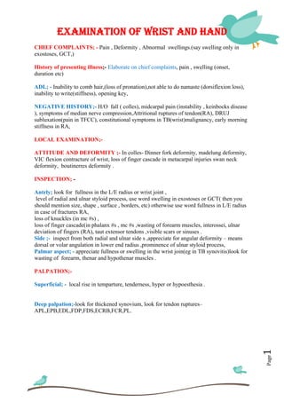
Examination of wrist and hand
- 1. Page1 EXAMINATION OF WRIST AND HAND CHIEF COMPLAINTS; - Pain , Deformity , Abnormal swellings.(say swelling only in exostoses, GCT,) History of presenting illness;- Elaborate on chief complaints, pain , swelling (onset, duration etc) ADL; - Inability to comb hair,(loss of pronation),not able to do namaste (dorsiflexion loss), inability to write(stiffness), opening key, NEGATIVE HISTORY;- H/O fall ( colles), midcarpal pain (instability , keinbocks disease ), symptoms of median nerve compression,Attritional ruptures of tendon(RA), DRUJ subluxation(pain in TFCC), constitutional symptoms in TB(wrist)malignancy, early morning stiffness in RA, LOCAL EXAMINATION;- ATTITUDE AND DEFORMITY ;- In colles- Dinner fork deformity, madelung deformity, VIC flexion contracture of wrist, loss of finger cascade in metacarpal injuries swan neck deformity, boutinerres deformity . INSPECTION; - Antrly; look for fullness in the L/E radius or wrist joint , level of radial and ulnar styloid process, use word swelling in exostoses or GCT( then you should mention size, shape , surface , borders, etc) otherwise use word fullness in L/E radius in case of fractures RA, loss of knuckles (in mc #s) , loss of finger cascade(in phalanx #s , mc #s ,wasting of forearm muscles, interossei, ulnar deviation of fingers (RA), taut extensor tendons ,visible scars or sinuses . Side ;- inspect from both radial and ulnar side s ,appreciate for angular deformity – means dorsal or volar angulation in lower end radius ,prominence of ulnar styloid process, Palmar aspect; - appreciate fullness or swelling in the wrist join(eg in TB synovitis)look for wasting of forearm, thenar and hypothenar muscles . PALPATION;- Superficial; - local rise in temparture, tenderness, hyper or hypoesthesia . Deep palpation;-look for thickened synovium, look for tendon ruptures– APL,EPB,EDL,FDP,FDS,ECRB,FCR,PL.
- 2. Page2 Bony palpation;- Palpate lower end radius and ulna , look for the following findings – angulation,displacement(dorsal , lateral ), irregularity, broadening, thickening, Crepitus , step sign (step up sign –colles#, stepdown sign –smiths#) Joint line palpation;- pass finger along the lowerend radius the first depression u see is joint line , look for tenderness (RA, keinbocks , I/A #s) Relative position of two styloid process and DRUJ subluxation (do piano sign). Palpation of carpal bones- scaphoid , lunate, etc Palpation of metacarpals, phalanges, MOVEMENTS :- check for the movements and also note-is it painful, Crepitus,l oss of movement, lag, critical arc. Palmar flexion 0-90deg FCR, FCU MEDIAN NERV FDP,FDS(ASSIST) Dorsiflexion 0-80 deg ECRL, ECRB, RADIAL EDC,EI Ulnar deviation 25-35 deg FCU ULNAR Radial deviation 15-25 deg FCR,ECRB MEDIAN,RADI APL,EPB Pronation 0-90 deg PRO,TERES, QUAD ANTR INTEROSS NER Supination 0-90 deg BICEPS,SUPIN C5-C6 MEASUREMENTS ;- Linear and circumferential . Linear;- forearm- tip of lateral epicondyle to tip of radial styloid by convention Circumferential – take a fixed mark from a bony point where there is maximum bulk of muscle ,and take circumference of the FA to check for wasting. DEFORMITY ASSESMENT :- look for Dorsal angulation and radial deviation NEUROVASCULAR AND LYMPHATIC EXAMINATION;- Look for distal pulses, radial , ulnar, and the nerves, and regional lymph node examination. SPECIAL TESTS;- Phalens and reverse phalens test for CTS. Grind test;- for MCP &IP (trapeziometacarpal arthritis) Finkelsteinn test- for De quervians . DRUJ instability;- piano key sign and DRUJ compression test . Carpal instability –Scapholunate instability- Watson shift test,scapholunate ballotment. Linotrequitral instability-Ballotment test /reagen test. DIAGNOSIS;-ANATOMICAL- which part is involved joint, metacarpal, phalanx,carpal bones PATHOLOGICAL- synovitis, arthritis, dislocation,instability, malunion . INVESTIGATIONS;- LABORATORY – Hb,Wbc, TcDc,ESR,CRP RADIOLOGICAL –plain x ray of wrist AP & Lat, hand x ray-AP & Oblique SPECIAL INVESTIGATION-MRI-in carpal instability.