OBJECTIVE:
To develop a repeatable model for studying colonization with streptomycin-resistant Escherichia coli O157:H7 in adult cattle.
ANIMALS:
5 adult mixed-breed beef cattle.
PROCEDURES:
Cattle were surgically cannulated in the duodenum, treated daily with streptomycin (33 mg/kg) via the duodenal cannula prior to and during experimental colonizations, and colonized with 10(10) CFUs of streptomycin-resistant E coli O157:H7 via the duodenal cannula. Colonization of rectal mucus and shedding in feces were monitored. Antimicrobials were administered to eliminate the colonizing strain so that 5 repeated colonization experiments could be performed. A comprehensive analysis of colonization was performed at necropsy.
RESULTS:
Streptomycin treatment resulted in improved experimental colonization variables, compared with untreated controls, during initiation (days 2 to 6) and early maintenance (days 7 to 12) of colonization. Elimination of the colonizing strain followed by 5 repeated colonizations in the same animals indicated the repeatability of the protocol. Positive results of bacteriologic culture of feces 7 and 12 days after colonization were obtained in 100% and 84% of samples, respectively, across all animals and trials. At necropsy, highest magnitude recovery was in terminal rectal mucus.
CONCLUSIONS AND CLINICAL RELEVANCE:
The model was highly repeatable and novel with respect to streptomycin treatment, use of duodenal cannulas, and repeated colonizations of the same animals. Its use in adult cattle, from which most bovine-derived food originates, is critical to the study of preharvest food safety. The findings have implications for understanding intermittency of shedding in the field and for proposed vaccine-based interventions.
Evaluation of a model for o157 h7 colonization in strep treated adult cattle
1. 1914 AJVR, Vol 67, No. 11, November 2006
Escherichia coli O157:H7 is an intestinal pathogen of
humans that causes hemorrhagic colitis and the
occasionally fatal sequela known as hemolytic-uremic
syndrome,1,2
which is mediated by Shiga toxin.1
Since
1982, when E coli O157:H7 was first associated with
human disease following consumption of undercooked
hamburger,3
the epidemiologic link to food contamina-
tion has been established. Contamination of ground
beef and other food products, including leafy vegeta-
bles, processed meats, and milk, usually occurs via
exposure to bovine feces.2
Bovine rectal mucosa is the
primary reservoir of this pathogen,4
but cattle are not
usually affected by the infection, a trait possibly con-
ferred by their lack of intestinal expression of the Shiga
toxin receptor.5
Intimin6
and long polar fimbriae,7
viru-
lence factors of E coli O157:H7, are known to be
important factors in colonization. Although progress
has occurred, a comprehensive understanding of the
interaction between E coli O157:H7 and cattle is still
lacking. The occurrence, magnitude, and duration of
shedding of E coli O157:H7 by cattle are highly vari-
able in field and experimental situations.8-13
Because
such variability suggests that unknown host and envi-
ronmental factors are at play, adult cattle models for
E coli O157:H7 are needed to study colonization.
In the natural setting, factors affecting initial colo-
nization of naïve animals are unknown, but horizontal
transmission is presumed and is observed experimen-
tally.11,13,14
With experimental infection, age-related dif-
ferences in pathogenicity are observed. Neonatal calves
(≤ 3 weeks) have diarrhea, attaching and effacing
histopathologic changes that are most severe in the
youngest calves.15
Conflicting results regarding the
occurrence16
or the absence9
of pathogenicity of E coli
O157:H7 following experimental colonization in young
calves have been reported. Adherence of E coli O157:H7
was reported in 1 yearling steer,4
but pathogenicity is
usually not observed in adults.4,9,10,13,15
Despite the lack of
detectable pathogenicity, persistent colonization and
shedding of E coli O157:H7 by adult cattle is the pri-
Received April 4, 2006.
Accepted May 9, 2006.
From the Departments of Veterinary Pathobiology (Snider, Sims, Blair, Clinkenbeard) and Veterinary Clinical Sciences (Washburn), Center for
Veterinary Health Sciences, Oklahoma State University, Stillwater, OK 74078; the Department of Botany and Microbiology, College of Arts
and Sciences, University of Oklahoma, Norman, OK 73019 (Fabich, Conway); and the Department of Cell and Molecular Biology, College of
Environmental and Life Sciences, University of Rhode Island, Kingston, RI 02881 (Cohen). Dr. Washburn’s present address is Department of
Large Animal Clinical Sciences, College of Veterinary Medicine and Biomedical Sciences, Texas A&M University, College Station, TX 77843.
This manuscript represents a portion of a thesis submitted by Dr. Snider to the Oklahoma State University Graduate School as partial fulfill-
ment of the requirements for the PhD degree.
The authors thank Patricia Clinkenbeard and Jennifer Miller for technical assistance.
Address correspondence to Dr. Snider.
Evaluation of a model for Escherichia coli
O157:H7 colonization in streptomycin-treated
adult cattle
Timothy A. Snider, DVM, PhD; Andrew J. Fabich, BS; Kevin E. Washburn, DVM, MS; Will P. Sims, BS;
Jeffrey L. Blair, DVM; Paul S. Cohen, PhD; Tyrrell Conway, PhD; Kenneth D. Clinkenbeard, DVM, PhD
Objective—To develop a repeatable model for study-
ing colonization with streptomycin-resistant
Escherichia coli O157:H7 in adult cattle.
Animals—5 adult mixed-breed beef cattle.
Procedures—Cattle were surgically cannulated in the
duodenum, treated daily with streptomycin (33
mg/kg) via the duodenal cannula prior to and during
experimental colonizations, and colonized with 1010
CFUs of streptomycin-resistant E coli O157:H7 via the
duodenal cannula. Colonization of rectal mucus and
shedding in feces were monitored. Antimicrobials
were administered to eliminate the colonizing strain
so that 5 repeated colonization experiments could be
performed. A comprehensive analysis of colonization
was performed at necropsy.
Results—Streptomycin treatment resulted in
improved experimental colonization variables, com-
pared with untreated controls, during initiation (days 2
to 6) and early maintenance (days 7 to 12) of colo-
nization. Elimination of the colonizing strain followed
by 5 repeated colonizations in the same animals indi-
cated the repeatability of the protocol. Positive results
of bacteriologic culture of feces 7 and 12 days after
colonization were obtained in 100% and 84% of sam-
ples, respectively, across all animals and trials. At
necropsy, highest magnitude recovery was in termi-
nal rectal mucus.
Conclusions and Clinical Relevance—The model
was highly repeatable and novel with respect to
streptomycin treatment, use of duodenal cannulas,
and repeated colonizations of the same animals. Its
use in adult cattle, from which most bovine-derived
food originates, is critical to the study of preharvest
food safety. The findings have implications for under-
standing intermittency of shedding in the field and for
proposed vaccine-based interventions. (Am J Vet Res
2006;67:1914–1920)
ABBREVIATIONS
ABSL-2 Animal biological safety level 2
SMAC Sorbitol-MacConkey
2. mary reason for contamination of ground beef, an
important vehicle for E coli O157:H7 infection in
humans.2
Because adult cattle are the primary source of
contaminated ground beef, modeling adult colonization
by E coli O157:H7 should be a priority.
Fecal shedding by orally colonized adults is short-
lived and of lower magnitude, compared with that in
young calves.9
Low-level, short-duration shedding by
adult cattle has also been observed in other experi-
ments,10,13
which is consistent with what is seen in the
field.17
The reasons for differing shedding patterns
between adults and young calves are thought to be
attributable to diet, age-related differences in rumen
function, or immune response.18
To more fully explore
the variables affecting colonization, robust adult mod-
els are needed.
We hypothesized that problems associated with
poor persistence and inconsistent colonization may be
explained by colonization resistance, which refers to
the ability of the intestinal microflora to resist colo-
nization by an invading bacterium.19
The large intestine
microflora includes hundreds of bacterial species,
characterized by stable populations controlled by com-
petition for limited nutrients and resistant to invading
species unless perturbed by an exogenous factor or
unless the invading bacterium can compete for a cer-
tain nutrient.20
This, the essence of Freter’s nutrient-
niche hypothesis,20
has not been addressed in prior
bovine E coli O157:H7 colonization models, but has
been addressed in a murine model. In that model, mice
treated continuously with orally administered strepto-
mycin are stably colonized with streptomycin-resistant
E coli O157:H7.21
Streptomycin, an aminoglycoside
antimicrobial with poor oral absorption, is satisfactory
for treatment of susceptible, gram-negative enteric
flora.22
In mice, without affecting the overall numbers
of anaerobes,23
streptomycin treatment decreases the
facultative anaerobic bacteria from 108
CFUs/g of feces
to 102
CFUs/g of feces, allowing the invading E coli
O157:H7 strain to colonize the open niche.21
Results of
another study24
suggest that E coli O157:H7 colonizes
the large intestinal mucus in this niche, which influ-
enced our decision to sample rectal mucus, as well as
feces.
With this background of murine models,21,24
we
proposed to similarly develop a streptomycin-treated
adult cattle model. We were, however, concerned that
streptomycin could affect the rumen microflora, which
controls normal digestive and fermentation processes,25
resulting in indigestion and anorexia. To avoid adverse
affects of streptomycin on the rumen microflora, the
drug was administered through a duodenal cannula, a
device that can be easily maintained for up to 2 years
in cattle, providing ready access to the lower portion of
the gastrointestinal tract for sampling or dispensing.26
Thus, the cannula provided for postgastric administra-
tion of streptomycin as well as instillation of the initial
experimental bacterial bolus.
The purpose of the study reported here was to
develop a repeatable model for studying colonization
with streptomycin-resistant E coli O157:H7 in adult
cattle. We intended to determine the success of colo-
nization via a duodenal cannula, the effect of strepto-
mycin treatment on colonization consistency, the dif-
ference between fecal shedding and rectal mucus
recovery, and the possibility of using the same animals
for repeated colonization trials, which lowers costs
associated with use of the adult animal model.
Materials and Methods
Research cattle and environment—Five 8-month-old
mixed-breed beef cattle (4 steers, 1 heifer; each approx 227
kg of body weight) were purchased and allowed to acclima-
tize to their initial environment, the Oklahoma State
University Veterinary Teaching Hospital. Animal experi-
ments and usage were approved by the Oklahoma State
University Institutional Animal Care and Use Committee.
Duodenal cannulas were placed 15 cm caudad to the pyloric
junction and anchored and exteriorized between the last 2
ribs by use of a published technique.27
Cattle were treated
after surgery with penicillin G procainea
(20,000 U/kg, IM, q
24 h) and ceftiofur sodiumb
(0.5 mg/kg, SC, q 24 h) for 1
week; povidone-iodinec
rinses were used daily for the cannu-
la site for 2 weeks. During experimental colonizations, cattle
were transferred to and maintained in an ABSL-2 large animal
facility; each animal was placed in a pen alone. Research and
animal care personnel followed strict ABSL-2 safeguards in
the animal facility and laboratories in accordance with a pro-
tocol approved by the Oklahoma State University
Institutional Biosafety Committee. Cattle were fed a pelleted
total mixed ration composed of corn, ground alfalfa hay, and
mineral supplements twice daily. Water was provided free
choice. At termination of experiments, cattle with negative
results of enrichment culture for E coli O157:H7 were tem-
porarily transferred to an outdoor facility and maintained
similarly while the ABSL-2 facility was cleaned.
Bacterial strain and inoculation preparation—The
E coli O157:H7 (EDL 933) strain used was streptomycin and
nalidixic acid resistant.24
Bacteria were grown overnight in
250 mL of Luria brothd
to an optical density600 of 1.5 to 1.6.
For inoculation, 100 mL of cell suspension was pelleted and
washed 3 times with PBS solution and resuspended in 50 mL
of PBS solution. Tenfold dilutions of the final inocula were
prepared and plated with spreading on selective SMAC agare
media to accurately assess the inoculation concentration.
Inoculation and streptomycin treatment—To test the
effect of streptomycin on experimental colonization, 5 study
cattle were randomly assigned by use of a coin toss to a strep-
tomycin-treated group (n = 3) and a nontreated control
group (2) and colonized per cannula identically. After 18
days of sampling, the colonization was terminated by admin-
istration of antimicrobials, a washout period of 21 days
ensued, and the group assignments were reversed. All proce-
dures were repeated so that 5 complete trials (repetitions)
were performed. Following those 2 trials, 3 additional trials,
each 15 days in length, were performed with streptomycin in
continuous use on all 5 cattle; washout periods ranged from
6 days to 4 weeks. For inoculation, an endotracheal tubef
was
placed in the opened cannula, followed by cuff inflation for
sealing. Each animal received 10 mL of the prepared inocu-
lum via the medicinal access port of the tube followed by a
5-mL flush with PBS solution via the same port, followed by
a 40- to 120-mL flush with PBS solution via the main lumen
of the tube.f
No loss of the inoculating bolus was ever
observed. For cattle that received streptomycing
treatment
daily, a 1 g/mL solution of streptomycin sulfateg
in water was
prepared and administered via the cannula at 33 mg/kg of
body weight. On the morning of colonization initiation,
streptomycin treatment was delayed approximately 12 hours
until the evening sampling. Cattle receiving streptomycin
daily began receiving the treatment regimen 3 days prior to
AJVR, Vol 67, No. 11, November 2006 1915
3. 1916 AJVR, Vol 67, No. 11, November 2006
colonization and continued to receive treatment until the
colonization was terminated.
Sampling and media—Samples of feces and rectal
mucus were collected at 0, 0.5, 1, 2, 3, 5, 7, 9, 12, 15, and 18
days after inoculation. Fecal samples ranged from 5 to 20 g
and were collected directly from the terminal portion of the
rectum. Feces were weighed and suspended at a 1:10 ratio in
1% tryptoneh
with mixing and placed on ice for 1 hour prior
to plating. Mucus samples were acquired from the rectum via
palpation; briefly, following manual evacuation of the feces,
mucus was collected from the ventral half of the most caudal
30 cm of the rectum by scraping with the lid from a sterile
syringe casing. Fecal contamination of mucus was minimized
by physical removal of feces, where possible, or reacquiring
the sample if fecal contamination was grossly apparent.
Mucus was scraped until 200 to 500 mg was acquired; mucus
was suspended at a 1:2 ratio in 1% tryptone,h
vortexed vigor-
ously, and placed on ice for 1 hour prior to plating. Feces and
mucus samples were processed into 10-fold dilution series by
diluting 100 µL of initial sample into 900 µL of sterile PBS
solution, vortexing, and plating 100 µL of each dilution onto
agar media by even spreading with an alcohol-flamed spread-
ing tool. Bacterial plates were incubated 12 to 18 hours at
37o
C prior to assessment and counting. Diluted feces and
mucus samples were plated onto SMACe
agar and SMAC agar
with the antimicrobials streptomycing
(40 µg/mL) and
nalidixic acidi
(50 µg/mL). On the antimicrobial-selective
media, sorbitol-negative (white) colonies were enumerated
daily and, where colony morphology was unusual, subjected
to confirmatory testing with a commercially available
immunoassay.j
All colonies were also enumerated daily on
the SMAC medium without antimicrobials, which gave an
estimate of the total concentration of streptomycin-resistant,
facultative anaerobic bacteria in samples. Enrichment cul-
tures were used for all daily samplings. Briefly, 100 to 150 µL
of primary sample was placed in tryptic soy brothk
with strep-
tomycing
(40 µg/mL) and grown at 37o
C for 16 hours with
shaking (210 oscillations/min). When the inoculated strain
was not recovered from primary samples, the enrichment
samples were subjected to dilution series and plated similar-
ly. Enrichment samples yielded qualitative data of presence
(enrichment positive) or absence (enrichment negative) of
isolates. For purposes of computation of means, enrichment
data were transformed to a semiquantitative estimate of 50
CFUs/g of feces or 10 CFUs/g of mucus, which was 50% of
the lowest limit of detection for each of the primary sampling
regimens. Rectal biopsy specimens, obtained at days 0, 3, 9,
and 15 of each colonization trial, were procured from the
dorsal half of the terminal 3 cm of rectum via an equine uter-
ine biopsy instrumentl
and fixed in neutral-buffered 10% for-
malin. Tissues were routinely processed, paraffin embedded,
sectioned, stained with H&E, and examined via light
microscopy.
Termination of colonization—At the conclusion of an
experiment, cattle were treated with 2 antimicrobials to which
the colonizing strain was determined to be susceptible.
Minimum inhibitory concentrations of neomycin (≤ 4 µg/mL)
and ceftiofur (≤ 0.5 µg/mL) were determined by use of a com-
mercially available antimicrobial test kitm
and by following the
manufacturer’s directions. Neomycin sulfaten
was adminis-
tered daily for 3 days via the cannula at 10 mg/kg. Ceftiofur
sodiumb
was administered SC daily for 3 days at 0.5 mg/kg.
Enrichment cultures of samples obtained the last day of such
treatment and 2 subsequent days were grown, plated, and
assessed to determine elimination of the colonizing strain.
Terminal study, euthanasia, and necropsy methods—
In a final colonization trial, the 5 cattle were colonized and
euthanized 1 day after colonization (n = 1), 3 days after col-
onization (2), and 7 days after colonization (2). To prevent
unintentional early colonization, cattle were penned accord-
ing to their planned date of euthanasia and separated by a
minimum of 20 feet. During this terminal study, 1 steer that
had been designated for euthanasia 3 days after colonization
developed sudden, severe, free gas bloat on the third day and
died before treatment could be administered. This was pre-
sumed to be attributable to individual predisposition to bloat
and dominance during feeding with the copenned smaller
steer. The other 4 cattle were sedated with xylazineo
and
euthanized with a captive-bolt gun. Necropsy was performed
by use of a standard protocol and was performed by an
American College of Veterinary Pathologists board-certified
pathologist. Contents from the rumen, abomasum, duode-
num, ileum, cecum, and proximal portion of the colon and
feces were diluted and plated identically as described for
feces. Gall bladder contents and mucus from duodenum,
ileum, cecum, proximal portion of the colon, and terminal
portion of the rectum were diluted and plated identically as
described for rectal mucus. Tissues examined by light
microscopy were fixed in neutral-buffered 10% formalin,
processed through graded concentrations of alcohol, sec-
tioned at 5 µm, stained with H&E, and cover slipped. The
final trial, primarily descriptive and qualitative, was con-
ducted with the knowledge that a valid statistical analysis of
these groups was not possible.
Statistical analysis—The nonparametric Wilcoxon
signed rank test28
was used in a 1-tailed fashion to test the
hypothesis that streptomycin-treated cattle would have sig-
nificantly (P < 0.05) better colonization variables, compared
with untreated control cattle. Data are given as mean ± SD.
In comparing treated with untreated cattle, accurate assess-
ment of colonization consistency and persistency was per-
formed by use of not only numeric data, but also the phases
of colonization, which were divided into 3 functional stages:
initiation (days 2 to 6; sampling on days 2, 3, and 5), early
maintenance (days 7 to 12; sampling on days 7, 9, and 12),
and late maintenance (days 13 to 18; sampling on days 15
and 18). Support for this concept was provided by studies by
Sheng et al13
and Rice et al,29
in which culture-positive results
obtained for ≤ 1 week indicated lack of a stable association
between the host and bacterium.
Results
Cattle health—All cattle remained healthy
throughout the course of the 6 repeated colonizations.
Some cattle had transient, moderate bloat during the
termination of colonization trials. This was presumed
to be secondary to the effects of the 2 antimicrobials
that were administered. The cannula of 1 steer became
broken and displaced during the course of 1 experi-
ment and was replaced. Sporadic diarrhea was seen in
some cattle but was not correlated with any coloniza-
tion event. All cattle had moderate average daily gains
of 0.71 ± 0.05 kg over the 221 days of these studies. No
attaching and effacing histopathologic changes were
recognized in any animal during the course of the col-
onizations or the necropsy study (4 rectal biopsy spec-
imens examined per animal per trial).
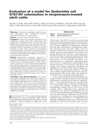
Effects of streptomycin—Recovery of the input
strain in a time-relative manner for feces and mucus
was determined (Figure 1). Streptomycin treatment
increased E coli O157:H7 shedding in mucus during
initiation (P = 0.01) and early maintenance (P = 0.02),
but not late maintenance. Streptomycin treatment
increased E coli O157:H7 shedding in feces only during
4. AJVR, Vol 67, No. 11, November 2006 1917
initiation (P = 0.01), but not during early and late
maintenance.
Persistence and magnitude of bacterial recov-
ery in mucus and feces—Greater persistence of
recovery was evident in mucus than feces during the
2-phased trial (trials 1 and 2) that tested the effect of
streptomycin (Figure 1). In determination of persis-
tence of colonization, days of persistence were
defined as the last sample day on which positive
results were obtained unless ≥ 2 intervening samples
yielded negative results between 2 final samples that
yielded positive results. The latter situation was
negated, as it possibly implied a reinfection from
another animal. In cattle treated with streptomycin,
duration of E coli O157:H7 persistence (ie, detection
in samples) was 16.8 ± 2.7 days in mucus and 14.0 ±
4.6 days in feces. In cattle that were not treated,
duration of E coli O157:H7 persistence was 15.6 ±
2.5 days in mucus and 9.8 ± 5.4 days in feces. In
analyses of the other 3 trials in which streptomycin
was in continuous use (Table 1; trials 3, 4, 5), the
input strain was recovered in mucus for 13.13 ± 3.25
days and in feces for 10.93 ± 3.97 days (P = 0.02).
Higher magnitude colonization was seen in mucus,
compared with feces, on day 2 (P = 0.025), day 3 (P
= 0.005), and day 5 (P < 0.05), but was not signifi-
cantly different between feces and mucus on days 7,
9, 12, and 15.
Repeatability of colonization—Five full-length
trials of 15 or 18 days were conducted on the same 5
cattle. For bacterial recovery from mucus across all 5
trials, 92% of all samples yielded positive results at day
9, 84% of all samples yielded positive results at day 12,
and 72% of all samples yielded positive results at day
15. No substantial diminished colonization magnitude
or persistence was detected in any subsequent trial
(Table 1).
Sites of colonization—In a final colonization
trial, cattle were euthanized and necropsied with a
goal to describe the temporo-spatial distribution of the
colonizing strain of E coli O157:H7. The E coli were
not detected in the abomasum or rumen (Table 2), but
were recovered from the gall bladder in 3 of the 5 cat-
tle. Generally, the highest concentrations of bacteria
were detected in the rectal mucus samples. However,
this was not true for 1 steer that yielded positive
results of enrichment cultures for many samples even
though it was euthanized 24 hours after colonization.
This animal inadvertently received a 1-log order of
magnitude lower colonization bolus (as determined
by CFU determination on the inoculum), which could
explain the reduced magnitude of recovery. The chief
general observation was that, within 7 days, the ter-
minal portion of the rectum became the primary site
of localization, with ileal mucus being a secondary site
of recovery. It was also evident that the colonizing
bolus was cleared from the duodenum quickly and
was only detected in the duodenum of the animal
euthanized 1 day after colonization. Histologic speci-
mens of the gastrointestinal tract (10 sites) and gall
bladder (1 site) revealed no evidence of attaching and
effacing histopathologic changes.
Figure 1—Results of bacteriologic culture of feces (A) and rectal
mucus (B) for Escherichia coli O157:H7 in cattle (n = 5) that were
or were not treated with streptomycin and were inoculated with
1010
CFUs via cannula into the duodenum. Bars represent SE of
the log10 values. Values at time 0 represent samples acquired 12
hours after inoculation.
Table 1—Concentrations (CFUs/g of feces or mucus) of Escherichia coli O157:H7 in feces and rectal
mucus of 5 streptomycin-treated adult cattle on various days after inoculation through a duodenal can-
nula in 3 trials.
Day 7 Day 12 Day 15
Variable Feces Mucus Feces Mucus Feces Mucus
Cattle* 1 5 2 1 0 1
Bacterial concentration† 0–101
101
–102
0–102
0–102
0 0–102
Cattle* 5 5 5 5 4 5
Bacterial concentration† 101
–102
101
–103
101
–103
101
–102
0–102
101
–102
Cattle* 5 5 2 5 1 5
Bacterial concentration† 101
–103
102
–103
0–103
101
–103
0–101
101
–102
*No. of cattle with positive results of bacteriologic culture. †Range of values for all 5 cattle.
5. 1918 AJVR, Vol 67, No. 11, November 2006
Discussion
The primary goal of developing an adult cattle
model for the study of colonization by E coli O157:H7
is a model that consistently yields reliable and repeat-
able establishment of colonization. The streptomycin-
treated adult cattle model described here had several
positive attributes that were consistent with this goal
and were novel in 3 respects: initiation of colonization
via the duodenal cannula, facilitation of occupation of
that niche by use of the continuous streptomycin treat-
ment, and use of the same cattle for repeated coloniza-
tion experiments. In particular, we believe this model
will be useful for studying the role of various factors in
colonization of cattle by use of streptomycin-resistant
mutant strains for these factors.
The most important benefit of the model is its reli-
ability and repeatability in adult cattle. Adult cattle are
more difficult to experimentally colonize than calves,
and this difficulty is experienced in the areas of shed-
ding concentrations and proportions of culture-posi-
tive study animals 1 week after colonization.8-10,12,13
The
shedding concentrations of these streptomycin-treated
cattle at 7 and 15 days after inoculation (101
to 104
CFUs/g and 101
to 103
CFUs/g, respectively) were sim-
ilar to those found in other adult cattle colonization
models. Furthermore, the model appears comparable
to other adult models with respect to proportions of
culture-positive study cattle 1 week after colonization.
In this model, 100% of the cattle were actively shed-
ding at day 12 in 4 trials; in the fifth trial, 100% were
actively shedding through 7 days. Also, colonization
persisted in all 5 animals for 15 days in at least 2 of the
5 trial repetitions. By comparison, Cray and Moon9
reported that 9 of 9 orally colonized adults were active-
ly shedding at 14 days. Sheng et al13
reported that 6 of
10 orally colonized cattle were shedding on day 12,
and 8 of 8 rectally colonized cattle were shedding at
day 14. Additionally, 12 of 18 adult cattle administered
bacteria PO were shedding at day 12 to 14 in a study
by Grauke et al.10
Further difficulty in adult coloniza-
tion was reported for studies by Ohya et al12
and
Buchko et al,8
in which 4 of 8 calves and 9 of 18 year-
ling steers, respectively, yielded negative results of bac-
teriologic culture of feces by 7 and 9 days, respectively.
Furthermore, in the analysis of animal-specific shed-
ding in studies by Grauke et al10
and Sheng et al,13
cul-
ture-positive cattle at 25 days after infection and
beyond were the same cattle that typically yielded pos-
itive results at days 12 to 14. Therefore, it appears that
positive results of culture at days 12 to 14 are a strong
predictor of long-term persistence of infection.
Although longer colonizations were not a goal of the
present study and were not attempted in these cattle,
this trend indicates that long-term colonization is like-
ly achievable with this protocol.
Reproducible colonizations of the same individual
cattle were achieved in the study reported here.
Successful colonization of the same 5 cattle 6 times
without substantial variations of shedding concentra-
tions and proportions colonized was achieved.
Reproducibly colonizing the same cattle has been
reported,9,10
but in those studies, cattle were reinoculat-
ed once instead of 5 times as in the study reported
here. In 1 study,9
there was a significant decrease in
duration of colonization after reinoculation, but in the
other study,10
no differences in shedding patterns after
reinoculation were detected. Furthermore, reinocula-
tion in those studies9,10
was performed after a lengthy
interval to allow for natural elimination of E coli
O157:H7. Attaching and effacing histopathologic
changes15,30
were not detected in the experiments
reported here, and this raises the possibility that,
although multiple colonizations occurred, a host
response was not induced. However, lack of such
lesions or response could be caused by sample timing
or the use of light microscopy instead of immunofluo-
rescence4
or electron microscopy.15
Such absence is also
consistent with a report9
of other experimentally
infected adult cattle and the findings typically seen in
naturally infected cattle.4
Furthermore, results of 1
study9
suggest that E coli O157:H7 cannot be detected
attached to epithelium in histologic sections when
shedding concentration is < 106
CFUs/g of feces, a
threshold established previously.31
Finally, it seems rea-
sonable to consider that the immune response of cattle
to E coli O157:H7 is manifested as intermittent shed-
ding, but Johnson et al18
determined that the serologic
responses of cattle were not correlated with elimina-
Table 2—Sites of isolation of E coli O157:H7 (CFUs/g of sample) on various days after inoculation
through a duodenal cannula in 5 streptomycin-treated adult cattle that were euthanized on various
days.
Day 1 Day 3 Day 7
Site Steer Heifer Steer Steer Steer
Rumen, abomasum 0, 0 0, 0 0, 0 0, 0 0, 0
Gall bladder E+ E+ 0 0 E+
Duodenum contents 1.0 X 10²
0 0 0 0
Duodenum mucus E+ 0 0 0 0
Ileum contents 4.0 X 10²
3.0 X 10²
2.1 X 10³
7.0 X 10²
8.0 X 10²
Ileum mucus E+ 2.0 X 101
1.2 X 10³
1.0 X 10²
1.1 X 10³
Cecum contents 1.0 X 10²
0 0 7.0 X 10²
E+
Cecum mucus E+ E+ 4.0 X 101
8.0 X 101
E+
Colon contents E+ 1.0 X 10²
0 1.0 X 10²
2.0 X 10²
Colon mucus E+ E+ 2.0 X 101
4.0 X 101
6.0 X 10²
Feces E+ 2.0 X 10²
1.4 X 10³
2.0 X 10²
1.0 X 10²
Rectal mucus E+ 8.2 X 10²
6.0 X 101
4.7 X 10³
6.2 X 10²
0 = Not detected by use of primary or enrichment cultures. E+ = Positive results obtained by use of enrich-
ment culture only.
6. tion or reinfection with E coli O157:H7. As a result of
their findings, they speculated that persistence and
reinfection in the face of an immune response con-
tributed to persistent infection in the herd and that
vaccine-based interventions could be of questionable
efficacy.18
The reproducible colonizations attained in
the present study seem to add additional weight to that
argument and may also have broader implications for
epidemiologic surveillance and food safety. For surveil-
lance of herds, this confirmed potential for repro-
ducible colonization largely restricts such data to
point-in-time relevance and renders such surveillance
powerless to predict future trends. For food safety, sim-
ilarly, our findings suggest that samples must be
obtained from every animal at the point of slaughter.
Obtaining samples from a fraction of a herd or from
animals 1 day before slaughter would likely be poorly
predictive of which cattle were actively shedding E coli
O157:H7 at the time of slaughter.
Administration of the colonizing bolus via the
duodenal cannula conferred the principal advantage of
avoiding ruminal dilution of the bolus. Whether or not
dilution of the inocula in ruminal contents (estimated
to be 100 to 120 L in these cattle) affects colonization
success is not known and was not tested here.
However, the data acquired from the streptomycin-
treated adult cattle were more similar to that of the rec-
tal administration model than the oral or ruminal col-
onization models.9,10,13
Thus, results of the present
study and those obtained by use of the rectal coloniza-
tion model indirectly indicate that success of experi-
mental colonization is negatively affected by transit
through the upper portion of the gastrointestinal tract.
To further support this assertion, conditions simulat-
ing rumen fluid of well-fed cattle negatively affect
growth of E coli O157:H7.32
Streptomycin caused a substantial reduction of
susceptible facultative anaerobic bacteria by 6 log-
orders of magnitude in a murine E coli O157:H7 colo-
nization model.21
However, although the results indi-
cated a beneficial effect of streptomycin on success of
colonization with E coli O157:H7, such a magnitude of
reduction was not observed in the streptomycin-treat-
ed cattle, in which reduction by 2 log-orders of magni-
tude (106
to 104
CFUs/g) was usually observed. The
reasons for this difference could be a higher proportion
of naturally streptomycin-resistant microflora in cattle
than in mice, the cumulative acquisition of resistance
by facultative anaerobes in the present study, or the
diminishing effect on the efficacy of the drug caused by
the acidity of the bovine rectum.22
In sampling rectal mucus as well as feces, it was
determined that rectal mucus samples yielded higher
magnitudes of shedding during early colonization and
that use of rectal mucus also resulted in more extend-
ed recovery of the organism. Mucus was processed dif-
ferently than feces because initial vortexing was
required to counteract the viscosity of the former but
introduced problems with the latter. Although this
could have affected recovery numbers, it should not
have affected the analysis of colonization longevity or
shedding occurrence in cattle and likely had a negligi-
ble effect on the quantitative data during early colo-
nization, when the difference was large. Although our
mucus sampling and resultant improved recovery of
E coli O157:H7 were broadly similar to the rectoanal
mucosal swab culture method described by Rice et al,29
the basis for and implementation of mucus sampling in
our study were 3-fold: generic E coli reside in the
mucus layer of the intestine and metabolize mucin-
derived sugars33,34
; murine cecal and colonic mucus
support growth of E coli O157:H7 strain EDL933,21
the
strain used in this study; and the mucus mass required
and protection of the biopsy sites in the dorsal portion
of the rectum necessitated sampling the ventral 30 cm
of the distal portion of the rectum. It should be further
stated that the mucus sampling technique necessarily
included mucosal epithelium, and its potential as a
confounder has been considered. Because E coli
O157:H7 cannot be detected attached to epithelium in
histologic sections when shedding concentrations are
< 106
CFUs/g of feces,9
it seems reasonable to suggest
that the inclusion of epithelium in our mucus samples
had a largely negligible effect on bacterial counts. It
might also suggest that the most promising locale for
interruption of the bovine–E coli O157:H7 relationship
is within the mucus, not at the mucosal surface.
In the terminal (euthanasia) colonization trial,
site-specific localization and magnitude of E coli
O157:H7 colonization were assessed at necropsy. Low-
level, widespread dilution of the bolus occurred at day
1 in intestinal contents. At days 3 and 7, higher mag-
nitude recovery both distally and within mucus was
typically observed. This indicated that there was not
just transient passage of the bolus, but that a stable
colonization of bacteria in the mucosa of the distal por-
tion of the intestine, accompanied by apparent replica-
tion, was occurring. Furthermore, the highest magni-
tude of bacterial recovery was typically in the mucus of
the terminal portion of the rectum, lending additional
support to the finding of Naylor et al4
that the terminal
rectal mucosa is a site of tropism for E coli O157:H7.
However, the consistent recovery of E coli O157:H7
from ileal mucus or feces was partially at odds with the
previous study,4
although cattle in that study were
younger and euthanized later, at 3 to 4 weeks after col-
onization. The mucosa of the terminal portion of the
rectum and ileum is rich in lymphoid tissue,4
and this
may be important for the confirmed and suspected
areas of tropism for E coli O157:H7. Recovery from the
gall bladder of the input strain was accomplished in 3
cattle in the study reported here, a finding that corrob-
orates previous work by Stoffregen et al.35
Although models of E coli O157:H7 colonization in
adult cattle are most relevant to the shedding and food
contamination problem, use of adult cattle is costly. The
purchase price and maintenance costs of adult cattle are
substantially higher than similar costs of studies with
young calves, and such costs could result in small sam-
ple sizes. Because sample sizes are generally smaller, this
places greater emphasis on consistency to achieve
appropriate statistical power. A further disadvantage of
this model is its labor intensity, chiefly caused by the
daily streptomycin treatment, which is not administered
in cattle in other colonization models.4,9,10,13,15,16
However,
the chief compensation for these disadvantages was the
AJVR, Vol 67, No. 11, November 2006 1919
7. 1920 AJVR, Vol 67, No. 11, November 2006
consistently reliable and reproducible colonizations of
adult cattle, which allowed testing of a hypothesis with
only 5 animals. The model is not presented as a replace-
ment for existing models, but provides a useful alterna-
tive for other E coli O157:H7 investigations, especially
those in which competitive exclusion, vaccine strategies,
or single-gene knockouts are being tested or used.
a. Pen-Aqueous, Agripharm, Grapevine, Tex.
b. Naxcel injectable, Pfizer Inc, Exton, Pa.
c. Betadine solution, Purdue Frederick, Stamford, Conn.
d. Luria broth, Becton-Dickinson, Franklin Lakes, NJ.
e. Sorbitol-MacConkey agar, Remel, Lenexa, Kan.
f. EMT endotracheal tube, Bound Tree Medical, Dublin, Ohio.
g. Streptomycin sulfate, Sigma Chemical Co, St Louis, Mo.
h. Bacto Tryptone, Becton-Dickinson, Franklin Lakes, NJ.
i. Nalidixic acid, Sigma Chemical Co, St Louis, Mo.
j. Immunocard-STAT O157, Meridian Bioscience, Cincinnati, Ohio.
k. Tryptic soy broth, Hardy Diagnostics, Santa Maria, Calif.
l. Jackson uterine biopsy forceps, Jorgensen Laboratories,
Loveland, Colo.
m. Sensititre Veterinary Antimicrobial Susceptibility Test, TREK
Diagnostic Systems, Cleveland, Ohio.
n. Biosol, Pharmacia & Upjohn, Kalamazoo, Mich.
o. Rompun, Bayer Animal Health, Shawnee Mission, Kan.
References
1. Paton JC, Paton AW. Pathogenesis and diagnosis of Shiga
toxin-producing Escherichia coli infections. Clin Microbiol Rev
1998;11:450–479.
2. Nataro JP, Kaper JB. Diarrheagenic Escherichia coli. Clin
Microbiol Rev 1998;11:142–201.
3. Riley LW, Remis RS, Helgerson SD, et al. Hemorrhagic coli-
tis associated with a rare Escherichia-coli serotype. N Engl J Med
1983;308:681–685.
4. Naylor SW, Low JC, Besser TE, et al. Lymphoid follicle-
dense mucosa at the terminal rectum is the principal site of colo-
nization of enterohemorrhagic Escherichia coli O157:H7 in the
bovine host. Infect Immun 2003;71:1505–1512.
5. Pruimboom-Brees IM, Morgan TW, Ackermann MR, et al.
Cattle lack vascular receptors for Escherichia coli O157:H7 Shiga tox-
ins. Proc Natl Acad Sci U S A 2000;97:10325–10329.
6. Cornick NA, Booher SL, Moon HW. Intimin facilitates col-
onization by Escherichia coli O157:H7 in adult ruminants. Infect
Immun 2002;70:2704–2707.
7. Jordan DM, Cornick N, Torres AG, et al. Long polar fimbri-
ae contribute to colonization by Escherichia coli O157:H7 in vivo.
Infect Immun 2004;72:6168–6171.
8. Buchko SJ, Holley RA, Olson WO, et al. The effect of dif-
ferent grain diets on fecal shedding of Escherichia coli O157:H7 by
steers. J Food Prot 2000;63:1467–1474.
9. Cray WC Jr, Moon HW. Experimental infection of calves
and adult cattle with Escherichia coli O157:H7. Appl Environ
Microbiol 1995;61:1586–1590.
10. Grauke LJ, Kudva IT, Yoon JW, et al. Gastrointestinal tract
location of Escherichia coli O157:H7 in ruminants. Appl Environ
Microbiol 2002;68:2269–2277.
11. Matthews L, Low JC, Gally DL, et al. Heterogeneous shed-
ding of Escherichia coli O157 in cattle and its implications for con-
trol. Proc Natl Acad Sci U S A 2006;103:547–552.
12. Ohya T, Marubashi T, Ito H. Significance of fecal volatile
fatty acids in shedding of Escherichia coli O157 from calves: experi-
mental infection and preliminary use of a probiotic product. J Vet
Med Sci 2000;62:1151–1155.
13. Sheng H, Davis MA, Knecht HJ, et al. Rectal administration
of Escherichia coli O157:H7: novel model for colonization of rumi-
nants. Appl Environ Microbiol 2004;70:4588–4595.
14. Faith NG, Shere JA, Brosch R, et al. Prevalence and clonal
nature of Escherichia coli O157:H7 on dairy farms in Wisconsin. Appl
Environ Microbiol 1996;62:1519–1525.
15. Dean-Nystrom EA, Bosworth BT, Cray WC Jr, et al.
Pathogenicity of Escherichia coli O157:H7 in the intestines of neona-
tal calves. Infect Immun 1997;65:1842–1848.
16. Dean-Nystrom EA, Bosworth BT, Moon HW. Pathogenesis
of Escherichia coli O157:H7 in weaned calves. Adv Exp Med Biol
1999;473:173–177.
17. Wells JG, Shipman LD, Greene KD, et al. Isolation of
Escherichia coli serotype O157:H7 and other Shiga-like-toxin-pro-
ducing E. coli from dairy cattle. J Clin Microbiol 1991;29:985–989.
18. Johnson RP, Cray WC Jr, Johnson ST. Serum antibody
responses of cattle following experimental infection with Escherichia
coli O157:H7. Infect Immun 1996;64:1879–1883.
19. Vollaard EJ, Clasener HA. Colonization resistance.
Antimicrob Agents Chemother 1994;38:409–414.
20. Freter R. Mechanisms that control the microflora in the
large intestine. In: Hentges DJ, ed. Human intestinal microflora in
health and disease. New York: Academic Press Inc, 1983;33–54.
21. Wadolkowski EA, Burris JA, O’Brien AD. Mouse model for
colonization and disease caused by enterohemorrhagic Escherichia
coli O157:H7. Infect Immun 1990;58:2438–2445.
22. Huber WG. Aminoglycosides, macrolides, lincosamides,
polymyxins, chloramphenicol, and other antibacterial drugs. In:
Booth NH, McDonald LE, eds. Veterinary pharmacology and thera-
peutics. 6th ed. Ames, Iowa: Iowa State University Press,
1988;822–826.
23. Hentges DJ, Que JU, Casey SW, et al. The influence of strep-
tomycin on colonization resistance in mice. Microecol Ther
1984;14:53–62.
24. Miranda RL, Conway T, Leatham MP, et al. Glycolytic and
gluconeogenic growth of Escherichia coli O157:H7 (EDL933) and E.
coli K-12 (MG1655) in the mouse intestine. Infect Immun
2004;72:1666–1676.
25. Garry FB. Indigestion in ruminants. In: Smith BP, ed. Large
animal internal medicine. 2nd ed. St Louis: Mosby, 1996;824–857.
26. Harmon DL, Richards CJ. Considerations for gastrointesti-
nal cannulations in ruminants. J Anim Sci 1997;75:2248–2255.
27. Streeter MN, Barron SJ, Wagner DG, et al. Technical note: a
double L intestinal cannula for cattle. J Anim Sci 1991;69:2601–2607.
28. Steel RGD, Torrie JH, Dickey DA. Nonparametric statistics.
In: Principles and procedures of statistics: a biometrical approach. 3rd
ed. New York: McGraw-Hill Book Co, 1997;569–570.
29. Rice DH, Sheng HQ, Wynia SA, et al. Rectoanal mucosal
swab culture is more sensitive than fecal culture and distinguishes
Escherichia coli O157:H7-colonized cattle and those transiently
shedding the same organism. J Clin Microbiol 2003;41:4924–4929.
30. Wray C, McLaren I, Pearson GR. Occurrence of “attaching
and effacing” lesions in the small intestine of calves experimentally
infected with bovine isolates of verocytotoxic E coli. Vet Rec 1989;
125:365–368.
31. Bertschinger HU, Moon HW, Whipp SC. Association of
Escherichia coli with the small intestinal epithelium. I. Comparison
of enteropathogenic and nonenteropathogenic porcine strains in
pigs. Infect Immun 1972;5:595–605.
32. Rasmussen MA, Cray WC Jr, Casey TA, et al. Rumen con-
tents as a reservoir of enterohemorrhagic Escherichia coli. FEMS
Microbiol Lett 1993;114:79–84.
33. Chang DE, Smalley DJ, Tucker DL, et al. Carbon nutrition
of Escherichia coli in the mouse intestine. Proc Natl Acad Sci U S A
2004;101:7427–7432.
34. Wadolkowski EA, Laux DC, Cohen PS. Colonization of the
streptomycin-treated mouse large intestine by a human fecal Escherichia
coli strain: role of growth in mucus. Infect Immun 1988;56:1030–1035.
35. Stoffregen WC, Pohlenz JF, Dean-Nystrom EA. Escherichia
coli O157:H7 in the gallbladders of experimentally infected calves.
J Vet Diagn Invest 2004;16:79–83.