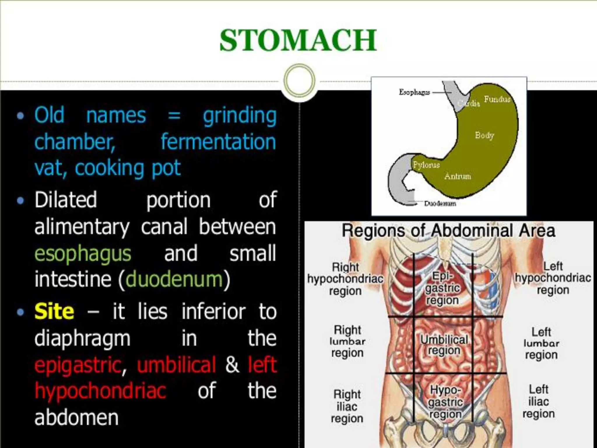
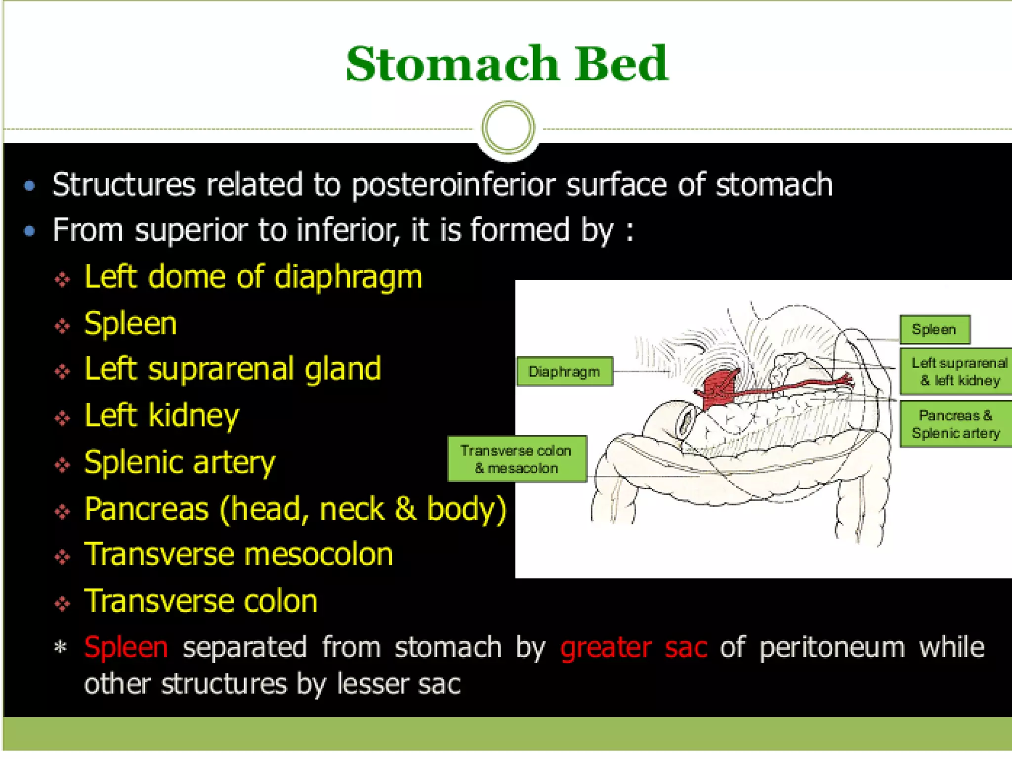
• Ingestion
– Taking in food through the mouth
• Propulsion (movement of
food)
– Swallowing
– Peristalsis – propulsion by alternate
contraction &relaxation
• Mechanical digestion
– Chewing
– Churning in stomach
– Mixing by segmentation
• Chemical digestion
– Complex food molecules (carbohydrates, proteins and lipids)
broken down into chemical building blocks (simple sugars,
amino acids, and fatty acids and glycerol)
– Carried out by enzymes secreted by digestive glands into
lumen of the alimentary canal
• Absorption
– Transport of digested end products into blood
and lymph in wall of canal
• Defecation
– Elimination of indigestible substances from
body as feces
Four main layers. From inside (the
lumen) to outside the are:
o Mucosa
o Sub mucosa
o Muscularis (external)
o Serosa ( visceral peritoneum)
is the innermost, moist, epithelial membrane that
lines the entire digestive tract.
• (1) It secretes mucus, digestive enzymes, and hormones;
• (2) absorbs digestive end products into the blood; and
• (3) protects against infectious disease.
• Consists of a lining epithelium, a lamina propria, and a Muscularis
mucosa.
• Epithelium - simple columnar epithelium and goblet cells
• Lamina propria - areola C.T. with capillaries and lymphoid follicles
• Muscularis mucosa - thin layer, produces local movements of the
mucosa
is a moderately dense connective tissue
layer containing blood and lymphatic vessels, lymphoid follicles, and
nerve fibers.
❖ typically consists of smooth
muscle and is responsible for peristalsis and segmentation.
• Contains the my enteric plexus of Auerbach, the other major intrinsic
nerve plexus. Located between the two layers of smooth muscle,
controls motility of the G.I. tract.
, the protective outer layer of the intraperitoneal
organs, is the visceral peritoneum
1. Enteric Nervous System
2. Autonomic Nervous System
3. Sensory Nervous System
• It is Intrinsic system of GIT extending from esophagus up to
anus.
• Division Two:
• Lies between longitudinal and circular muscle layer.
❖ Functions: Control GIT movements.
• Lies in submucosa.
❖ Functions:
• Control GIT secretions and blood flow.
• Sub serves sensory functions by receiving signals from GIT
Epithelium and from stretch receptors of GIT wall.
• Activity is control by sympathetic and
parasympathetic
nervous system.
Vagus Nerve innervates GIT from esophagus up
to proximal 2/3 of transverse colon.
Pelvic Parasympathetic Nerve:(S2-S4)Innervates
GIT from distal 1/3 of transverse colon.
Increase peristalsis and tone.
Relaxes Sphincters.
Increase digestive secretions.
:
• Preganglionic sympathetic originate in
T5 TO L2 segment of spinal cord, Pass
thru sympathetic chain, synapse with
• post ganglionic neuron in celiac and
mesenteric and hypo gastric ganglia.
Postganglionic fibers innervates in GIT.
• Decrease peristalsis and tone
• Contract sphincters
• Decrease digestive secretion
➢ Sensory nerve endings originate in the gastrointestinal
epithelium or gut wall and send afferent fibers to both
plexuses of the enteric system, as well as
(1) to the prevertebral ganglia of the sympathetic nervous
system,
(2) to the spinal cord, and
(3) in the Vagus nerves all the way to the brain stem.
➢ These sensory nerves can elicit local reflexes within the
gut wall itself and still other reflexes that are relayed to
the gut from either the prevertebral ganglia or the basal
regions of the brain.
• Two types of movements occur in
the gastrointestinal tract:
1. Mixing movements: (
Segmentation Contraction)
2. Propulsive movements:
(Peristalsis)
❖ First the intestinal wall is in relaxing state when it is distended with
chyme (food which is mixed with secretion)
❖ The distension of intestinal wall causes contraction in intestine and
these contraction divides the intestine into segments
❖ Each segment is 1-2cm long As one set of segmentation contraction
relaxes, a new set often begins but the contraction this time appears
at new sites between the previous contraction.
❖ These contraction help in chopping of chyme and promotes mixing
of food particles with the secretion of small intestine.
❖ Segmentation contraction occurs 12time/min in duodenum and
8time/min in the ileum
❖ These contraction last for 5-6sec, they occur through out the
digestive period
❖ These segmentation contraction are controlled by the Myenteric
plexuses of Autonomic Nervous System.
➢ Chyme is propelled or pushed through small
intestine by peristaltic waves.
➢ These waves are produced and response to stretch
.This is called Myenteric Reflex.
➢ They occur in any part of small intestine and move
at a velocity of 0.2-2cm/sec. They usually dies off
after travel this distance.
➢ From here it starts a new peristalsis state and move
a chyme in forward direction, thus several peristaltic
waves occur one after the other and push the small
intestine contents at the distal end of the small
intestine.
➢ Therefore due to the slow movement of peristalsis
waves usually 3-5hours are required for passage of
chyme from pyrolysis to the ileocecal valve.
➢ These peristaltic wave don't occur alone but are
superimposed upon segmental movement of small
intestine. Thus both segmental and peristalsis
movements occurs simultaneously.
Gastro intestinal track

Gastro intestinal track

  • 5.
    • Ingestion – Takingin food through the mouth • Propulsion (movement of food) – Swallowing – Peristalsis – propulsion by alternate contraction &relaxation • Mechanical digestion – Chewing – Churning in stomach – Mixing by segmentation • Chemical digestion – Complex food molecules (carbohydrates, proteins and lipids) broken down into chemical building blocks (simple sugars, amino acids, and fatty acids and glycerol) – Carried out by enzymes secreted by digestive glands into lumen of the alimentary canal • Absorption – Transport of digested end products into blood and lymph in wall of canal • Defecation – Elimination of indigestible substances from body as feces
  • 6.
    Four main layers.From inside (the lumen) to outside the are: o Mucosa o Sub mucosa o Muscularis (external) o Serosa ( visceral peritoneum)
  • 7.
    is the innermost,moist, epithelial membrane that lines the entire digestive tract. • (1) It secretes mucus, digestive enzymes, and hormones; • (2) absorbs digestive end products into the blood; and • (3) protects against infectious disease. • Consists of a lining epithelium, a lamina propria, and a Muscularis mucosa. • Epithelium - simple columnar epithelium and goblet cells • Lamina propria - areola C.T. with capillaries and lymphoid follicles • Muscularis mucosa - thin layer, produces local movements of the mucosa is a moderately dense connective tissue layer containing blood and lymphatic vessels, lymphoid follicles, and nerve fibers. ❖ typically consists of smooth muscle and is responsible for peristalsis and segmentation. • Contains the my enteric plexus of Auerbach, the other major intrinsic nerve plexus. Located between the two layers of smooth muscle, controls motility of the G.I. tract. , the protective outer layer of the intraperitoneal organs, is the visceral peritoneum
  • 9.
    1. Enteric NervousSystem 2. Autonomic Nervous System 3. Sensory Nervous System
  • 10.
    • It isIntrinsic system of GIT extending from esophagus up to anus. • Division Two: • Lies between longitudinal and circular muscle layer. ❖ Functions: Control GIT movements. • Lies in submucosa. ❖ Functions: • Control GIT secretions and blood flow. • Sub serves sensory functions by receiving signals from GIT Epithelium and from stretch receptors of GIT wall.
  • 11.
    • Activity iscontrol by sympathetic and parasympathetic nervous system. Vagus Nerve innervates GIT from esophagus up to proximal 2/3 of transverse colon. Pelvic Parasympathetic Nerve:(S2-S4)Innervates GIT from distal 1/3 of transverse colon. Increase peristalsis and tone. Relaxes Sphincters. Increase digestive secretions.
  • 13.
    : • Preganglionic sympatheticoriginate in T5 TO L2 segment of spinal cord, Pass thru sympathetic chain, synapse with • post ganglionic neuron in celiac and mesenteric and hypo gastric ganglia. Postganglionic fibers innervates in GIT. • Decrease peristalsis and tone • Contract sphincters • Decrease digestive secretion
  • 14.
    ➢ Sensory nerveendings originate in the gastrointestinal epithelium or gut wall and send afferent fibers to both plexuses of the enteric system, as well as (1) to the prevertebral ganglia of the sympathetic nervous system, (2) to the spinal cord, and (3) in the Vagus nerves all the way to the brain stem. ➢ These sensory nerves can elicit local reflexes within the gut wall itself and still other reflexes that are relayed to the gut from either the prevertebral ganglia or the basal regions of the brain.
  • 15.
    • Two typesof movements occur in the gastrointestinal tract: 1. Mixing movements: ( Segmentation Contraction) 2. Propulsive movements: (Peristalsis)
  • 16.
    ❖ First theintestinal wall is in relaxing state when it is distended with chyme (food which is mixed with secretion) ❖ The distension of intestinal wall causes contraction in intestine and these contraction divides the intestine into segments ❖ Each segment is 1-2cm long As one set of segmentation contraction relaxes, a new set often begins but the contraction this time appears at new sites between the previous contraction. ❖ These contraction help in chopping of chyme and promotes mixing of food particles with the secretion of small intestine. ❖ Segmentation contraction occurs 12time/min in duodenum and 8time/min in the ileum ❖ These contraction last for 5-6sec, they occur through out the digestive period ❖ These segmentation contraction are controlled by the Myenteric plexuses of Autonomic Nervous System.
  • 18.
    ➢ Chyme ispropelled or pushed through small intestine by peristaltic waves. ➢ These waves are produced and response to stretch .This is called Myenteric Reflex. ➢ They occur in any part of small intestine and move at a velocity of 0.2-2cm/sec. They usually dies off after travel this distance. ➢ From here it starts a new peristalsis state and move a chyme in forward direction, thus several peristaltic waves occur one after the other and push the small intestine contents at the distal end of the small intestine. ➢ Therefore due to the slow movement of peristalsis waves usually 3-5hours are required for passage of chyme from pyrolysis to the ileocecal valve. ➢ These peristaltic wave don't occur alone but are superimposed upon segmental movement of small intestine. Thus both segmental and peristalsis movements occurs simultaneously.