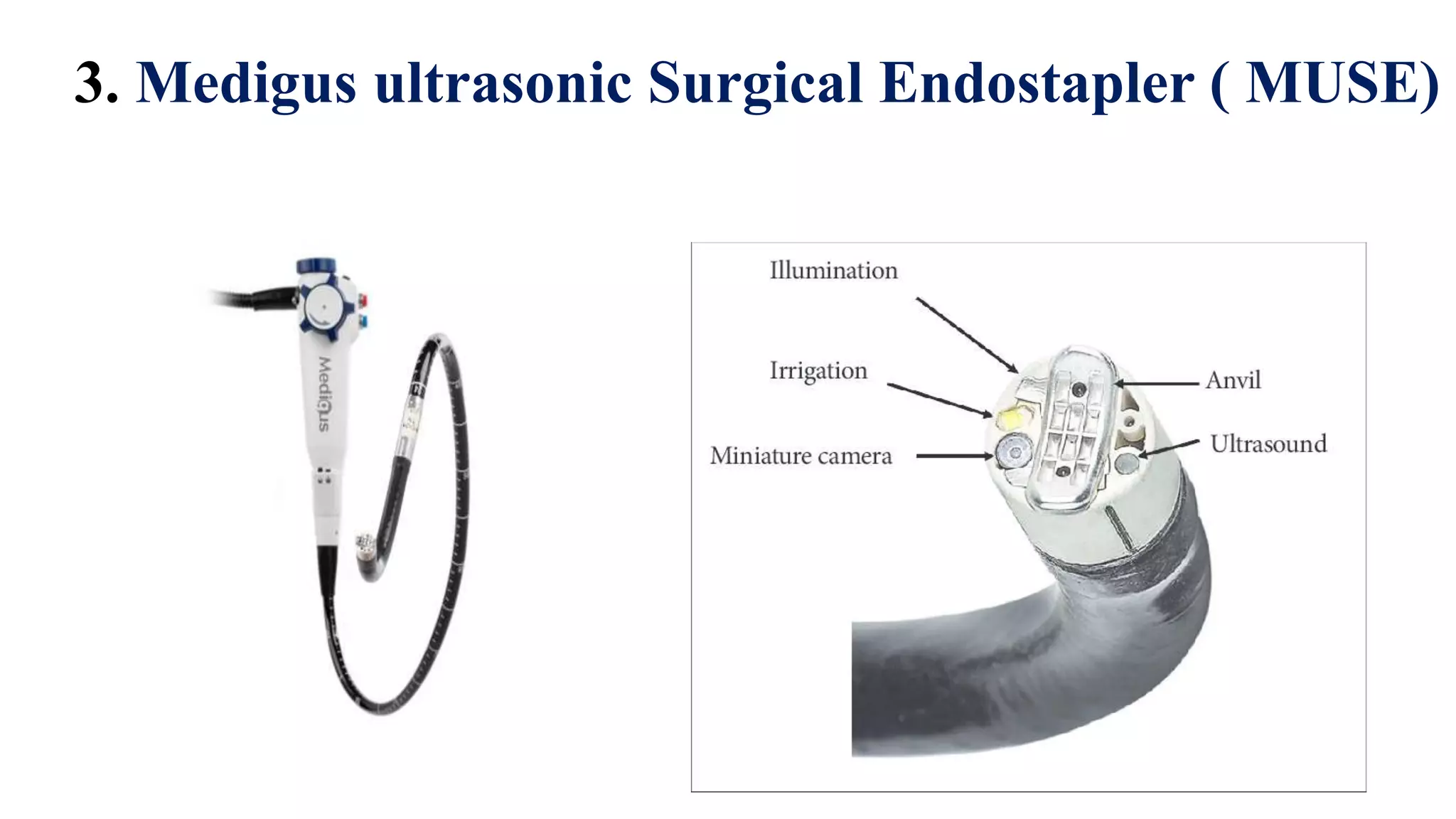
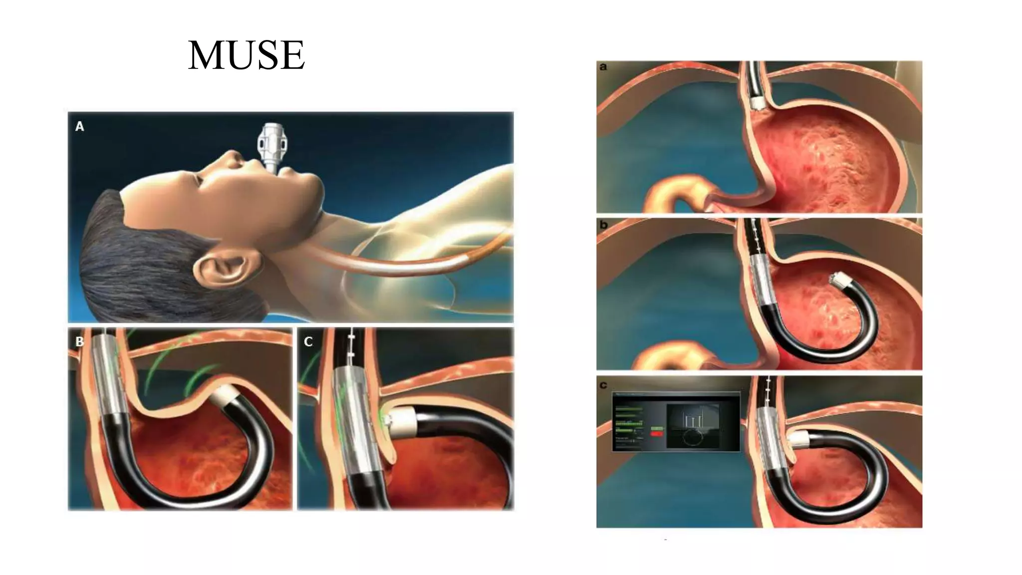
Seminar on
Endoluminal treatment in GERD
Presenter
Dr. Biswajit Deka
PGT
Deptt of surgery , SMCH
Introduction
• Gastroesophageal reflux disease (GERD) is due to the failure of the
gastroesophageal barrier.
• The disease has dramatically increased in the last 2 decades.
• The optimal treatment of GERD continues to be debated.
• The rising number of patients having daily reflux-related
symptoms and their impact on quality of life has lead to a better
understanding of the pathophysiology of this condition and its
appropriate management.
• New anti-secretory medications, laparoscopic techniques, and
novel endoluminal devices have been introduced ; the outcomes
are encouraging, but the cost is a matter of concern.
Anatomical & physiological background
• The LES and the geometric profile of the cardia prevent GERD
and are the targets of surgical and endoluminal GERD
procedures.
• The LES is characterized by its length, relative position to the
diaphragm, and pressure.
• A decrease in pressure, overall or intra-abdominal length
predisposes to reflux.
• In severe GERD, the “high-pressure zone” (LES) is virtually non-
existent or greatly reduced, and reflux in this instance is understandable.
• However, the cause of reflux in milder disease with a normal lower
oesophageal sphincter resting pressure (LESP) is under considerable
debate.
• It is believed that transient LES relaxations (tLESR), ie, intermittent
spontaneous decreases in LESP, are responsible for reflux events.
• Recent electrophysiological data suggest that the relevant vagal
afferent fibres terminate with specialized intra-ganglionic laminar
endings (IGLEs).
• These deformity-sensitive transducers are lined in series with
muscle fibres at the cardia and fundus and are believed to mediate
both fundic receptive relaxation and elicitation of tLESRs.
• In light of the above-described pathophysiologic factors,
endoscopic therapies should prevent reflux in one or more of the
following ways:
(1) alter the compliance of the cardia and prevent tLES shortening or
relaxation.
(2) increase baseline LES tone.
(3) increase baseline LES length.
Indications & contra-indication
• Refractory symptoms despite maximized medical treatment
• Contra-indication to medication therapy.
• Conscious sedation- (No GA) - target large population
• Contra-indication: anatomical ( hiatal hernia longer than 2cm
& wider than 3cm )
Pre-op preparation
• Complete esophageal work up –
UGI endoscopy
Esophageal manometry
 Evaluate for other disorder with GERD like symptoms
 Extent of esophagitis
 Barrett’s esophagus
Anatomical abnormality
Types
1. EsophyX – create incisionless fundoplication, FDA- 2007
2. Stretta – utilizes RF to remodel the LES by contracting
collagen and creating a barrier to gastric pressure.
3. Medigus ultrasonic Surgical Endostapler – creates an anterior
partial wrap
4. Injection / implantation – inert substance
5. Mucosal excision
1. EsophyX : Transoral Incisionless Fundoplication
• Deployed in the stomach with an endoscope
• Creates a full thickness plication of GEJ using H-shaped polypropylene
fasteners.
• Several revisions : TIF-1 : produce a gastric wrap at the level of GEJ.
• TIF-2 : place the fasteners above GEJ to create a gastroesophageal flap
valve that replicates laparoscopic fundoplication ; TIF 2.0 – current
technique
EsophyX
EsophyX
Procedure
• Patient under GA , left lateral position
• Endoscopy - rule out hiatal hernia
• 2 physicians reqd ; Device introduced with CO2 insufflation.
• Device retroflexed- greater & lesser curvatures identified.
• Helical screw deployed at GEJ to retract the tissue.
• A paddle- shaped tissue mold is then used to deploy
polypropylene fasteners to create a ‘crimp’.
• The tech. performed circumferentially to create a 270 degree
fundoplication. (TIF-1: 2000-2500 fundoplication)
• Device removed- endoscopy done – to assess plication ,
bleeding or perforation
Fig: A) A bad valve B) Fastener delivery C) Repair with Helical Retractor D)
Repaired tissue E) Repaired tissue (with wrap) F) Repaired valve with fasteners
Post-op care
• Kept under observation
• Can be discharged the following day
• Omeprazole 40mg x 14 days : to promote mucosal healing
• Liquid diet x 2 weeks
• Soft food x 3-7 weeks
• Normal diet after 2 months
Complications and their management
• Very few
• Bleeding – Self limiting
BT occasionally
• Perforation – laparoscopic transabdominal drainage
partial fundoplication over damaged esophagus
2. STRETTA
• An inflatable balloon catheter system with 4 electrode needle
sheaths
• Radiofrequency delivered to electrodes – heat the adjacent cells
• Target temp- 65o to 85oC x 1 min cycles.
• RF- induce fibrosis of LES- reduce tissue compliance and transient
LES relaxation.
Radiofrequency device
Technique
• Deep sedation & endoscopy
• Hiatal hernia >2cm & severe esophagitis – excluded
• Using a guidewire exchange tech- STRATTA catheter introduced
• Placed 2cm proximal to Z-line
• Balloon inflated, electrode needles deployed into esophageal wall
• RF energy delivered x 1min
• Needle retracted – balloon deflated- catheter rotated 45 degree
• Tech repeated , cover a length 2cm above &1.5cm below Z-line
• Mucosa- cooled with water irrigation
Post-op period
• Day care procedure
• Minor bleeding
• Pain
• Perforation , deaths – not reported yet
3. Medigus ultrasonic Surgical Endostapler ( MUSE)
• It’s a stand alone unit – operated by a single user
• Designed in a shape of an endoscope, with a video camera, light
source, ultrasonic transducer and a stapling mechanism at the tip.
• Device is advanced down the esophagus through a endoscopic over
tube with 17mm internal diameter
• Retroflexed- to identify the site to place staplers
• Fundic tissue compressed against esophagus and USG transducer
measure tissue thickness – once it reaches 1.4-1.6mm, stapler is
fired.
• Procedure repeated to mimic a partial anterior fundoplication
MUSE
MUSE
Post-op period.
• Observation for 72 hrs.
• UGI endoscopy on POD-1 to evaluate for leaks
• Complication – insufficient data: Bleeding
Esophageal perforation
Pneumomediastinum
Pneumothorax
Chest pain , sore throat
4. Injection/Implantation Techniques
• The goal: to deliver a biologically inert, injectable substance into
different depths of the LES region.
• Submucosal injections increase the volume of the LES, and
injection into the muscularis propria results in granulation and
fibrous capsule formation.
• From the mid 1980s, several biopolymers were tested in animals
and obtained FDA approval
1. Enteryx : an ethylene vinyl alcohol copolymer with tantalum
dissolved in dimethyl sulfide, not in use
2. Gatekeeper Reflux Repair System was a dehydrated hydrogel
prosthesis implanted into the submucosa of the cardia/LES ,
removed from the market because of lack of efficacy.
3. Durasphere : a new sterile, biocompatible injectable bulking
agent composed of pyrolytic carbon-coated graphite beads
containing zirconium oxide, suspended in a water-based, absorbable
polysaccharide carrier gel.
5. ANTI-REFLUX MUCOSECTOMY (ARMS)
• Based on the principle that after mucosal resection, the mucosal healing
results in scar formation.
• Shrinkage and remodelling of gastric cardia flap valve; thereby, reducing
reflux events.
• It involves resection of gastric (about 2 cm) & oesophageal mucosa (about 1
cm) in crescentic fashion; field marked by electrocautery .
• Next , mucosal resection is performed along the lesser curvature
• Subsequently, a solution of saline mixed with indigo-carmine
dye is injected submucosally to raise a wheal.
• A gap equal to twice the scope diameter is left along the greater
curvature side.
• Circumferential resection of the mucosa is avoided to prevent
stricture formation as was noted in initial few cases of this series.
• Any bleeding during the procedure is controlled by coagrasper
(A) Submucosal injection of saline
with indigo-carmine at gastric cardia.
(B) Application of snare over the
mucosa with cap-endoscopic mucosal
resection technique.
(C) Completion of near
circumferential (2/3) resection of
gastric mucosa.
(D) Actively bleeding spurter during
mucosectomy procedure.
(E) Effective control of bleeding
vessel with coagrasper
• The advantages of ARMS: no requirement of any propriety
devices and no end-prostheses are left in situ.
• However, no randomized studies ; durability of response is
unknown.
• The amount of mucosa to be resected for optimal results is not
known and needs further evaluation.
• Patients with large hiatal hernia are not suitable candidates
• Anti-reflux surgery is recommended for patients with refractory or
complicated GERD and provides excellent symptom control in 85% to 90% of
cases
• These anti-reflux procedures can be categorised into ablation, injection or
implantation, fixation and mucosal excision.
• Endoluminal intervention for GERD is relatively new and still immature but is
a promising option for GERD patients. Morbidities and mortality should be
reduced, and repeat procedures may be easier to perform.
Conclusion
THANK YOU …

Endoluminal therapy in GERD

  • 1.
    Seminar on Endoluminal treatmentin GERD Presenter Dr. Biswajit Deka PGT Deptt of surgery , SMCH
  • 2.
    Introduction • Gastroesophageal refluxdisease (GERD) is due to the failure of the gastroesophageal barrier. • The disease has dramatically increased in the last 2 decades. • The optimal treatment of GERD continues to be debated.
  • 3.
    • The risingnumber of patients having daily reflux-related symptoms and their impact on quality of life has lead to a better understanding of the pathophysiology of this condition and its appropriate management. • New anti-secretory medications, laparoscopic techniques, and novel endoluminal devices have been introduced ; the outcomes are encouraging, but the cost is a matter of concern.
  • 4.
    Anatomical & physiologicalbackground • The LES and the geometric profile of the cardia prevent GERD and are the targets of surgical and endoluminal GERD procedures. • The LES is characterized by its length, relative position to the diaphragm, and pressure. • A decrease in pressure, overall or intra-abdominal length predisposes to reflux.
  • 5.
    • In severeGERD, the “high-pressure zone” (LES) is virtually non- existent or greatly reduced, and reflux in this instance is understandable. • However, the cause of reflux in milder disease with a normal lower oesophageal sphincter resting pressure (LESP) is under considerable debate. • It is believed that transient LES relaxations (tLESR), ie, intermittent spontaneous decreases in LESP, are responsible for reflux events.
  • 6.
    • Recent electrophysiologicaldata suggest that the relevant vagal afferent fibres terminate with specialized intra-ganglionic laminar endings (IGLEs). • These deformity-sensitive transducers are lined in series with muscle fibres at the cardia and fundus and are believed to mediate both fundic receptive relaxation and elicitation of tLESRs.
  • 8.
    • In lightof the above-described pathophysiologic factors, endoscopic therapies should prevent reflux in one or more of the following ways: (1) alter the compliance of the cardia and prevent tLES shortening or relaxation. (2) increase baseline LES tone. (3) increase baseline LES length.
  • 9.
    Indications & contra-indication •Refractory symptoms despite maximized medical treatment • Contra-indication to medication therapy. • Conscious sedation- (No GA) - target large population • Contra-indication: anatomical ( hiatal hernia longer than 2cm & wider than 3cm )
  • 10.
    Pre-op preparation • Completeesophageal work up – UGI endoscopy Esophageal manometry  Evaluate for other disorder with GERD like symptoms  Extent of esophagitis  Barrett’s esophagus Anatomical abnormality
  • 11.
    Types 1. EsophyX –create incisionless fundoplication, FDA- 2007 2. Stretta – utilizes RF to remodel the LES by contracting collagen and creating a barrier to gastric pressure. 3. Medigus ultrasonic Surgical Endostapler – creates an anterior partial wrap 4. Injection / implantation – inert substance 5. Mucosal excision
  • 12.
    1. EsophyX :Transoral Incisionless Fundoplication • Deployed in the stomach with an endoscope • Creates a full thickness plication of GEJ using H-shaped polypropylene fasteners. • Several revisions : TIF-1 : produce a gastric wrap at the level of GEJ. • TIF-2 : place the fasteners above GEJ to create a gastroesophageal flap valve that replicates laparoscopic fundoplication ; TIF 2.0 – current technique
  • 13.
  • 14.
  • 15.
    Procedure • Patient underGA , left lateral position • Endoscopy - rule out hiatal hernia • 2 physicians reqd ; Device introduced with CO2 insufflation. • Device retroflexed- greater & lesser curvatures identified. • Helical screw deployed at GEJ to retract the tissue.
  • 16.
    • A paddle-shaped tissue mold is then used to deploy polypropylene fasteners to create a ‘crimp’. • The tech. performed circumferentially to create a 270 degree fundoplication. (TIF-1: 2000-2500 fundoplication) • Device removed- endoscopy done – to assess plication , bleeding or perforation
  • 17.
    Fig: A) Abad valve B) Fastener delivery C) Repair with Helical Retractor D) Repaired tissue E) Repaired tissue (with wrap) F) Repaired valve with fasteners
  • 18.
    Post-op care • Keptunder observation • Can be discharged the following day • Omeprazole 40mg x 14 days : to promote mucosal healing • Liquid diet x 2 weeks • Soft food x 3-7 weeks • Normal diet after 2 months
  • 19.
    Complications and theirmanagement • Very few • Bleeding – Self limiting BT occasionally • Perforation – laparoscopic transabdominal drainage partial fundoplication over damaged esophagus
  • 20.
    2. STRETTA • Aninflatable balloon catheter system with 4 electrode needle sheaths • Radiofrequency delivered to electrodes – heat the adjacent cells • Target temp- 65o to 85oC x 1 min cycles. • RF- induce fibrosis of LES- reduce tissue compliance and transient LES relaxation.
  • 21.
  • 22.
    Technique • Deep sedation& endoscopy • Hiatal hernia >2cm & severe esophagitis – excluded • Using a guidewire exchange tech- STRATTA catheter introduced • Placed 2cm proximal to Z-line • Balloon inflated, electrode needles deployed into esophageal wall
  • 23.
    • RF energydelivered x 1min • Needle retracted – balloon deflated- catheter rotated 45 degree • Tech repeated , cover a length 2cm above &1.5cm below Z-line • Mucosa- cooled with water irrigation
  • 24.
    Post-op period • Daycare procedure • Minor bleeding • Pain • Perforation , deaths – not reported yet
  • 25.
    3. Medigus ultrasonicSurgical Endostapler ( MUSE)
  • 26.
    • It’s astand alone unit – operated by a single user • Designed in a shape of an endoscope, with a video camera, light source, ultrasonic transducer and a stapling mechanism at the tip. • Device is advanced down the esophagus through a endoscopic over tube with 17mm internal diameter
  • 27.
    • Retroflexed- toidentify the site to place staplers • Fundic tissue compressed against esophagus and USG transducer measure tissue thickness – once it reaches 1.4-1.6mm, stapler is fired. • Procedure repeated to mimic a partial anterior fundoplication
  • 28.
  • 29.
  • 30.
    Post-op period. • Observationfor 72 hrs. • UGI endoscopy on POD-1 to evaluate for leaks • Complication – insufficient data: Bleeding Esophageal perforation Pneumomediastinum Pneumothorax Chest pain , sore throat
  • 31.
    4. Injection/Implantation Techniques •The goal: to deliver a biologically inert, injectable substance into different depths of the LES region. • Submucosal injections increase the volume of the LES, and injection into the muscularis propria results in granulation and fibrous capsule formation. • From the mid 1980s, several biopolymers were tested in animals and obtained FDA approval
  • 32.
    1. Enteryx :an ethylene vinyl alcohol copolymer with tantalum dissolved in dimethyl sulfide, not in use 2. Gatekeeper Reflux Repair System was a dehydrated hydrogel prosthesis implanted into the submucosa of the cardia/LES , removed from the market because of lack of efficacy. 3. Durasphere : a new sterile, biocompatible injectable bulking agent composed of pyrolytic carbon-coated graphite beads containing zirconium oxide, suspended in a water-based, absorbable polysaccharide carrier gel.
  • 33.
    5. ANTI-REFLUX MUCOSECTOMY(ARMS) • Based on the principle that after mucosal resection, the mucosal healing results in scar formation. • Shrinkage and remodelling of gastric cardia flap valve; thereby, reducing reflux events. • It involves resection of gastric (about 2 cm) & oesophageal mucosa (about 1 cm) in crescentic fashion; field marked by electrocautery . • Next , mucosal resection is performed along the lesser curvature
  • 34.
    • Subsequently, asolution of saline mixed with indigo-carmine dye is injected submucosally to raise a wheal. • A gap equal to twice the scope diameter is left along the greater curvature side. • Circumferential resection of the mucosa is avoided to prevent stricture formation as was noted in initial few cases of this series. • Any bleeding during the procedure is controlled by coagrasper
  • 35.
    (A) Submucosal injectionof saline with indigo-carmine at gastric cardia. (B) Application of snare over the mucosa with cap-endoscopic mucosal resection technique. (C) Completion of near circumferential (2/3) resection of gastric mucosa. (D) Actively bleeding spurter during mucosectomy procedure. (E) Effective control of bleeding vessel with coagrasper
  • 36.
    • The advantagesof ARMS: no requirement of any propriety devices and no end-prostheses are left in situ. • However, no randomized studies ; durability of response is unknown. • The amount of mucosa to be resected for optimal results is not known and needs further evaluation. • Patients with large hiatal hernia are not suitable candidates
  • 37.
    • Anti-reflux surgeryis recommended for patients with refractory or complicated GERD and provides excellent symptom control in 85% to 90% of cases • These anti-reflux procedures can be categorised into ablation, injection or implantation, fixation and mucosal excision. • Endoluminal intervention for GERD is relatively new and still immature but is a promising option for GERD patients. Morbidities and mortality should be reduced, and repeat procedures may be easier to perform. Conclusion
  • 38.