endocytosis and exocytosis is a procss of cell eating and drinnking. it is a mazor tool for self defence to an individual cell. there are some molecular mechanism for this process described in given notes.
explain the types and the formation of vesicles.for downloading the presentation ,more presentations , infographics and blogs visit :
https://studyscienceblog.wordpress.com
explain the types and the formation of vesicles.for downloading the presentation ,more presentations , infographics and blogs visit :
https://studyscienceblog.wordpress.com
General overview of Plasma/ Cell membrane.
Definition of Plasma/ Cell membrane
Structure of Plasma membrane
1. Sandwitch model ORDanielli- Davson Model
2. Fluid mosaic model
Plasma Membrane Proteins
Chemical Composition of Plasma/ Cell Membrane
Movement across the Cell Membrane
Channels through cell membrane
Exocytosis is the process of moving materials from within a cell to the exterior of the cell. This process requires energy and is therefore a type of active transport. Exocytosis is an important process of plant and animal cells as it performs the opposite function of endocytosis. In endocytosis, substances that are external to a cell are brought into the cell.
In exocytosis, membrane-bound vesicles containing cellular molecules are transported to the cell membrane. The vesicles fuse with the cell membrane and expel their contents to the exterior of the cell. The process of exocytosis can be summarized in a few steps.
Vesicles containing molecules are transported from within the cell to the cell membrane.
The vesicle membrane attaches to the cell membrane.
Fusion of the vesicle membrane with the cell membrane releases the vesicle contents outside the cell.
There are three common pathways of exocytosis. One pathway, constitutive exocytosis, involves the regular secretion of molecules. This action is performed by all cells. Constitutive exocytosis functions to deliver membrane proteins and lipids to the cell's surface and to expel substances to the cell's exterior.
Regulated exocytosis relies on the presence of extracellular signals for the expulsion of materials within vesicles. Regulated exocytosis occurs commonly in secretory cells and not in all cell types. Secretory cells store products such as hormones, neurotransmitters, and digestive enzymes that are released only when triggered by extracellular signals. Secretory vesicles are not incorporated into the cell membrane but fuse only long enough to release their contents. Once the delivery has been made, the vesicles reform and return to the cytoplasm.
A third pathway for exocytosis in cells involves the fusion of vesicles with lysosomes. These organelles contain acid hydrolase enzymes that break down waste materials, microbes, and cellular debris. Lysosomes carry their digested material to the cell membrane where they fuse with the membrane and release their contents into the extracellular matrix.
Active transport
Types of Active Transport
Primary Active transport
Sodium-Potassium pump
secondary Active transport
uniport, Symport, Antiport
Endocytosis
Types of endocytosis
Phagocytosis
Pinocytosis
Receptor-mediated endocytosis
Exocytosis
Purposes of Endocytosis and Exocytosis
Passive Transport
Types of Passive Transport
Diffusion
Osmosis
Facilitated Diffusion
What causes diffusion?
Why is diffusion useful?
Supercritical fluid
Osmotic Solutions
Isotonic Solution
Hypertonic Solution
Hypotonic Solution
Types of Osmosis
Difference Between Endosmosis And Exosmosis
Significance of Osmosis
Factors Affecting Facilitated Diffusion
Importance of Facilitated Diffusion
Transmembrane Proteins
Channel Proteins and carrier protein
At the end of this session, the student should be able to:
a. Describe the representative functional systems of the cell and discuss their potential roles.
b. Describe the types of locomotion in non-muscle cells and discuss their mechanisms.
med_students0
this presentation providing about the cell .Cell is the basic living, structural, and functional unit of the body.
Cells are grouped together to form tissues, each of which has a specialized function, e.g.- Bone and blood tissue.
Different tissues are grouped together to form a organs, e.g. liver, stomach, and kidney etc.
Organs are grouped together to form a system, each of which performs a particular function responsible for maintaining homeostasis .
e.g. Urinary system, Respiratory system etc.
Autoimmune disease HEMOLYTIC ANEMIA AND DIABETESArchanaSoni3
An autoimmune disease is a condition in which your immune system mistakenly attacks your body.
The immune system normally guards against germs like bacteria and viruses. When it senses these foreign invaders, it sends out an army of fighter cells to attack them.
Normally, the immune system can tell the difference between foreign cells and your own cells.
In an autoimmune disease, the immune system mistakes part of your body — like your joints or skin — as foreign. It releases proteins called autoantibodies that attack healthy cells.
Some autoimmune diseases target only one organ. Type 1 diabetes damages the pancreas. Other diseases, like lupus, affect the whole body.
Cancer cell metabolism: special Reference to Lactate PathwayAADYARAJPANDEY1
Normal Cell Metabolism:
Cellular respiration describes the series of steps that cells use to break down sugar and other chemicals to get the energy we need to function.
Energy is stored in the bonds of glucose and when glucose is broken down, much of that energy is released.
Cell utilize energy in the form of ATP.
The first step of respiration is called glycolysis. In a series of steps, glycolysis breaks glucose into two smaller molecules - a chemical called pyruvate. A small amount of ATP is formed during this process.
Most healthy cells continue the breakdown in a second process, called the Kreb's cycle. The Kreb's cycle allows cells to “burn” the pyruvates made in glycolysis to get more ATP.
The last step in the breakdown of glucose is called oxidative phosphorylation (Ox-Phos).
It takes place in specialized cell structures called mitochondria. This process produces a large amount of ATP. Importantly, cells need oxygen to complete oxidative phosphorylation.
If a cell completes only glycolysis, only 2 molecules of ATP are made per glucose. However, if the cell completes the entire respiration process (glycolysis - Kreb's - oxidative phosphorylation), about 36 molecules of ATP are created, giving it much more energy to use.
IN CANCER CELL:
Unlike healthy cells that "burn" the entire molecule of sugar to capture a large amount of energy as ATP, cancer cells are wasteful.
Cancer cells only partially break down sugar molecules. They overuse the first step of respiration, glycolysis. They frequently do not complete the second step, oxidative phosphorylation.
This results in only 2 molecules of ATP per each glucose molecule instead of the 36 or so ATPs healthy cells gain. As a result, cancer cells need to use a lot more sugar molecules to get enough energy to survive.
introduction to WARBERG PHENOMENA:
WARBURG EFFECT Usually, cancer cells are highly glycolytic (glucose addiction) and take up more glucose than do normal cells from outside.
Otto Heinrich Warburg (; 8 October 1883 – 1 August 1970) In 1931 was awarded the Nobel Prize in Physiology for his "discovery of the nature and mode of action of the respiratory enzyme.
WARNBURG EFFECT : cancer cells under aerobic (well-oxygenated) conditions to metabolize glucose to lactate (aerobic glycolysis) is known as the Warburg effect. Warburg made the observation that tumor slices consume glucose and secrete lactate at a higher rate than normal tissues.
Multi-source connectivity as the driver of solar wind variability in the heli...Sérgio Sacani
The ambient solar wind that flls the heliosphere originates from multiple
sources in the solar corona and is highly structured. It is often described
as high-speed, relatively homogeneous, plasma streams from coronal
holes and slow-speed, highly variable, streams whose source regions are
under debate. A key goal of ESA/NASA’s Solar Orbiter mission is to identify
solar wind sources and understand what drives the complexity seen in the
heliosphere. By combining magnetic feld modelling and spectroscopic
techniques with high-resolution observations and measurements, we show
that the solar wind variability detected in situ by Solar Orbiter in March
2022 is driven by spatio-temporal changes in the magnetic connectivity to
multiple sources in the solar atmosphere. The magnetic feld footpoints
connected to the spacecraft moved from the boundaries of a coronal hole
to one active region (12961) and then across to another region (12957). This
is refected in the in situ measurements, which show the transition from fast
to highly Alfvénic then to slow solar wind that is disrupted by the arrival of
a coronal mass ejection. Our results describe solar wind variability at 0.5 au
but are applicable to near-Earth observatories.
Richard's entangled aventures in wonderlandRichard Gill
Since the loophole-free Bell experiments of 2020 and the Nobel prizes in physics of 2022, critics of Bell's work have retreated to the fortress of super-determinism. Now, super-determinism is a derogatory word - it just means "determinism". Palmer, Hance and Hossenfelder argue that quantum mechanics and determinism are not incompatible, using a sophisticated mathematical construction based on a subtle thinning of allowed states and measurements in quantum mechanics, such that what is left appears to make Bell's argument fail, without altering the empirical predictions of quantum mechanics. I think however that it is a smoke screen, and the slogan "lost in math" comes to my mind. I will discuss some other recent disproofs of Bell's theorem using the language of causality based on causal graphs. Causal thinking is also central to law and justice. I will mention surprising connections to my work on serial killer nurse cases, in particular the Dutch case of Lucia de Berk and the current UK case of Lucy Letby.
The increased availability of biomedical data, particularly in the public domain, offers the opportunity to better understand human health and to develop effective therapeutics for a wide range of unmet medical needs. However, data scientists remain stymied by the fact that data remain hard to find and to productively reuse because data and their metadata i) are wholly inaccessible, ii) are in non-standard or incompatible representations, iii) do not conform to community standards, and iv) have unclear or highly restricted terms and conditions that preclude legitimate reuse. These limitations require a rethink on data can be made machine and AI-ready - the key motivation behind the FAIR Guiding Principles. Concurrently, while recent efforts have explored the use of deep learning to fuse disparate data into predictive models for a wide range of biomedical applications, these models often fail even when the correct answer is already known, and fail to explain individual predictions in terms that data scientists can appreciate. These limitations suggest that new methods to produce practical artificial intelligence are still needed.
In this talk, I will discuss our work in (1) building an integrative knowledge infrastructure to prepare FAIR and "AI-ready" data and services along with (2) neurosymbolic AI methods to improve the quality of predictions and to generate plausible explanations. Attention is given to standards, platforms, and methods to wrangle knowledge into simple, but effective semantic and latent representations, and to make these available into standards-compliant and discoverable interfaces that can be used in model building, validation, and explanation. Our work, and those of others in the field, creates a baseline for building trustworthy and easy to deploy AI models in biomedicine.
Bio
Dr. Michel Dumontier is the Distinguished Professor of Data Science at Maastricht University, founder and executive director of the Institute of Data Science, and co-founder of the FAIR (Findable, Accessible, Interoperable and Reusable) data principles. His research explores socio-technological approaches for responsible discovery science, which includes collaborative multi-modal knowledge graphs, privacy-preserving distributed data mining, and AI methods for drug discovery and personalized medicine. His work is supported through the Dutch National Research Agenda, the Netherlands Organisation for Scientific Research, Horizon Europe, the European Open Science Cloud, the US National Institutes of Health, and a Marie-Curie Innovative Training Network. He is the editor-in-chief for the journal Data Science and is internationally recognized for his contributions in bioinformatics, biomedical informatics, and semantic technologies including ontologies and linked data.
Slide 1: Title Slide
Extrachromosomal Inheritance
Slide 2: Introduction to Extrachromosomal Inheritance
Definition: Extrachromosomal inheritance refers to the transmission of genetic material that is not found within the nucleus.
Key Components: Involves genes located in mitochondria, chloroplasts, and plasmids.
Slide 3: Mitochondrial Inheritance
Mitochondria: Organelles responsible for energy production.
Mitochondrial DNA (mtDNA): Circular DNA molecule found in mitochondria.
Inheritance Pattern: Maternally inherited, meaning it is passed from mothers to all their offspring.
Diseases: Examples include Leber’s hereditary optic neuropathy (LHON) and mitochondrial myopathy.
Slide 4: Chloroplast Inheritance
Chloroplasts: Organelles responsible for photosynthesis in plants.
Chloroplast DNA (cpDNA): Circular DNA molecule found in chloroplasts.
Inheritance Pattern: Often maternally inherited in most plants, but can vary in some species.
Examples: Variegation in plants, where leaf color patterns are determined by chloroplast DNA.
Slide 5: Plasmid Inheritance
Plasmids: Small, circular DNA molecules found in bacteria and some eukaryotes.
Features: Can carry antibiotic resistance genes and can be transferred between cells through processes like conjugation.
Significance: Important in biotechnology for gene cloning and genetic engineering.
Slide 6: Mechanisms of Extrachromosomal Inheritance
Non-Mendelian Patterns: Do not follow Mendel’s laws of inheritance.
Cytoplasmic Segregation: During cell division, organelles like mitochondria and chloroplasts are randomly distributed to daughter cells.
Heteroplasmy: Presence of more than one type of organellar genome within a cell, leading to variation in expression.
Slide 7: Examples of Extrachromosomal Inheritance
Four O’clock Plant (Mirabilis jalapa): Shows variegated leaves due to different cpDNA in leaf cells.
Petite Mutants in Yeast: Result from mutations in mitochondrial DNA affecting respiration.
Slide 8: Importance of Extrachromosomal Inheritance
Evolution: Provides insight into the evolution of eukaryotic cells.
Medicine: Understanding mitochondrial inheritance helps in diagnosing and treating mitochondrial diseases.
Agriculture: Chloroplast inheritance can be used in plant breeding and genetic modification.
Slide 9: Recent Research and Advances
Gene Editing: Techniques like CRISPR-Cas9 are being used to edit mitochondrial and chloroplast DNA.
Therapies: Development of mitochondrial replacement therapy (MRT) for preventing mitochondrial diseases.
Slide 10: Conclusion
Summary: Extrachromosomal inheritance involves the transmission of genetic material outside the nucleus and plays a crucial role in genetics, medicine, and biotechnology.
Future Directions: Continued research and technological advancements hold promise for new treatments and applications.
Slide 11: Questions and Discussion
Invite Audience: Open the floor for any questions or further discussion on the topic.
This pdf is about the Schizophrenia.
For more details visit on YouTube; @SELF-EXPLANATORY;
https://www.youtube.com/channel/UCAiarMZDNhe1A3Rnpr_WkzA/videos
Thanks...!
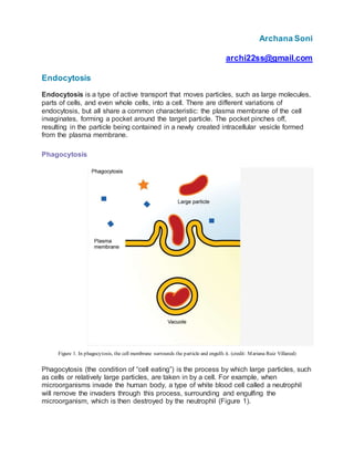
1. Archana Soni
archi22ss@gmail.com
Endocytosis
Endocytosis is a type of active transport that moves particles, such as large molecules,
parts of cells, and even whole cells, into a cell. There are different variations of
endocytosis, but all share a common characteristic: the plasma membrane of the cell
invaginates, forming a pocket around the target particle. The pocket pinches off,
resulting in the particle being contained in a newly created intracellular vesicle formed
from the plasma membrane.
Phagocytosis
Figure 1. In phagocytosis, the cell membrane surrounds the particle and engulfs it. (credit: Mariana Ruiz Villareal)
Phagocytosis (the condition of “cell eating”) is the process by which large particles, such
as cells or relatively large particles, are taken in by a cell. For example, when
microorganisms invade the human body, a type of white blood cell called a neutrophil
will remove the invaders through this process, surrounding and engulfing the
microorganism, which is then destroyed by the neutrophil (Figure 1).
2. In preparation for phagocytosis, a portion of the inward-facing surface of the plasma
membrane becomes coated with a protein called clathrin, which stabilizes this section of
the membrane. The coated portion of the membrane then extends from the body of the
cell and surrounds the particle, eventually enclosing it. Once the vesicle containing the
particle is enclosed within the cell, the clathrin disengages from the membrane and the
vesicle merges with a lysosome for the breakdown of the material in the newly formed
compartment (endosome). When accessible nutrients from the degradation of the
vesicular contents have been extracted, the newly formed endosome merges with the
plasma membrane and releases its contents into the extracellular fluid. The endosomal
membrane again becomes part of the plasma membrane.
Pinocytosis
Figure 2. In pinocytosis, the cell membrane invaginates, surrounds a small volume of fluid, and pinches off. (credit: Mariana Ruiz
Villareal)
A variation of endocytosis is called pinocytosis. This literally means “cell drinking” and
was named at a time when the assumption was that the cell was purposefully taking in
extracellular fluid. In reality, this is a process that takes in molecules, including water,
which the cell needs from the extracellular fluid. Pinocytosis results in a much smaller
vesicle than does phagocytosis, and the vesicle does not need to merge with a
lysosome (Figure 2).
A variation of pinocytosis is called potocytosis. This process uses a coating protein,
called caveolin, on the cytoplasmic side of the plasma membrane, which performs a
similar function to clathrin. The cavities in the plasma membrane that form the vacuoles
have membrane receptors and lipid rafts in addition to caveolin.
3. The vacuoles or vesicles formed in caveolae (singular caveola) are smaller than those
in pinocytosis. Potocytosis is used to bring small molecules into the cell and to transport
these molecules through the cell for their release on the other side of the cell, a process
called transcytosis.
Receptor-Mediated Endocytosis
Figure 3. In receptor-mediated endocytosis, uptake of substances by the cell is targeted to a single type of substance that binds to
the receptor on the external surface of the cell membrane. (credit: modification of work by Mariana Ruiz Villareal)
A targeted variation of endocytosis employs receptor proteins in the plasma membrane
that have a specific binding affinity for certain substances (Figure 3).
In receptor-mediated endocytosis, as in phagocytosis, clathrin is attached to the
cytoplasmic side of the plasma membrane. If uptake of a compound is dependent on
receptor-mediated endocytosis and the process is ineffective, the material will not be
removed from the tissue fluids or blood. Instead, it will stay in those fluids and increase
in concentration.
Some human diseases are caused by the failure of receptor-mediated endocytosis. For
example, the form of cholesterol termed low-density lipoprotein or LDL (also referred to
as “bad” cholesterol) is removed from the blood by receptor-mediated endocytosis. In
the human genetic disease familial hypercholesterolemia, the LDL receptors are
defective or missing entirely. People with this condition have life-threatening levels of
cholesterol in their blood, because their cells cannot clear LDL particles from their
blood.
4. Although receptor-mediated endocytosis is designed to bring specific substances that
are normally found in the extracellular fluid into the cell, other substances may gain
entry into the cell at the same site. Flu viruses, diphtheria, and cholera toxin all have
sites that cross-react with normal receptor-binding sites and gain entry into cells.
See receptor-mediated endocytosis in action, and click on different parts for a focused
animation.
Exocytosis
The reverse process of moving material into a cell is the process of exocytosis.
Exocytosis is the opposite of the processes discussed in the last section in that its
purpose is to expel material from the cell into the extracellular fluid. Waste material is
enveloped in a membrane and fuses with the interior of the plasma membrane. This
fusion opens the membranous envelope on the exterior of the cell, and the waste
material is expelled into the extracellular space (Figure 4). Other examples of cells
releasing molecules via exocytosis include the secretion of proteins of the extracellular
matrix and secretion of neurotransmitters into the synaptic cleft by synaptic vesicles.
Figure 4. In exocytosis, vesicles containing substances fuse with the plasma membrane. The contents are then released to the
exterior of the cell. (credit: modification of work by Mariana Ruiz Villareal)
5. A summary of the cellular transport methods discussed is contained in Table 1, which
also includes the energy requirements and materials transported by each.
Table 1. Methods of Transport, Energy Requirements,and Types of Material Transported
Transport Method Active/Passive Material Transported
Diffusion Passive Small-molecular weight material
Osmosis Passive Water
Facilitated
transport/diffusion
Passive Sodium, potassium, calcium, glucose
Primary active transport Active Sodium, potassium, calcium
Secondary active transport Active Amino acids, lactose
Phagocytosis Active Large macromolecules, whole cells, or cellular st
Pinocytosis and potocytosis Active Small molecules (liquids/water)
Receptor-mediated
endocytosis
Active Large quantities of macromolecules
Exocytosis Active
Waste materials, proteins for the extracellular ma
neurotransmitters
IN SUMMARY: ENDOCYTOSIS AND EXOCYTOSIS
Cells perform three main types of endocytosis. Phagocytosis is the process by which cells ingest
large particles, including other cells, by enclosing the particles in an extension of the cell
membrane and budding off a new vacuole. During pinocytosis, cells take in molecules such as
water from the extracellular fluid. Finally, receptor-mediated endocytosis is a targeted version of
endocytosis where receptor proteins in the plasma membrane ensure only specific, targeted
substances are brought into the cell.
Exocytosis in many ways is the reverse process from endocytosis. Here cells expel material
through the fusion of vesicles with the plasma membrane and subsequent dumping of their
content into the extracellular fluid.
6. Comparison
Exocytosis Endocytosis
It results is expelling molecules outside the
cell.
It helps to ingest molecules towards the cell
interior.
This process leads to the destruction of
vesicles.
This process leads to creation of vesicles.
There is a discharge of enzymes, hormones,
proteins, and glucose. All these constituents
are used for the functioning of other body
parts.
By this process, nutrients, food particles, and
proteins are received by the body cells. Apart
from this, some bacteria and pathogens can
also gain entry into the body through this
process.
Example 1: Neurotransmitters released from
the neuron cells.
Example 1: The body cells engulf pathogens
and destroy them.
Example 2: In case of an infection, the cells
communicate among themselves, and
strengthen the immune system of the body
by the process of exocytosis.
Example 2: Endocytosis is used in case of cell
migration and adhesion related functions.
This process helps in expelling wastes from
the body.
This process serves as a signal receptor.