Programmed cell death
•Download as DOCX, PDF•
0 likes•196 views

detailed notes on programmed cell death or apoptosis, very essential topic for life science students

Recommended
More Related Content
What's hot
What's hot (19)
Similar to Programmed cell death
Similar to Programmed cell death (20)
More from ArchanaSoni3
More from ArchanaSoni3 (19)
Recently uploaded
Recently uploaded (20)
Programmed cell death
- 1. Archana Soni archi22ss@gmail.com Programmed Cell Death (Apoptosis) In a multicellular organism cell numbers is tightly regulated by controlling the rate of cell division, and the rate of cell death. Cells which are not needed or damaged are killed by a process of programmed cell death called apoptosis. Apoptosis takes place in normal body in bone marrow and intestine. During embryonic development apoptosis helps in sculpting organs eg. Development of mouse claws, Disappearance of tadpole tail. In adult tissues, cell death exactly balances cell division, otherwise organs will shrink or expand. Apoptosis Is Mediated by an Intracellular Proteolytic Cascade Cells that die as a result of acute injury typically swell and burst, spilling over its contents and initiating an inflammatory response that affects its neighbours. This process is called necrosis. However when a cell undergoes apoptosis it shrinks and condenses. The cytoskeleton collapses, the nuclear envelope disassembles, and the nuclear DNA breaks up into fragments. Most importantly, the cell surface is altered, displaying properties that cause the dying cell to be rapidly phagocytosed, either by a neighboring cell or by a macrophage before any leakage of its contents occurs. This not only avoids the damaging consequences of cell necrosis but also allows the organic components of the dead cell to be recycled by the cell that ingests it. The intracellular machinery of apoptosis depends on a family of proteases that have a cysteine at their active site and cleave their target proteins at specific aspartic acids. They are therefore called caspases. Caspases are synthesized in the cell as inactive precursors, or procaspases, which are usually activated by cleavage at aspartic acids by other caspases. Once activated, caspases cleave, and thereby activate, other procaspases, resulting in an amplifying proteolytic cascade. Some of the activated caspases then cleave other key proteins in the cell. Some cleave the nuclear lamins, for example, causing the irreversible breakdown of the nuclear lamina; another cleaves a protein that normally holds a DNA-degrading enzyme (a DNAse) in an inactive form, freeing the DNAse to cut up the DNA in the cell nucleus. In this way, the cell dismantles itself quickly and neatly, and its corpse is rapidly taken up and digested by another cell. Apoptosis is activated in an all or none fashion, and is a self amplifying and irreversible process.
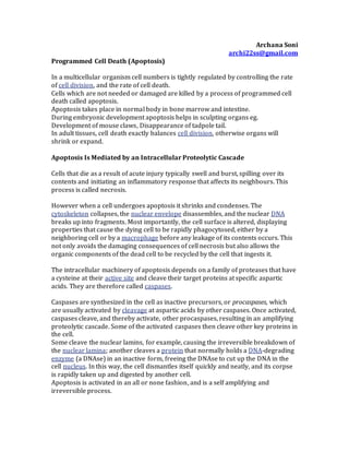
- 2. Figure: The caspase cascade involved in apoptosis
- 3. Procaspases Are Activated by Binding to Adaptor Proteins Cells contain inactive procaspases. Activation of procaspases depends on adaptor proteins that bring multiple copies of specific procaspases, known as initiator procaspases, close together in a complex or aggregate. In some cases the initiator procaspases have protease activity which help them cleave and activate each other. In other cases close packing of procaspases causes conformation change which help them activate other procaspases. Within moments, the activated caspase at the top of the cascade cleaves downstream procaspases to amplify the death signal and spread it throughout the cell. Procaspase activation can be triggered from outside the cell by the activation of death receptors on the cell surface. Killer lymphocytes, for example, can induce apoptosis by producing a protein called Fas ligand, which binds to the death receptor protein Fas on the surface of the target cell. The clustered Fas proteins then recruit intracellular adaptor proteins that bind and aggregate procaspase-8 molecules, which cleave and activate one another. The activated caspase-8 molecules then activate downstream procaspases to induce apoptosis. Some stressed or damaged cells kill themselves by producing both the Fas ligand and the Fas protein, thereby triggering an intracellular caspase cascade. Figure: Induction of apoptosis by either extracellular or intracellular stimuli
- 4. When cells are damaged or stressed, they can also kill themselves by triggering procaspase aggregation and activation from within the cell. In the best understood pathway, mitochondria are induced to release the electron carrier protein cytochrome c into the cytosol, where it binds and activates an adaptor protein called Apaf-1. This mitochondrial pathway of procaspase activation is recruited in most forms of apoptosis to initiate or to accelerate and amplify the caspase cascade. DNAdamage,can trigger apoptosis. This response usually requires p53, which can activate the transcription of genes that encode proteins that promote the release of cytochrome c from mitochondria. These proteins belong to the Bcl-2 family. Bcl-2 Family Proteins and IAP Proteins Are the Main Intracellular Regulators of the Cell Death Program Some Bcl family proteins like Bcl-2 inhibit apoptosis by blocking the release of cytochromes from mitochondria, while others activate apoptosis by activating procaspases. Bad proteins promote apoptosis by inactivating the death-inhibiting members of Bcl family. Bax and Bak, stimulate the release of cytochrome c from mitochondria and promote apoptosis. Another important family of intracellular apoptosis regulators is the IAP (inhibitor of apoptosis) family. These proteins are thought to inhibit apoptosis in two ways: they bind to some procaspases to prevent their activation, and they bind to caspases to inhibit their activity. When mitochondria release cytochromec to activate Apaf-1, they also release a protein that blocks IAPs, thereby greatly increasing the efficiency of the death activation process. The intracellular cell death program is also regulated by extracellular signals, which can either activate apoptosis or inhibit it. These signal molecules mainly act by regulating the levels or activity of members of the Bcl-2 and IAP families. Reference: Molecular Biology of the cell. Alberts B.